|
Non-X Histiocytoses
Non-X histiocytoses are a clinically well-defined group of cutaneous syndromes characterized by infiltrates of monocytes/macrophages, as opposed to X-type histiocytoses in which the infiltrates contain Langerhans cells. Conditions included in this group are: :* Juvenile xanthogranuloma :* Benign cephalic histiocytosis :* Generalized eruptive histiocytoma :* Xanthoma disseminatum :* Progressive nodular histiocytosis :* Papular xanthoma :* Hereditary progressive mucinous histiocytosis :* Reticulohistiocytosis :* Indeterminate cell histiocytosis :* Sea-blue histiocytosis :* Erdheim–Chester disease See also * X-type histiocytosis * Histiocytosis In medicine, histiocytosis is an excessive number of histiocytes (tissue macrophages), and the term is also often used to refer to a group of rare diseases which share this sign as a characteristic. Occasionally and confusingly, the term "histioc ... References Monocyte- and macrophage-related cutaneous conditions Histioc ... [...More Info...] [...Related Items...] OR: [Wikipedia] [Google] [Baidu] |
Monocytes
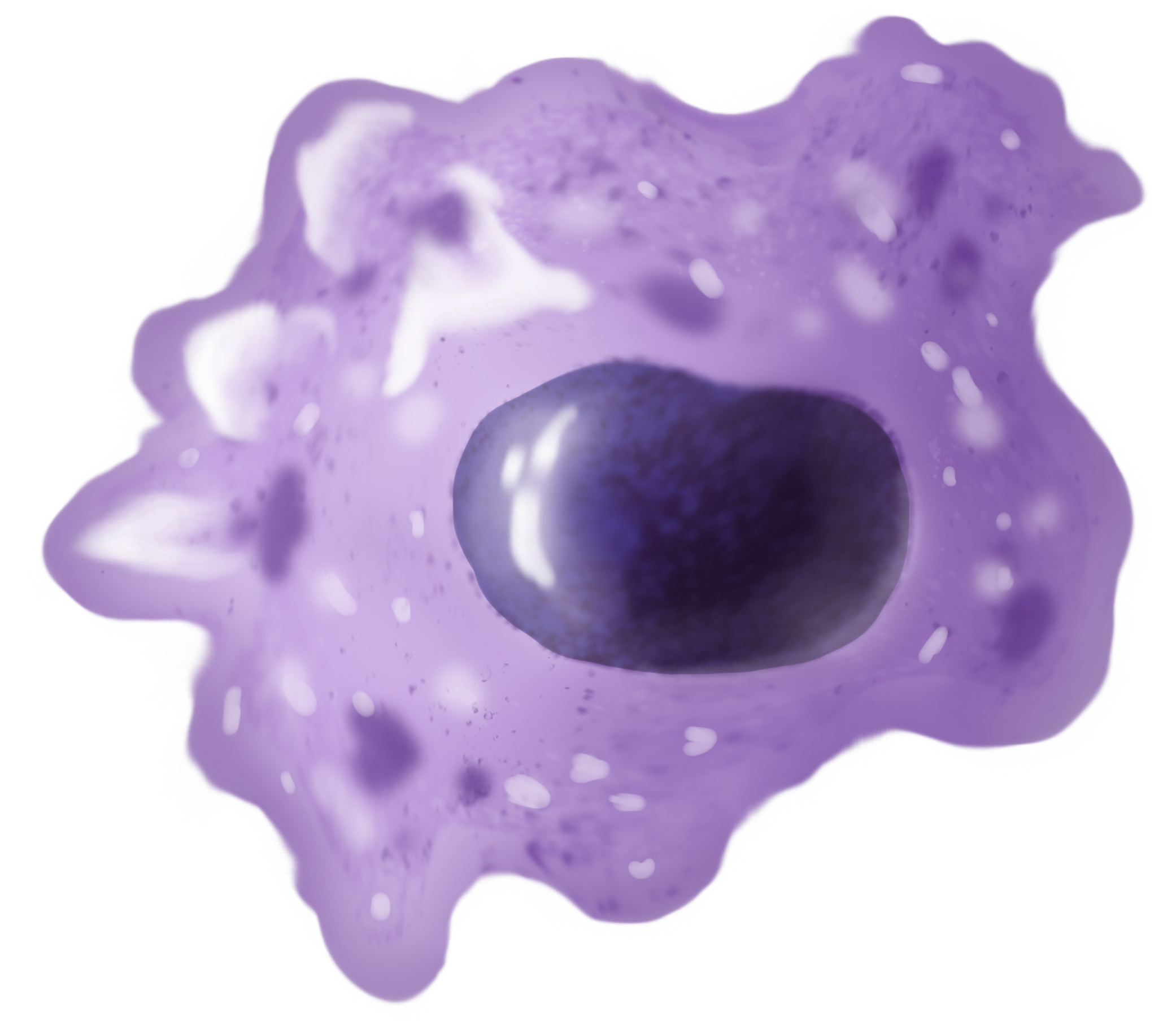
Monocytes are a type of leukocyte or white blood cell. They are the largest type of leukocyte in blood and can differentiate into macrophages and conventional dendritic cells. As a part of the vertebrate innate immune system monocytes also influence adaptive immune responses and exert tissue repair functions. There are at least three subclasses of monocytes in human blood based on their phenotypic receptors. Structure Monocytes are amoeboid in appearance, and have nongranulated cytoplasm. Thus they are classified as agranulocytes, although they might occasionally display some azurophil granules and/or vacuoles. With a diameter of 15–22 μm, monocytes are the largest cell type in peripheral blood. Monocytes are mononuclear cells and the ellipsoidal nucleus is often lobulated/indented, causing a bean-shaped or kidney-shaped appearance. Monocytes compose 2% to 10% of all leukocytes in the human body. Development Monocytes are produced by the bone marrow from precursors c ... [...More Info...] [...Related Items...] OR: [Wikipedia] [Google] [Baidu] |
Hereditary Progressive Mucinous Histiocytosis
Hereditary progressive mucinous histiocytosis is a very rare, benign, non-Langerhans' cell histiocytosis. An autosomal dominant or X-linked hereditary disease described on the skin, it has been found almost exclusively in women. One case of the disease in a male patient has been reported. See also * Non-X histiocytosis Non-X histiocytoses are a clinically well-defined group of cutaneous syndromes characterized by infiltrates of monocytes/macrophages, as opposed to X-type histiocytoses in which the infiltrates contain Langerhans cells. Conditions included in thi ... References External links Monocyte- and macrophage-related cutaneous conditions {{Cutaneous-condition-stub ... [...More Info...] [...Related Items...] OR: [Wikipedia] [Google] [Baidu] |
Histiocytosis
In medicine, histiocytosis is an excessive number of histiocytes (tissue macrophages), and the term is also often used to refer to a group of rare diseases which share this sign as a characteristic. Occasionally and confusingly, the term "histiocytosis" is sometimes used to refer to individual diseases. According to the Histiocytosis Association of America, 1 in 200,000 children in the United States are born with histiocytosis each year. HAA also states that most of the people diagnosed with histiocytosis are children under the age of 10, although the disease can afflict adults. The disease usually occurs from birth to age 15. Histiocytosis (and malignant histiocytosis) are both important in veterinary as well as human pathology. Diagnosis Histiocytosis is a rare disease, thus its diagnosis may be challenging. A variety of tests may be used, including: * Imaging ** CT scans of various organs such as lung, heart and kidneys. ** MRI of the brain, pituitary gland, heart, among ... [...More Info...] [...Related Items...] OR: [Wikipedia] [Google] [Baidu] |
X-type Histiocytosis
X-type histiocytoses are a clinically well-defined group of cutaneous syndromes characterized by infiltrates of Langerhans cells, as opposed to Non-X histiocytosis in which the infiltrates contain monocytes/macrophages. Conditions included in this group are: :* Congenital self-healing reticulohistiocytosis :* Langerhans cell histiocytosis See also * Non-X histiocytosis * Histiocytosis In medicine, histiocytosis is an excessive number of histiocytes (tissue macrophages), and the term is also often used to refer to a group of rare diseases which share this sign as a characteristic. Occasionally and confusingly, the term "histioc ... References Monocyte- and macrophage-related cutaneous conditions Histiocytosis {{Cutaneous-condition-stub ... [...More Info...] [...Related Items...] OR: [Wikipedia] [Google] [Baidu] |
Erdheim–Chester Disease
Erdheim–Chester disease (ECD) is an extremely rare disease characterized by the abnormal multiplication of a specific type of white blood cells called histiocytes, or tissue macrophages (technically, this disease is termed a non- Langerhans-cell histiocytosis). It was declared a histiocytic neoplasm by the World Health Organization in 2016. Onset typically is in middle age, although younger patients have been documented. The disease involves an infiltration of lipid-laden macrophages, multinucleated giant cells, an inflammatory infiltrate of lymphocytes and histiocytes in the bone marrow, and a generalized sclerosis of the long bones. Signs and symptoms Long bone involvement is almost universal in ECD patients and is bilateral and symmetrical in nature. More than 50% of cases have some sort of extraskeletal involvement. This can include kidney, skin, brain and lung involvement, and less frequently retroorbital tissue, pituitary gland and heart involvement is observed. Bon ... [...More Info...] [...Related Items...] OR: [Wikipedia] [Google] [Baidu] |
Sea-blue Histiocytosis
Sea-blue histiocytosis is a cutaneous condition that may occur as a familial inherited syndrome or as an acquired secondary or systemic infiltrative process. Causes It can be associated with the gene APOE. It can also be acquired. Sea-blue histiocyte syndrome is seen in patients receiving fat emulsion as a part of long-term parenteral nutrition (TPN) for intestinal failure. Pathophysiology The high lipid content in the blood leads to excessive cytoplasm loading of lipids within histiocytes. The subsequent incomplete degradation of these lipids leads to the formation of cytoplasmic lipid pigments. High lipid content may also cause membrane abnormality of the hemopoietic cells which is recognized by macrophages and therefore, increased accumulation within the bone marrow. These lipid laden histiocytes appear blue with May-Giemsa /PAS stain hence the name of Sea-Blue Histocyte Syndrome. Sea-blue histiocytosis is also seen in lipid disorders. Diagnosis See also * Non-X histioc ... [...More Info...] [...Related Items...] OR: [Wikipedia] [Google] [Baidu] |
Indeterminate Cell Histiocytosis
Indeterminate cell histiocytosis is a cutaneous condition felt to be caused by dermal precursors of Langerhans cells. See also * Non-X histiocytosis * List of cutaneous conditions Many skin conditions affect the human integumentary system—the organ system covering the entire surface of the body and composed of skin, hair, nails, and related muscle and glands. The major function of this system is as a barrier agai ... References External links Monocyte- and macrophage-related cutaneous conditions {{Cutaneous-condition-stub ... [...More Info...] [...Related Items...] OR: [Wikipedia] [Google] [Baidu] |
Reticulohistiocytosis
Reticulohistiocytosis is a cutaneous condition of which there are two distinct forms: :* Reticulohistiocytoma :* Multicentric reticulohistiocytosis See also * Non-X histiocytosis Non-X histiocytoses are a clinically well-defined group of cutaneous syndromes characterized by infiltrates of monocytes/macrophages, as opposed to X-type histiocytoses in which the infiltrates contain Langerhans cells. Conditions included in thi ... References Monocyte- and macrophage-related cutaneous conditions {{Cutaneous-condition-stub ... [...More Info...] [...Related Items...] OR: [Wikipedia] [Google] [Baidu] |
Papular Xanthoma
Papular xanthoma is a cutaneous condition that is a rare form of non-X histiocytosis. See also * Non-X histiocytosis * List of cutaneous conditions Many skin conditions affect the human integumentary system—the organ system covering the entire surface of the body and composed of skin, hair, nails, and related muscle and glands. The major function of this system is as a barrier agai ... References Monocyte- and macrophage-related cutaneous conditions {{Cutaneous-condition-stub ... [...More Info...] [...Related Items...] OR: [Wikipedia] [Google] [Baidu] |
Macrophages
Macrophages (abbreviated as M φ, MΦ or MP) ( el, large eaters, from Greek ''μακρός'' (') = large, ''φαγεῖν'' (') = to eat) are a type of white blood cell of the immune system that engulfs and digests pathogens, such as cancer cells, microbes, cellular debris, and foreign substances, which do not have proteins that are specific to healthy body cells on their surface. The process is called phagocytosis, which acts to defend the host against infection and injury. These large phagocytes are found in essentially all tissues, where they patrol for potential pathogens by amoeboid movement. They take various forms (with various names) throughout the body (e.g., histiocytes, Kupffer cells, alveolar macrophages, microglia, and others), but all are part of the mononuclear phagocyte system. Besides phagocytosis, they play a critical role in nonspecific defense (innate immunity) and also help initiate specific defense mechanisms (adaptive immunity) by recruiting other immune ... [...More Info...] [...Related Items...] OR: [Wikipedia] [Google] [Baidu] |
Progressive Nodular Histiocytosis
Progressive nodular histiocytosis is a cutaneous condition clinically characterized by the development of two types of skin lesions: superficial papules and deeper larger subcutaneous nodules. See also * Non-X histiocytosis Non-X histiocytoses are a clinically well-defined group of cutaneous syndromes characterized by infiltrates of monocytes/macrophages, as opposed to X-type histiocytoses in which the infiltrates contain Langerhans cells. Conditions included in thi ... References Monocyte- and macrophage-related cutaneous conditions {{Cutaneous-condition-stub ... [...More Info...] [...Related Items...] OR: [Wikipedia] [Google] [Baidu] |
Xanthoma Disseminatum
Xanthoma disseminatum is a rare cutaneous condition that preferentially affects males in childhood, characterized by the insidious onset of small, yellow-red to brown papules and nodules that are discrete and disseminated. It is a histiocytosis syndrome. See also * Non-X histiocytosis Non-X histiocytoses are a clinically well-defined group of cutaneous syndromes characterized by infiltrates of monocytes/macrophages, as opposed to X-type histiocytoses in which the infiltrates contain Langerhans cells. Conditions included in thi ... * List of cutaneous conditions References External links Monocyte- and macrophage-related cutaneous conditions {{Cutaneous-condition-stub ... [...More Info...] [...Related Items...] OR: [Wikipedia] [Google] [Baidu] |
.png)
.jpg)
