|
Neurokinin B
Neurokinin B (NKB) belongs in the family of tachykinin peptides. Neurokinin B is implicated in a variety of human functions and pathways such as the secretion of gonadotropin-releasing hormone. Additionally, NKB is associated with pregnancy in females and maturation in young adults. Reproductive function is highly dependent on levels of both neurokinin B and also the G-protein coupled receptor ligand kisspeptin. The first NKB studies done attempted to resolve why high levels of the peptide may be implicated in pre-eclampsia during pregnancy. NKB, kisspeptin, and dynorphin together are found in the arcuate nucleus (ARC) known as the KNDy subpopulation. This subpopulation is targeted by many steroid hormones and works to form a network that feeds back to GnRH pulse generator. Synthesis Neurokinin B is found in humans as a ten-peptide chain (decapeptide) attached to a terminal amide group. The peptide formula is H-Asp-Met-His-Asp-Phe-Phe-Val-Gly-Leu-Met-NH2 (DMHDFFVGLM-NH2). Neuroki ... [...More Info...] [...Related Items...] OR: [Wikipedia] [Google] [Baidu] |
Tachykinin Peptides
Tachykinin peptides are one of the largest families of neuropeptides, found from amphibians to mammals. They were so named due to their ability to rapidly induce contraction of gut tissue. The tachykinin family is characterized by a common C-terminal sequence, Phe-X-Gly- Leu- Met-NH2, where X is either an Aromatic or an Aliphatic amino acid. The genes that produce tachykinins encode precursor proteins called preprotachykinins, which are chopped apart into smaller peptides by posttranslational proteolytic processing. The genes also code for multiple splice forms that are made up of different sets of peptides. Tachykinins excite neurons, evoke behavioral responses, are potent vasodilators, and contract (directly or indirectly) many smooth muscles. Tachykinins are from ten to twelve residues long. The two human tachykinin genes are called TAC1 and TAC3 for historical reasons, and are equivalent to Tac1 and Tac2 of the mouse, respectively. TAC1 encodes neurokinin A (formerly kno ... [...More Info...] [...Related Items...] OR: [Wikipedia] [Google] [Baidu] |
Spinal Cord
The spinal cord is a long, thin, tubular structure made up of nervous tissue, which extends from the medulla oblongata in the brainstem to the lumbar region of the vertebral column (backbone). The backbone encloses the central canal of the spinal cord, which contains cerebrospinal fluid. The brain and spinal cord together make up the central nervous system (CNS). In humans, the spinal cord begins at the occipital bone, passing through the foramen magnum and then enters the spinal canal at the beginning of the cervical vertebrae. The spinal cord extends down to between the first and second lumbar vertebrae, where it ends. The enclosing bony vertebral column protects the relatively shorter spinal cord. It is around long in adult men and around long in adult women. The diameter of the spinal cord ranges from in the cervical and lumbar regions to in the thoracic area. The spinal cord functions primarily in the transmission of nerve signals from the motor cortex to the ... [...More Info...] [...Related Items...] OR: [Wikipedia] [Google] [Baidu] |
Ovariectomized Rat
An ovariectomized rodent (OVX) is a female rat or mouse whose ovaries have been removed. Currently there is no single animal model that identically represents the stages of osteoporosis in humans although there are some animals that are relatively close and can be used for the purpose of comparison. Both small animals and large animals are used depending on which aspects of the osteoporotic condition are being studied. Such animals include rats, rabbits, and sheep. Of these animal models, the ovariectomised rat model remains the most popular choice as it has been validated to represent the most important clinical features of estrogen deficiency-induced (or postmenopausal) bone loss in the adult human, particularly during the early stages of osteoporosis. These include: increased rate of bone turnover with resorption exceeding formation; an initial rapid phase of bone loss followed by a much slower phase; greater loss of cancellous bone than cortical bone; reduced intestinal calcium ... [...More Info...] [...Related Items...] OR: [Wikipedia] [Google] [Baidu] |
Animal Testing On Rodents
Rodents are commonly used in animal testing, particularly mice and rats, but also guinea pigs, hamsters, gerbils and others. Mice are the most commonly used vertebrate species, due to their availability, size, low cost, ease of handling, and fast reproduction rate. __TOC__ Statistics In the UK in 2015, there were 3.33 million procedures on rodents (80% of total procedures that year). The most common species used were mice (3.03 million procedures, or 73% of total) and rats (268,522, or 6.5%). Other rodents species included guinea pigs (21,831 / 0.7%), hamsters (1,500 / 0.04%) and gerbils (278 / 0.01%). In the U.S., the numbers of rats and mice used are not reported, but estimates range from around 11 million to approximately 100 million. In 2000, the Federal Research Division, Library of Congress, published the results of an analysis of its Rats/Mice/and Birds Database: Researchers, Breeders, Transporters, and Exhibitors. Rodent types Mice Mice are the most commonly used ... [...More Info...] [...Related Items...] OR: [Wikipedia] [Google] [Baidu] |
Trophoblast
The trophoblast (from Greek : to feed; and : germinator) is the outer layer of cells of the blastocyst. Trophoblasts are present four days after fertilization in humans. They provide nutrients to the embryo and develop into a large part of the placenta. They form during the first stage of pregnancy and are the first cells to differentiate from the fertilized egg to become extraembryonic structures that do not directly contribute to the embryo. After gastrulation, the trophoblast is contiguous with the ectoderm of the embryo and is referred to as the trophectoderm. After the first differentiation, the cells in the human embryo lose their totipotency and are no longer totipotent stem cells because they cannot form a trophoblast. They are now pluripotent stem cells. Structure The trophoblast proliferates and differentiates into two cell layers at approximately six days after fertilization for humans. Function Trophoblasts are specialized cells of the placenta that play ... [...More Info...] [...Related Items...] OR: [Wikipedia] [Google] [Baidu] |
Hypertrophic Decidual Vasculopathy High Mag
Hypertrophy is the increase in the volume of an organ or tissue due to the enlargement of its component cells. It is distinguished from hyperplasia, in which the cells remain approximately the same size but increase in number.Updated by Linda J. Vorvick. 8/14/1Hyperplasia/ref> Although hypertrophy and hyperplasia are two distinct processes, they frequently occur together, such as in the case of the hormonally-induced proliferation and enlargement of the cells of the uterus during pregnancy. Eccentric hypertrophy is a type of hypertrophy where the walls and chamber of a hollow organ undergo growth in which the overall size and volume are enlarged. It is applied especially to the left ventricle of heart. Sarcomeres are added in series, as for example in dilated cardiomyopathy (in contrast to hypertrophic cardiomyopathy, a type of concentric hypertrophy, where sarcomeres are added in parallel). Gallery File:*+ * Photographic documentation on sexual education - Hypertrophy of brea ... [...More Info...] [...Related Items...] OR: [Wikipedia] [Google] [Baidu] |
Placenta
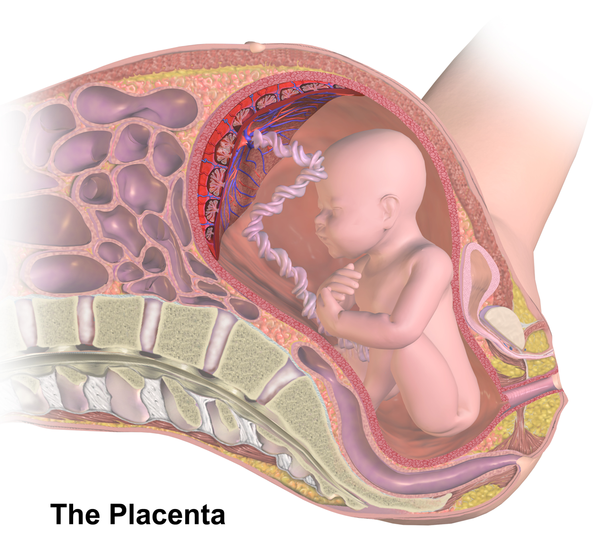
The placenta is a temporary embryonic and later fetal organ that begins developing from the blastocyst shortly after implantation. It plays critical roles in facilitating nutrient, gas and waste exchange between the physically separate maternal and fetal circulations, and is an important endocrine organ, producing hormones that regulate both maternal and fetal physiology during pregnancy. The placenta connects to the fetus via the umbilical cord, and on the opposite aspect to the maternal uterus in a species-dependent manner. In humans, a thin layer of maternal decidual ( endometrial) tissue comes away with the placenta when it is expelled from the uterus following birth (sometimes incorrectly referred to as the 'maternal part' of the placenta). Placentas are a defining characteristic of placental mammals, but are also found in marsupials and some non-mammals with varying levels of development. Mammalian placentas probably first evolved about 150 million to 200 millio ... [...More Info...] [...Related Items...] OR: [Wikipedia] [Google] [Baidu] |
Amniotic Fluid
The amniotic fluid is the protective liquid contained by the amniotic sac of a gravid amniote. This fluid serves as a cushion for the growing fetus, but also serves to facilitate the exchange of nutrients, water, and biochemical products between mother and fetus. For humans, the amniotic fluid is commonly called water or waters (Latin liquor amnii). Development Amniotic fluid is present from the formation of the gestational sac. Amniotic fluid is in the amniotic sac. It is generated from maternal plasma, and passes through the fetal membranes by osmotic and hydrostatic forces. When fetal kidneys begin to function around week 16, fetal urine also contributes to the fluid. In earlier times, it was believed that the amniotic fluid was composed entirely of fetal urine. The fluid is absorbed through the fetal tissue and skin. After 22 to 25 week of pregnancy, keratinization of an embryo's skin occurs. When this process completes around the 25th week, the fluid is primarily absor ... [...More Info...] [...Related Items...] OR: [Wikipedia] [Google] [Baidu] |
Pre-eclampsia
Pre-eclampsia is a disorder of pregnancy characterized by the onset of high blood pressure and often a significant amount of protein in the urine. When it arises, the condition begins after 20 weeks of pregnancy. In severe cases of the disease there may be red blood cell breakdown, a thrombocytopenia, low blood platelet count, impaired liver function, kidney dysfunction, edema, swelling, pulmonary edema, shortness of breath due to fluid in the lungs, or visual disturbances. Pre-eclampsia increases the risk of undesirable outcomes for both the mother and the fetus. If left untreated, it may result in seizures at which point it is known as eclampsia. Risk factors for pre-eclampsia include obesity, prior hypertension, older age, and diabetes mellitus. It is also more frequent in a woman's first pregnancy and if she is carrying twins. The underlying mechanism involves abnormal placentation, formation of blood vessels in the placenta amongst other factors. Most cases are diagnosed ... [...More Info...] [...Related Items...] OR: [Wikipedia] [Google] [Baidu] |
Androgen Receptor
The androgen receptor (AR), also known as NR3C4 (nuclear receptor subfamily 3, group C, member 4), is a type of nuclear receptor that is activated by binding any of the androgenic hormones, including testosterone and dihydrotestosterone in the cytoplasm and then translocating into the nucleus. The androgen receptor is most closely related to the progesterone receptor, and progestins in higher dosages can block the androgen receptor. The main function of the androgen receptor is as a DNA-binding transcription factor that regulates gene expression; however, the androgen receptor has other functions as well. Androgen-regulated genes are critical for the development and maintenance of the male sexual phenotype. Function Effect on development In some cell types, testosterone interacts directly with androgen receptors, whereas, in others, testosterone is converted by 5-alpha-reductase to dihydrotestosterone, an even more potent agonist for androgen receptor activatio ... [...More Info...] [...Related Items...] OR: [Wikipedia] [Google] [Baidu] |
Progesterone Receptor
The progesterone receptor (PR), also known as NR3C3 or nuclear receptor subfamily 3, group C, member 3, is a protein found inside cells. It is activated by the steroid hormone progesterone. In humans, PR is encoded by a single ''PGR'' gene residing on chromosome 11q22, it has two isoforms, PR-A and PR-B, that differ in their molecular weight. The PR-B is the positive regulator of the effects of progesterone, while PR-A serve to antagonize the effects of PR-B. Mechanism Progesterone is necessary to induce the progesterone receptors. When no binding hormone is present the carboxyl terminal inhibits transcription. Binding to a hormone induces a structural change that removes the inhibitory action. Progesterone antagonists prevent the structural reconfiguration. After progesterone binds to the receptor, restructuring with dimerization follows and the complex enters the nucleus and binds to DNA. There transcription takes place, resulting in formation of messenger RNA that is t ... [...More Info...] [...Related Items...] OR: [Wikipedia] [Google] [Baidu] |
Estrogen Receptor
Estrogen receptors (ERs) are a group of proteins found inside cells. They are receptors that are activated by the hormone estrogen (17β-estradiol). Two classes of ER exist: nuclear estrogen receptors ( ERα and ERβ), which are members of the nuclear receptor family of intracellular receptors, and membrane estrogen receptors (mERs) ( GPER (GPR30), ER-X, and Gq-mER), which are mostly G protein-coupled receptors. This article refers to the former (ER). Once activated by estrogen, the ER is able to translocate into the nucleus and bind to DNA to regulate the activity of different genes (i.e. it is a DNA-binding transcription factor). However, it also has additional functions independent of DNA binding. As hormone receptors for sex steroids ( steroid hormone receptors), ERs, androgen receptors (ARs), and progesterone receptors (PRs) are important in sexual maturation and gestation. Proteomics There are two different forms of the estrogen receptor, usually referred to as α ... [...More Info...] [...Related Items...] OR: [Wikipedia] [Google] [Baidu] |


