|
Neurophilic
The development of the nervous system in humans, or neural development, or neurodevelopment involves the studies of embryology, developmental biology, and neuroscience. These describe the cellular and molecular mechanisms by which the complex nervous system forms in humans, develops during prenatal development, and continues to develop postnatally. Some landmarks of neural development in the embryo include: # The formation and differentiation of neurons from stem cell precursors (neurogenesis) # The migration of immature neurons from their birthplaces in the embryo to their final positions. # The outgrowth of axons from neurons and the guidance of the motile growth cone through the embryo towards postsynaptic partners. # The generation of synapses between axons and their postsynaptic partners. # The synaptic pruning that occurs in adolescence. # The lifelong changes in synapses which are thought to underlie learning and memory. Typically, these neurodevelopmental processes ... [...More Info...] [...Related Items...] OR: [Wikipedia] [Google] [Baidu] |
Cellular Migration
Cell migration is a central process in the development and maintenance of multicellular organisms. Tissue formation during embryonic development, wound healing and immune responses all require the orchestrated movement of cells in particular directions to specific locations. Cells often migrate in response to specific external signals, including chemical signals and mechanical signals. Errors during this process have serious consequences, including intellectual disability, vascular disease, tumor formation and metastasis. An understanding of the mechanism by which cells migrate may lead to the development of novel therapeutic strategies for controlling, for example, invasive tumour cells. Due to the highly viscous environment (low Reynolds number), cells need to continuously produce forces in order to move. Cells achieve active movement by very different mechanisms. Many less complex prokaryotic organisms (and sperm cells) use flagella or cilia to propel themselves. Eukaryo ... [...More Info...] [...Related Items...] OR: [Wikipedia] [Google] [Baidu] |
Embryology
Embryology (from Ancient Greek, Greek ἔμβρυον, ''embryon'', "the unborn, embryo"; and -λογία, ''-logy, -logia'') is the branch of animal biology that studies the Prenatal development (biology), prenatal development of gametes (sex cells), fertilization, and development of embryos and fetuses. Additionally, embryology encompasses the study of congenital disorders that occur before birth, known as teratology. Early embryology was proposed by Marcello Malpighi, and known as preformationism, the theory that organisms develop from pre-existing miniature versions of themselves. Aristotle proposed the theory that is now accepted, Epigenesis (biology), epigenesis. Epigenesis (biology), Epigenesis is the idea that organisms develop from seed or egg in a sequence of steps. Modern embryology developed from the work of Karl Ernst von Baer, though accurate observations had been made in Italy by anatomists such as Aldrovandi and Leonardo da Vinci in the Renaissance. Comparative ... [...More Info...] [...Related Items...] OR: [Wikipedia] [Google] [Baidu] |
Synaptic Plasticity
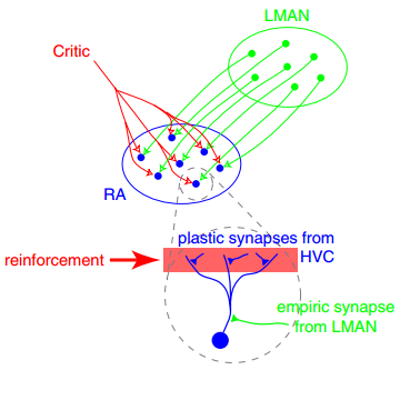
In neuroscience, synaptic plasticity is the ability of synapses to Chemical synapse#Synaptic strength, strengthen or weaken over time, in response to increases or decreases in their activity. Since memory, memories are postulated to be represented by vastly interconnected neural circuits in the brain, synaptic plasticity is one of the important neurochemical foundations of learning and memory (''see Hebbian theory''). Plastic change often results from the alteration of the number of neurotransmitter receptors located on a synapse. There are several underlying mechanisms that cooperate to achieve synaptic plasticity, including changes in the quantity of neurotransmitters released into a synapse and changes in how effectively cells respond to those neurotransmitters. Synaptic plasticity in both Excitatory synapse, excitatory and Inhibitory synapse, inhibitory synapses has been found to be dependent upon postsynaptic calcium release. Historical discoveries In 1973, Terje Lømo and ... [...More Info...] [...Related Items...] OR: [Wikipedia] [Google] [Baidu] |
Prosencephalon
In the anatomy of the brain of vertebrates, the forebrain or prosencephalon is the rostral (forward-most) portion of the brain. The forebrain controls body temperature, reproductive functions, eating, sleeping, and the display of emotions. Vesicles of the forebrain (prosencephalon), the midbrain (mesencephalon), and hindbrain (rhombencephalon) are the three primary brain vesicles during the early development of the nervous system. At the five-vesicle stage, the forebrain separates into the diencephalon (thalamus, hypothalamus, subthalamus, and epithalamus) and the telencephalon which develops into the cerebrum. The cerebrum consists of the cerebral cortex, underlying white matter, and the basal ganglia. In humans, by 5 weeks in utero it is visible as a single portion toward the front of the fetus. At 8 weeks in utero, the forebrain splits into the left and right cerebral hemispheres. When the embryonic forebrain fails to divide the brain into two lobes, it results in a ... [...More Info...] [...Related Items...] OR: [Wikipedia] [Google] [Baidu] |
Brain Vesicle
Brain vesicles are the bulge-like enlargements of the early development of the neural tube in vertebrates, which eventually give rise to the brain. Vesicle formation begins shortly after the rostral closure of the neural tube, at about embryonic day 9.0 in mice, or the fourth and fifth gestational week in humans. In zebrafish and chicken embryos, brain vesicles form by about 24 hours and 48 hours post- conception, respectively. Initially there are three primary brain vesicles: prosencephalon (i.e. forebrain), mesencephalon (i.e. midbrain) and rhombencephalon (i.e. hindbrain). These develop into five secondary brain vesicles – the prosencephalon is subdivided into the telencephalon and diencephalon, and the rhombencephalon into the metencephalon and myelencephalon. During these early vesicle stages, the walls of the neural tube contain neural stem cells in a region called the neuroepithelium or ventricular zone. These neural stem cells divide rapidly, driving grow ... [...More Info...] [...Related Items...] OR: [Wikipedia] [Google] [Baidu] |
Cerebrospinal Fluid
Cerebrospinal fluid (CSF) is a clear, colorless Extracellular fluid#Transcellular fluid, transcellular body fluid found within the meninges, meningeal tissue that surrounds the vertebrate brain and spinal cord, and in the ventricular system, ventricles of the brain. CSF is mostly produced by specialized Ependyma, ependymal cells in the choroid plexuses of the ventricles of the brain, and absorbed in the arachnoid granulations. It is also produced by ependymal cells in the lining of the ventricles. In humans, there is about 125 mL of CSF at any one time, and about 500 mL is generated every day. CSF acts as a shock absorber, cushion or buffer, providing basic mechanical and immune system, immunological protection to the brain inside the Human skull, skull. CSF also serves a vital function in the cerebral autoregulation of cerebral blood flow. CSF occupies the subarachnoid space (between the arachnoid mater and the pia mater) and the ventricular system around and inside t ... [...More Info...] [...Related Items...] OR: [Wikipedia] [Google] [Baidu] |
Neural Tube
In the developing chordate (including vertebrates), the neural tube is the embryonic precursor to the central nervous system, which is made up of the brain and spinal cord. The neural groove gradually deepens as the neural folds become elevated, and ultimately the folds meet and coalesce in the middle line and convert the groove into the closed neural tube. In humans, neural tube closure usually occurs by the fourth week of pregnancy (the 28th day after conception). Development The neural tube develops in two ways: primary neurulation and secondary neurulation. Primary neurulation divides the ectoderm into three cell types: * The internally located neural tube * The externally located epidermis * The neural crest cells, which develop in the region between the neural tube and epidermis but then migrate to new locations # Primary neurulation begins after the neural plate forms. The edges of the neural plate start to thicken and lift upward, forming the neural folds. The center ... [...More Info...] [...Related Items...] OR: [Wikipedia] [Google] [Baidu] |
Neural Plate
In embryology, the neural plate is a key Development of the human body, developmental structure that serves as the basis for the nervous system. Cranial to the primitive node of the embryonic primitive streak, Ectoderm, ectodermal tissue thickens and flattens to become the neural plate. The region anterior to the primitive node can be generally referred to as the neural plate. Cells take on a columnar appearance in the process as they continue to lengthen and narrow. The ends of the neural plate, known as the neural folds, push the ends of the plate up and together, folding into the neural tube, a structure critical to brain and spinal cord development. This process as a whole is termed primary neurulation. Signaling proteins are also important in neural plate development, and aid in Cellular differentiation, differentiating the tissue destined to become the neural plate. Examples of such proteins include bone morphogenetic proteins and cadherins. Expression of these proteins i ... [...More Info...] [...Related Items...] OR: [Wikipedia] [Google] [Baidu] |
Neuroectoderm
Neuroectoderm (or neural ectoderm or neural tube epithelium) consists of cells derived from the ectoderm. Formation of the neuroectoderm is the first step in the development of the nervous system. The neuroectoderm receives bone morphogenetic protein-inhibiting signals from proteins such as noggin, which leads to the development of the nervous system from this tissue. Histologically, these cells are classified as pseudostratified columnar cells. After recruitment from the ectoderm, the neuroectoderm undergoes three stages of development: transformation into the neural plate, transformation into the neural groove (with associated neural folds), and transformation into the neural tube. After formation of the tube, the brain forms into three sections; the hindbrain, the midbrain, and the forebrain. The types of neuroectoderm include: * Neural crest ** pigment cells in the skin **ganglia of the autonomic nervous system ** dorsal root ganglia. **facial cartilage ** aorticopulm ... [...More Info...] [...Related Items...] OR: [Wikipedia] [Google] [Baidu] |
Human Embryonic Development
Human embryonic development or human embryogenesis is the development and formation of the human embryo. It is characterised by the processes of cell division and cellular differentiation of the embryo that occurs during the early stages of development. In biological terms, the development of the human body entails growth from a one-celled zygote to an adult human being. Fertilization occurs when the sperm cell successfully enters and fuses with an egg cell (ovum). The genetic material of the sperm and egg then combine to form the single cell zygote and the germinal stage of development commences. Human embryonic development covers the first eight weeks of development, which have 23 stages, called Carnegie stages. At the beginning of the ninth week, the embryo is termed a fetus (spelled "foetus" in British English). In comparison to the embryo, the fetus has more recognizable external features and a more complete set of developing organs. Human embryology is the study o ... [...More Info...] [...Related Items...] OR: [Wikipedia] [Google] [Baidu] |
Germ Layer
A germ layer is a primary layer of cell (biology), cells that forms during embryonic development. The three germ layers in vertebrates are particularly pronounced; however, all eumetazoans (animals that are sister taxa to the sponges) produce two or three primary germ layers. Some animals, like cnidarians, produce two germ layers (the ectoderm and endoderm) making them diploblastic. Other animals such as bilaterians produce a third layer (the mesoderm) between these two layers, making them triploblastic. Germ layers eventually give rise to all of an animal's Tissue (biology), tissues and organ (anatomy), organs through the process of organogenesis. History Caspar Friedrich Wolff observed organization of the early embryo in leaf-like layers. In 1817, Heinz Christian Pander discovered three primordial germ layers while studying chick embryos. Between 1850 and 1855, Robert Remak had further refined the germ cell layer (''Keimblatt'') concept, stating that the external, internal ... [...More Info...] [...Related Items...] OR: [Wikipedia] [Google] [Baidu] |


