|
Lymphangiomatosis
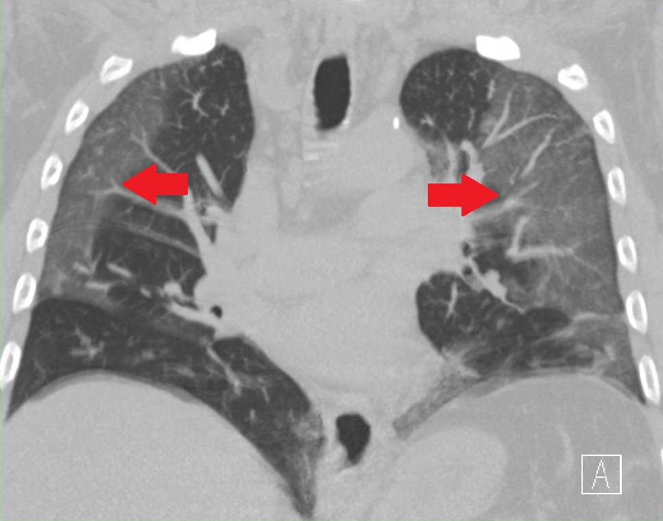
Lymphangiomatosis is a condition where a lymphangioma is not present in a single localised mass, but in a widespread or multifocal manner. It is a rare type of tumor which results from an abnormal development of the lymphatic system. It is thought to be the result of congenital errors of lymphatic development occurring prior to the 20th week of gestation. Lymphangiomatosis is a condition marked by the presence of cysts that result from an increase both in the size and number of thin-walled lymphatic channels that are abnormally interconnected and dilated.Pernick, Nat. "Soft Tissue Tumors Part 3 Muscle, Vascular, Nerve, Other Lymphangiomatosis." PathologyOutlines.com. PathologyOutlines.com, Inc., 10/17/2009. Web. 6 Sep 2011. http://www.pathologyoutlines.com/topic/softtissue3lymphangiomatosis.html. 75% of cases involve multiple organs. It typically presents by age 20 and, although it is technically benign, these deranged lymphatics tend to invade surrounding tissues and cause problem ... [...More Info...] [...Related Items...] OR: [Wikipedia] [Google] [Baidu] |
Lymphatic System
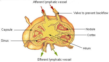
The lymphatic system, or lymphoid system, is an organ system in vertebrates that is part of the immune system, and complementary to the circulatory system. It consists of a large network of lymphatic vessels, lymph nodes, lymphatic or lymphoid organs, and lymphoid tissues. The vessels carry a clear fluid called lymph (the Latin word ''lympha'' refers to the deity of fresh water, "Lympha") back towards the heart, for re-circulation. Unlike the circulatory system that is a closed system, the lymphatic system is open. The human circulatory system processes an average of 20 litres of blood per day through capillary filtration, which removes plasma from the blood. Roughly 17 litres of the filtered blood is reabsorbed directly into the blood vessels, while the remaining three litres are left in the interstitial fluid. One of the main functions of the lymphatic system is to provide an accessory return route to the blood for the surplus three litres. The other main function is that ... [...More Info...] [...Related Items...] OR: [Wikipedia] [Google] [Baidu] |
Lymphangioma
Lymphangiomas are malformations of the lymphatic system characterized by lesions that are thin-walled cysts; these cysts can be macroscopic, as in a cystic hygroma, or microscopic. The lymphatic system is the network of vessels responsible for returning to the venous system excess fluid from tissues as well as the lymph nodes that filter this fluid for signs of pathogens. These malformations can occur at any age and may involve any part of the body, but 90% occur in children less than 2 years of age and involve the head and neck. These malformations are either congenital or acquired. Congenital lymphangiomas are often associated with chromosomal abnormalities such as Turner syndrome, although they can also exist in isolation. Lymphangiomas are commonly diagnosed before birth using fetal ultrasonography. Acquired lymphangiomas may result from trauma, inflammation, or lymphatic obstruction. Most lymphangiomas are benign lesions that result only in a soft, slow-growing, "doughy" ... [...More Info...] [...Related Items...] OR: [Wikipedia] [Google] [Baidu] |
Tumor
A neoplasm () is a type of abnormal and excessive growth of tissue. The process that occurs to form or produce a neoplasm is called neoplasia. The growth of a neoplasm is uncoordinated with that of the normal surrounding tissue, and persists in growing abnormally, even if the original trigger is removed. This abnormal growth usually forms a mass, when it may be called a tumor. ICD-10 classifies neoplasms into four main groups: benign neoplasms, in situ neoplasms, malignant neoplasms, and neoplasms of uncertain or unknown behavior. Malignant neoplasms are also simply known as cancers and are the focus of oncology. Prior to the abnormal growth of tissue, as neoplasia, cells often undergo an abnormal pattern of growth, such as metaplasia or dysplasia. However, metaplasia or dysplasia does not always progress to neoplasia and can occur in other conditions as well. The word is from Ancient Greek 'new' and 'formation, creation'. Types A neoplasm can be benign, potentially m ... [...More Info...] [...Related Items...] OR: [Wikipedia] [Google] [Baidu] |
Gorham's Disease
Gorham's disease (pronounced GOR-amz), also known as Gorham vanishing bone disease and phantom bone disease, is a very rare skeletal condition of unknown cause, characterized by the uncontrolled proliferation of distended, thin-walled vascular or lymphatic channels within bone, which leads to resorption and replacement of bone with angiomas and/or fibrosis.Gorham LW, Stout AP. Massive osteolysis (acute spontaneous absorption of bone, phantom bone, disappearing bone): its relation to hemangiomatosis. J Bone Joint Surg m1955;37-A:985-1004.Ross JL., Schinella R., and Shenkman L. Massive osteolysis: An unusual cause of bone destruction. The American Journal of Medicine 1978; 65(2): 367-372. Signs and symptoms The symptoms of Gorham's disease vary depending on the bones involved. It may affect any part of the skeleton, but the most common sites of disease are the shoulder, skull, pelvic girdle, jaw, ribs, and spine.MÖLLER, G., Priemel, M., Amling, M. Werner, M., Kuhlmey, A. S., D ... [...More Info...] [...Related Items...] OR: [Wikipedia] [Google] [Baidu] |
Chylothorax
A chylothorax is an abnormal accumulation of chyle, a type of lipid-rich lymph, in the space surrounding the lung. The lymphatics of the digestive system normally returns lipids absorbed from the small bowel via the thoracic duct, which ascends behind the esophagus to drain into the left brachiocephalic vein. If normal thoracic duct drainage is disrupted, either due to obstruction or rupture, chyle can leak and accumulate within the negative-pressured pleural space. In people on a normal diet, this fluid collection can sometimes be identified by its turbid, milky white appearance, since chyle contains emulsified triglycerides. Chylothorax is a rare but serious condition, as it signals leakage of the thoracic duct or one of its tributaries. There are many treatments, both surgical and conservative. About 2–3% of all fluid collections surrounding the lungs (pleural effusions) are chylothoraces. It is important to distinguish a chylothorax from a pseudochylothorax (a ... [...More Info...] [...Related Items...] OR: [Wikipedia] [Google] [Baidu] |
Lymphangiectasis
Lymphangiectasia, also known as "lymphangiectasis", is a pathologic dilation of lymph vessels.McGavin/ Zachary (2007), Pathologic Basis of Veterinary Disease When it occurs in the intestines of dogs, and more rarely humans, it causes a disease known as "intestinal lymphangiectasia". This disease is characterized by lymphatic vessel dilation, chronic diarrhea and loss of proteins such as serum albumin and globulin. It is considered to be a chronic form of protein-losing enteropathy. Signs and symptoms Chronic diarrhea is almost always seen with lymphangiectasia, but most other signs are linked to low blood protein levels (hypoproteinemia), which causes low oncotic pressure. These signs include ascites, pleural effusion, and edema of the limbs and trunk. Weight loss is seen with long-term disease. Cause Biopsy of the small intestine shows dilation of the lacteals of the villi and distension of the lymphatic vessels. Reduced lymph flow leads to a malabsorption syndrome of the ... [...More Info...] [...Related Items...] OR: [Wikipedia] [Google] [Baidu] |
Lymphangioleiomyomatosis
Lymphangioleiomyomatosis (LAM) is a rare, progressive and systemic disease that typically results in cystic lung destruction. It predominantly affects women, especially during childbearing years. The term sporadic LAM is used for patients with LAM not associated with tuberous sclerosis complex (TSC), while TSC-LAM refers to LAM that is associated with TSC. Signs and symptoms The average age of onset is the early to mid 30s. Exertional dyspnea (shortness of breath) and spontaneous pneumothorax (lung collapse) have been reported as the initial presentation of the disease in 49% and 46% of patients, respectively. Diagnosis is typically delayed 5 to 6 years. The condition is often misdiagnosed as asthma or chronic obstructive pulmonary disease. The first pneumothorax, or lung collapse, precedes the diagnosis of LAM in 82% of patients. The consensus clinical definition of LAM includes multiple symptoms: * Fatigue * Cough * Coughing up blood (rarely massive) * Chest pain * Chylous com ... [...More Info...] [...Related Items...] OR: [Wikipedia] [Google] [Baidu] |
Pulmonary Capillary Hemangiomatosis
Pulmonary capillary hemangiomatosis (PCH) is a disease affecting the blood vessels of the lungs, where abnormal capillary proliferation and venous fibrous intimal thickening result in progressive increase in vascular resistance. It is a rare cause of pulmonary hypertension, and occurs predominantly in young adults. Together with pulmonary veno-occlusive disease, PCH comprises WHO Group I' causes for pulmonary hypertension. Indeed, there is some evidence to suggest that PCH and pulmonary veno-occlusive disease are different forms of a similar disease process. Presentation These are non specific. Typical symptoms include dyspnea, cough, chest pain and fatigue.Chaisson NF, Dodson MW, Elliott CG (2016) Pulmonary Capillary Hemangiomatosis and Pulmonary Veno-occlusive Disease. Clin Chest Med 37(3):523-534 Genetics At least some cases appear to be due to mutations in the eukaryotic translation initiation factor 2-alpha kinase 4 (EIF2AK4) gene.Best DH, Sumner KL, Smith BP, Damjanovich- ... [...More Info...] [...Related Items...] OR: [Wikipedia] [Google] [Baidu] |
Kaposi’s Sarcoma
Kaposi's sarcoma (KS) is a type of cancer that can form masses in the skin, in lymph nodes, in the mouth, or in other organs. The skin lesions are usually painless, purple and may be flat or raised. Lesions can occur singly, multiply in a limited area, or may be widespread. Depending on the sub-type of disease and level of immune suppression, KS may worsen either gradually or quickly. Except for Classical KS where there is generally no immune suppression, KS is caused by a combination of immune suppression (such as due to HIV/AIDS) and infection by Human herpesvirus 8 (HHV8 – also called KS-associated herpesvirus (KSHV)). Four sub-types are described: classic, endemic, immunosuppression therapy-related (also called iatrogenic), and epidemic (also called AIDS-related). Classic KS tends to affect older men in regions where KSHV is highly prevalent (Mediterranean, Eastern Europe, Middle East), is usually slow-growing, and most often affects only the legs. Endemic KS is most comm ... [...More Info...] [...Related Items...] OR: [Wikipedia] [Google] [Baidu] |
Ground-glass Opacities
Ground-glass opacity (GGO) is a finding seen on chest x-ray (radiograph) or computed tomography (CT) imaging of the lungs. It is typically defined as an area of hazy opacification (x-ray) or increased attenuation (CT) due to air displacement by fluid, airway collapse, fibrosis, or a neoplastic process. When a substance other than air fills an area of the lung it increases that area's density. On both x-ray and CT, this appears more grey or hazy as opposed to the normally dark-appearing lungs. Although it can sometimes be seen in normal lungs, common pathologic causes include infections, interstitial lung disease, and pulmonary edema. Definition In both CT and chest radiographs, normal lungs appear dark due to the relative lower density of air compared to the surrounding tissues. When air is replaced by another substance (e.g. fluid or fibrosis), the density of the area increases, causing the tissue to appear lighter or more grey. Ground-glass opacity is most often used to ... [...More Info...] [...Related Items...] OR: [Wikipedia] [Google] [Baidu] |
Dermal And Subcutaneous Growths
The dermis or corium is a layer of skin between the epidermis (with which it makes up the cutis) and subcutaneous tissues, that primarily consists of dense irregular connective tissue and cushions the body from stress and strain. It is divided into two layers, the superficial area adjacent to the epidermis called the papillary region and a deep thicker area known as the reticular dermis.James, William; Berger, Timothy; Elston, Dirk (2005). ''Andrews' Diseases of the Skin: Clinical Dermatology'' (10th ed.). Saunders. Pages 1, 11–12. . The dermis is tightly connected to the epidermis through a basement membrane. Structural components of the dermis are collagen, elastic fibers, and extrafibrillar matrix.Marks, James G; Miller, Jeffery (2006). ''Lookingbill and Marks' Principles of Dermatology'' (4th ed.). Elsevier Inc. Page 8–9. . It also contains mechanoreceptors that provide the sense of touch and thermoreceptors that provide the sense of heat. In addition, hair follicles, sw ... [...More Info...] [...Related Items...] OR: [Wikipedia] [Google] [Baidu] |
Rare Diseases
A rare disease is any disease that affects a small percentage of the population. In some parts of the world, an orphan disease is a rare disease whose rarity means there is a lack of a market large enough to gain support and resources for discovering treatments for it, except by the government granting economically advantageous conditions to creating and selling such treatments. Orphan drugs are ones so created or sold. Most rare diseases are genetic and thus are present throughout the person's entire life, even if symptoms do not immediately appear. Many rare diseases appear early in life, and about 30% of children with rare diseases will die before reaching their fifth birthdays. With only four diagnosed patients in 27 years, ribose-5-phosphate isomerase deficiency is considered the rarest known genetic disease. No single cut-off number has been agreed upon for which a disease is considered rare. A disease may be considered rare in one part of the world, or in a particular gro ... [...More Info...] [...Related Items...] OR: [Wikipedia] [Google] [Baidu] |


