|
List Of Anatomical Variations
This article lists anatomical variations that are not deemed inherently pathological. {{incomplete list, date=December 2013 Accessory features Bones * Cervical rib * Fabella * Foramen tympanicum * Supracondylar process of the humerus * Sternal foramen * Stafne bone cavity * Episternal ossicles * Fossa navicularis magna * Transverse basilar fissure - or ''Saucer's fissure'' * Canalis basilaris medianus * Craniopharyngeal canal * Intermediate condylar canal * Foramen arcuale * Os odontoideum * Os acromiale * Ossiculum terminale (of dens) * Scapular foramina and tunnels Muscles * Accessory soleus muscle * Axillary arch * Epitrochleoanconeus muscle - or ''anconeous epitrochlearis'' * Extensor medii proprius muscle * Extensor digitorum brevis manus muscle * Extensor indicis et medii communis muscle * Extensor pollicis et indicis communis muscle * Extensor carpi radialis tertius muscle - or ''extensor carpi radialis accessorius'' * Linburg-Comstock variation - or conjoin ... [...More Info...] [...Related Items...] OR: [Wikipedia] [Google] [Baidu] |
Anatomical Variations
An anatomical variation, anatomical variant, or anatomical variability is a presentation of body structure with morphological features different from those that are typically described in the majority of individuals. Anatomical variations are categorized into three types including morphometric (size or shape), consistency (present or absent), and spatial (proximal/distal or right/left). Variations are seen as normal in the sense that they are found consistently among different individuals, are mostly without symptoms, and are termed anatomical variations rather than abnormalities. Anatomical variations are mainly caused by genetics and may vary considerably between different populations. The rate of variation considerably differs between single organs, particularly in muscles. Knowledge of anatomical variations is important in order to distinguish them from pathological conditions. A very early paper published in 1898, presented anatomic variations to have a wide range and sign ... [...More Info...] [...Related Items...] OR: [Wikipedia] [Google] [Baidu] |
Extensor Digitorum Brevis Manus
Extensor digitorum brevis manus is an extra or accessory muscle on the backside (dorsum) of the hand. It was first described by Albinus in 1758. The muscles lies in the fourth extensor compartment of the wrist, and is relatively rare. It has a prevalence of 4% in the general population according to a meta-analysis. This muscle is commonly misdiagnosed as a ganglion cysta, synovial nodule or cyst. Structure The extensor digitorum brevis manus usually originates from the dorsal aspect (backside) of the wrist, either from the joint capsule, the distal end (the most distant end) of the radius, the metacarpal, or from the radiocarpal ligament in the area of the fourth extensor compartment. Many variations of the muscle have been described in the literature. It could have up to four tendons with a single tendon inserting to the index or the middle finger being the two most common variations. At the insertion the tendon of the extensor digitorum brevis manus often joins the extensor i ... [...More Info...] [...Related Items...] OR: [Wikipedia] [Google] [Baidu] |
Tensor Fasciae Suralis Muscle
In mathematics, a tensor is an algebraic object that describes a multilinear relationship between sets of algebraic objects related to a vector space. Tensors may map between different objects such as vectors, scalars, and even other tensors. There are many types of tensors, including scalars and vectors (which are the simplest tensors), dual vectors, multilinear maps between vector spaces, and even some operations such as the dot product. Tensors are defined independent of any basis, although they are often referred to by their components in a basis related to a particular coordinate system. Tensors have become important in physics because they provide a concise mathematical framework for formulating and solving physics problems in areas such as mechanics (stress, elasticity, fluid mechanics, moment of inertia, ...), electrodynamics (electromagnetic tensor, Maxwell tensor, permittivity, magnetic susceptibility, ...), general relativity ( stress–energy tensor, cu ... [...More Info...] [...Related Items...] OR: [Wikipedia] [Google] [Baidu] |
Accessory Popliteus Muscle
Accessory may refer to: * Accessory (legal term), a person who assists a criminal In anatomy * Accessory bone * Accessory muscle * Accessory nucleus, in anatomy, a cranial nerve nucleus * Accessory nerve In arts and entertainment * Accessory (band), with members Dirk Steyer and Ivo Lottig * Video game accessory, a piece of hardware used in conjunction with a video game console for playing video games * ''Accessories'' (album), a compilation album from Dutch alternative rock band The Gathering * Accessory, a type of rulebook in ''Dungeons & Dragons'' and other role-playing games Other uses * Fashion accessory, an item used to complement a fashion or style * Accessory suite, a secondary dwelling on a parcel of land * Rental accessories and attachments, accessories used in the rental industry * Cable accessories for connecting and terminating cables * Accessory fruit An accessory fruit is a fruit in which some of the flesh is derived not from the floral ovary but from ... [...More Info...] [...Related Items...] OR: [Wikipedia] [Google] [Baidu] |
Transversus Nuchae Muscle
The transverse abdominal muscle (TVA), also known as the transverse abdominis, transversalis muscle and transversus abdominis muscle, is a muscle layer of the anterior and lateral (front and side) abdominal wall which is deep to (layered below) the internal oblique muscle. It is thought by most fitness instructors to be a significant component of the core. Structure The transverse abdominal, so called for the direction of its fibers, is the innermost of the flat muscles of the abdomen. It is positioned immediately inside of the internal oblique muscle. The transverse abdominal arises as fleshy fibers, from the lateral third of the inguinal ligament, from the anterior three-fourths of the inner lip of the iliac crest, from the inner surfaces of the cartilages of the lower six ribs, interdigitating with the diaphragm, and from the thoracolumbar fascia. It ends anteriorly in a broad aponeurosis (the Spigelian fascia), the lower fibers of which curve inferomedially (medially and ... [...More Info...] [...Related Items...] OR: [Wikipedia] [Google] [Baidu] |
Pterygoideus Proprius Muscle
Pterygoid, from the Greek for 'winglike', may refer to: * Pterygoid bone, a bone of the palate of many vertebrates * Pterygoid processes of the sphenoid bone ** Lateral pterygoid plate ** Medial pterygoid plate * Lateral pterygoid muscle The lateral pterygoid muscle (or external pterygoid muscle) is a muscle of mastication. It has two heads. It lies superior to the medial pterygoid muscle. It is supplied by pterygoid branches of the maxillary artery, and the lateral pterygoid ne ... * Medial pterygoid muscle * a branch of the Mandibular nerve {{Disambiguation ... [...More Info...] [...Related Items...] OR: [Wikipedia] [Google] [Baidu] |
Palmaris Profundus Muscle
Palmaris profundus (also known as ''musculus comitans nervi mediani'' or ''palmaris bitendinous'') is a rare anatomical variant in the anterior compartment of forearm. It was first described in 1908. It is usually found incidentally in cadaveric dissection or surgery. Structure Pirola et al. classified the muscle into subtypes depending on its origin: (1) from the radius, (2) from the flexor digitorum superficialis fascia, and (3) from the ulna. Though, other origins of the muscle were reported including the medial epicondyle of humerus, the palmaris longus and the flexor pollicis longus. It runs deep to the pronator teres and lateral to the flexor digitorum superficialis. Its tendon passes beneath the flexor retinaculum through the carpal tunnel before broadening out to insert to the deep part of palmar aponeurosis. In many cases, the muscle is contained within the same fascial sheath as the median nerve. To indicate this association, the term musculus comitans nervi media ... [...More Info...] [...Related Items...] OR: [Wikipedia] [Google] [Baidu] |
Psoas Minor Muscle
The psoas minor muscle ( or ; from grc, ψόᾱ, psóā, muscles of the loins) is a long, slender skeletal muscle. When present, it is located anterior to the psoas major muscle.Tank (2005), p 93Gray (2008), p 1372 Structure The psoas minor muscle originates from the vertical fascicles inserted on the last thoracic and first lumbar vertebrae. From there, it passes down onto the medial border of the psoas major, and is inserted to the innominate line and the iliopectineal eminence. Additionally, it attaches to and stretches the deep surface of the iliac fascia and occasionally its lowermost fibers reach the inguinal ligament.Bendavid (2001), p 58 It is posteriolateral to the iliopsoas muscle. Variations occur, however, and the insertion on the iliopubic eminence sometimes radiates into the iliopectineal arch.Platzer (2004), p 234 The psoas minor muscle receives oxygenated blood from the four lumbar arteries (inferior to the subcostal artery) and the lumbar branch of the iliol ... [...More Info...] [...Related Items...] OR: [Wikipedia] [Google] [Baidu] |
Sternalis Muscle
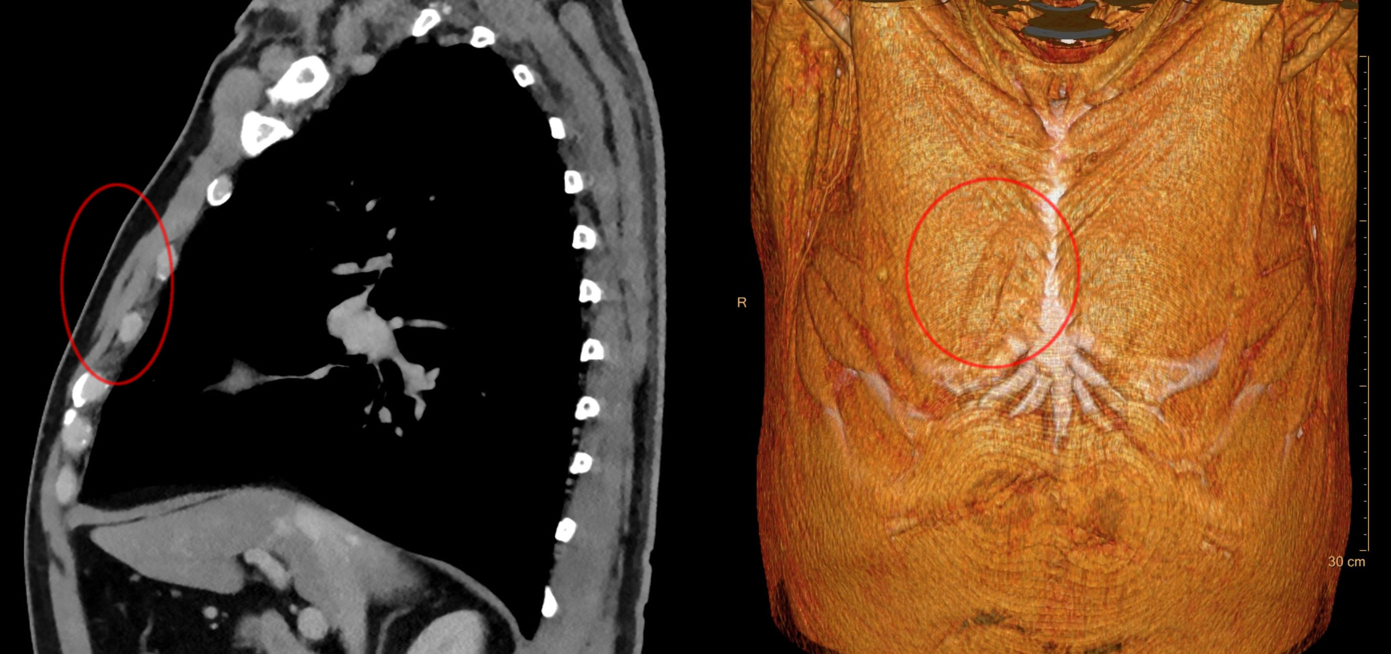
The sternalis muscle is an anatomical variation that lies in front of the sternal end of the pectoralis major parallel to the margin of the sternum. The sternalis muscle may be a variation of the pectoralis major or of the rectus abdominis. Structure The sternalis is a muscle that runs along the anterior aspect of the body of the sternum. It lies superficially and parallel to the sternum. Its origin and insertion are variable. The sternalis muscle often originates from the upper part of the sternum and can display varying insertions such as the pectoral fascia, lower ribs, costal cartilages, rectus sheath, aponeurosis of the abdominal external oblique muscle. There is still a great deal of disagreement about its innervation and its embryonic origin. In a review, it was reported that the muscle was innervated by the external or internal thoracic nerves in 55% of the cases, by the intercostal nerves in 43% of the cases, while the remaining cases were supplied by both nerves. How ... [...More Info...] [...Related Items...] OR: [Wikipedia] [Google] [Baidu] |
