|
Lobular Carcinoma
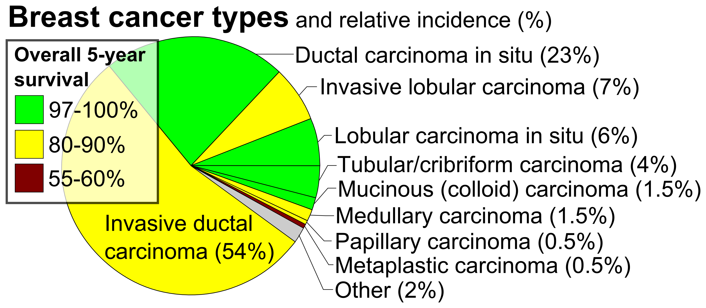
Lobular carcinoma is a form of tumor which primarily affects the lobules of a gland. It is sometimes considered equivalent to "terminal duct carcinoma". If not otherwise specified, it generally refers to breast cancer. Examples include: * Lobular carcinoma in situ Lobular carcinoma in situ (LCIS) is an incidental microscopic finding with characteristic cellular morphology and multifocal tissue patterns. The condition is a laboratory diagnosis and refers to unusual cells in the lobules of the breast. The lo ... * Invasive lobular carcinoma References External links Carcinoma {{neoplasm-stub ... [...More Info...] [...Related Items...] OR: [Wikipedia] [Google] [Baidu] |
Micrograph
A micrograph is an image, captured photographically or digitally, taken through a microscope or similar device to show a magnify, magnified image of an object. This is opposed to a macrograph or photomacrograph, an image which is also taken on a microscope but is only slightly magnified, usually less than 10 times. Micrography is the practice or art of using microscopes to make photographs. A photographic micrograph is a photomicrograph, and one taken with an electron microscope is an electron micrograph. A micrograph contains extensive details of microstructure. A wealth of information can be obtained from a simple micrograph like behavior of the material under different conditions, the phases found in the system, failure analysis, grain size estimation, elemental analysis and so on. Micrographs are widely used in all fields of microscopy. Types Photomicrograph A light micrograph or photomicrograph is a micrograph prepared using an optical microscope, a process referred to ... [...More Info...] [...Related Items...] OR: [Wikipedia] [Google] [Baidu] |
H&E Stain
Hematoxylin and eosin stain ( or haematoxylin and eosin stain or hematoxylin–eosin stain; often abbreviated as H&E stain or HE stain) is one of the principal tissue stains used in histology. It is the most widely used stain in medical diagnosis and is often the ''gold standard.'' For example, when a pathologist looks at a biopsy of a suspected cancer, the histological section is likely to be stained with H&E. H&E is the combination of two histological stains: hematoxylin and eosin. The hematoxylin stains cell nuclei a purplish blue, and eosin stains the extracellular matrix and cytoplasm pink, with other structures taking on different shades, hues, and combinations of these colors. Hence a pathologist can easily differentiate between the nuclear and cytoplasmic parts of a cell, and additionally, the overall patterns of coloration from the stain show the general layout and distribution of cells and provides a general overview of a tissue sample's structure. Thus, patte ... [...More Info...] [...Related Items...] OR: [Wikipedia] [Google] [Baidu] |
Lobe (anatomy)
In anatomy, a lobe is a clear anatomical division or extension of an organ (as seen for example in the brain, lung, liver, or kidney) that can be determined without the use of a microscope at the gross anatomy level. This is in contrast to the much smaller lobule, which is a clear division only visible under the microscope. Interlobar ducts connect lobes and interlobular ducts connect lobules. Examples of lobes *The four main lobes of the brain **the frontal lobe **the parietal lobe **the occipital lobe **the temporal lobe *The three lobes of the human cerebellum **the flocculonodular lobe **the anterior lobe **the posterior lobe *The two lobes of the thymus *The two and three lobes of the lungs ** Left lung: superior and inferior ** Right lung: superior, middle, and inferior *The four lobes of the liver ** Left lobe of liver ** Right lobe of liver ** Quadrate lobe of liver ** Caudate lobe of liver *The renal lobes of the kidney * Earlobes Examples of lobules *the ... [...More Info...] [...Related Items...] OR: [Wikipedia] [Google] [Baidu] |
Breast Cancer
Breast cancer is a cancer that develops from breast tissue. Signs of breast cancer may include a Breast lump, lump in the breast, a change in breast shape, dimpling of the skin, Milk-rejection sign, milk rejection, fluid coming from the nipple, a newly inverted nipple, or a red or scaly patch of skin. In those with Metastatic breast cancer, distant spread of the disease, there may be bone pain, swollen lymph nodes, shortness of breath, or yellow skin. Risk factors for developing breast cancer include obesity, a Sedentary lifestyle, lack of physical exercise, alcohol consumption, hormone replacement therapy during menopause, ionizing radiation, an early age at Menarche, first menstruation, having children late in life (or not at all), older age, having a prior history of breast cancer, and a family history of breast cancer. About five to ten percent of cases are the result of an inherited genetic predisposition, including BRCA mutation, ''BRCA'' mutations among others. Breast ... [...More Info...] [...Related Items...] OR: [Wikipedia] [Google] [Baidu] |
Lobular Carcinoma In Situ
Lobular carcinoma in situ (LCIS) is an incidental microscopic finding with characteristic cellular morphology and multifocal tissue patterns. The condition is a laboratory diagnosis and refers to unusual cells in the lobules of the breast. The lobules and acini of the terminal duct-lobular unit (TDLU), the basic functional unit of the breast, may become distorted and undergo expansion due to the abnormal proliferation of cells comprising the structure. These changes represent a spectrum of atypical epithelial lesions that are broadly referred to as lobular neoplasia (LN). One subset of LN can be defined as LCIS based on specific cellular traits and tissue changes seen histologically. These lesions are preceded by atypical lobular hyperplasia and may follow a linear progression to invasive lobular carcinoma (ILC), with specific genetic aberrations. This process coincides with the progression of ductal neoplasia to ductal carcinoma in situ and invasive carcinoma. Rarely, terminal ... [...More Info...] [...Related Items...] OR: [Wikipedia] [Google] [Baidu] |
Invasive Lobular Carcinoma
Invasive lobular carcinoma (ILC) is breast cancer arising from the lobules of the mammary glands. It accounts for 5–10% of invasive breast cancer. Rare cases of this carcinoma have been diagnosed in men (see male breast cancer). Types The histologic patterns include:Fletcher's diagnostic histopathology of tumors. 3rd Ed. p. 931-932. File:Histopathology of invasive lobular carcinoma, next to lobular carcinoma in situ, annotated.jpg, Histopathology of invasive lobular carcinoma (ILC), next to lobular carcinoma in situ (LCIS) File:Breast invasive lobular carcinoma (2).jpg, Invasive lobular carcinoma demonstrating a predominantly lobular growth pattern File:LobularBreastCancer.jpg, Lobular breast cancer. Single file cells and cell nests. File:Histopathology of subtle invasive lobular carcinoma, annotated.png, ILC may be subtle on low magnification (left). Higher magnification (right) shows invasive growth pattern and vesicular nuclei with prominent nucleoli. Prognosis Overall ... [...More Info...] [...Related Items...] OR: [Wikipedia] [Google] [Baidu] |


