|
Langerhans Cell
A Langerhans cell (LC) is a tissue-resident macrophage of the skin once thought to be a resident dendritic cell. These cells contain organelles called Birbeck granules. They are present in all layers of the epidermis and are most prominent in the stratum spinosum. They also occur in the papillary dermis, particularly around blood vessels, as well as in the oral mucosa, mucosa of the mouth, foreskin, and vaginal epithelium. They can be found in other tissues, such as lymph nodes, particularly in association with the condition Langerhans cell histiocytosis (LCH). Function In skin infections, the local Langerhans cells take up and process microbe, microbial antigens to become fully functional antigen-presenting cells. Generally, tissue-resident macrophages are involved in immune homeostasis and the uptake of Apoptosis, apoptotic bodies. However, Langerhans cells can also take on a dendritic cell-like phenotype and migrate to lymph nodes to interact with naive T-cells. Matrix meta ... [...More Info...] [...Related Items...] OR: [Wikipedia] [Google] [Baidu] |
Islets Of Langerhans
The pancreatic islets or islets of Langerhans are the regions of the pancreas that contain its endocrine (hormone-producing) cells, discovered in 1869 by German pathological anatomist Paul Langerhans. The pancreatic islets constitute 1–2% of the pancreas volume and receive 10–15% of its blood flow. The pancreatic islets are arranged in density routes throughout the human pancreas, and are important in the metabolism of glucose. Structure There are about 1 million islets distributed throughout the pancreas of a healthy adult human. While islets vary in size, the average diameter is about 0.2 mm.:928 Each islet is separated from the surrounding pancreatic tissue by a thin, fibrous, connective tissue capsule which is continuous with the fibrous connective tissue that is interwoven throughout the rest of the pancreas.:928 Microanatomy Hormones produced in the pancreatic islets are secreted directly into the blood flow by (at least) five types of cells. In rat islets, e ... [...More Info...] [...Related Items...] OR: [Wikipedia] [Google] [Baidu] |
Vaginal Epithelium
The vaginal epithelium is the inner lining of the vagina consisting of multiple layers of (Epithelium, squamous) cells. The basal membrane provides the support for the first layer of the epithelium-the basal layer. The intermediate layers lie upon the basal layer, and the superficial layer is the outermost layer of the epithelium. Anatomists have described the epithelium as consisting of as many as 40 distinct layers of cells. The mucus found on the epithelium is secreted by the cervix and uterus. The rugae of the epithelium create an involuted surface and result in a large surface area that covers 360 cm2. This large surface area allows the trans-epithelial absorption of some medications via the vaginal route. In the course of the Biological life cycle, reproductive cycle, the vaginal epithelium is subject to normal, cyclic changes, that are influenced by estrogen: with increasing circulating levels of the hormone, there is proliferation of epithelial cells along with an i ... [...More Info...] [...Related Items...] OR: [Wikipedia] [Google] [Baidu] |
Cell Proliferation
Cell proliferation is the process by which ''a cell grows and divides to produce two daughter cells''. Cell proliferation leads to an exponential increase in cell number and is therefore a rapid mechanism of tissue growth. Cell proliferation requires both cell growth and cell division to occur at the same time, such that the average size of cells remains constant in the population. Cell division can occur without cell growth, producing many progressively smaller cells (as in cleavage of the zygote), while cell growth can occur without cell division to produce a single larger cell (as in growth of neurons). Thus, cell proliferation is not synonymous with either cell growth or cell division, despite these terms sometimes being used interchangeably. Stem cells undergo cell proliferation to produce proliferating "transit amplifying" daughter cells that later differentiate to construct tissues during normal development and tissue growth, during tissue regeneration after da ... [...More Info...] [...Related Items...] OR: [Wikipedia] [Google] [Baidu] |
Embryo
An embryo ( ) is the initial stage of development for a multicellular organism. In organisms that reproduce sexually, embryonic development is the part of the life cycle that begins just after fertilization of the female egg cell by the male sperm cell. The resulting fusion of these two cells produces a single-celled zygote that undergoes many cell divisions that produce cells known as blastomeres. The blastomeres (4-cell stage) are arranged as a solid ball that when reaching a certain size, called a morula, (16-cell stage) takes in fluid to create a cavity called a blastocoel. The structure is then termed a blastula, or a blastocyst in mammals. The mammalian blastocyst hatches before implantating into the endometrial lining of the womb. Once implanted the embryo will continue its development through the next stages of gastrulation, neurulation, and organogenesis. Gastrulation is the formation of the three germ layers that will form all of the different parts of t ... [...More Info...] [...Related Items...] OR: [Wikipedia] [Google] [Baidu] |
Stratum Basale
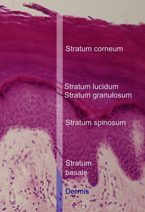
The stratum basale (basal layer, sometimes referred to as ''stratum germinativum'') is the deepest layer of the five layers of the epidermis, the external covering of skin in mammals. The stratum basale is a single layer of columnar or cuboidal basal cells. The cells are attached to each other and to the overlying stratum spinosum cells by desmosomes and hemidesmosomes. The nucleus is large, ovoid and occupies most of the cell. Some basal cells can act like stem cells with the ability to divide and produce new cells, and these are sometimes called basal keratinocyte stem cells. Others serve to anchor the epidermis glabrous skin (hairless), and hyper-proliferative epidermis (from a skin disease).McGrath, J.A.; Eady, R.A.; Pope, F.M. (2004). ''Rook's Textbook of Dermatology'' (Seventh Edition). Blackwell Publishing. Pages 3.7. . They divide to form the keratinocytes of the stratum spinosum, which migrate superficially. Other types of cells found within the stratum basale ... [...More Info...] [...Related Items...] OR: [Wikipedia] [Google] [Baidu] |
Matrix Metalloproteinase
Matrix metalloproteinases (MMPs), also known as matrix metallopeptidases or matrixins, are metalloproteinases that are calcium-dependent zinc-containing endopeptidases; other family members are adamalysins, serralysins, and astacins. The MMPs belong to a larger family of proteases known as the metzincin superfamily. Collectively, these enzymes are capable of degrading all kinds of extracellular matrix proteins, but also can process a number of Biological activity, bioactive molecules. They are known to be involved in the cleavage of cell surface Receptor (biochemistry), receptors, the release of apoptosis, apoptotic ligands (such as the FAS ligand), and chemokine/cytokine inactivation. MMPs are also thought to play a major role in cell behaviors such as cell proliferation, cell migration, migration (cell adhesion, adhesion/dispersion), Cellular differentiation, differentiation, angiogenesis, apoptosis, and Immune system, host defense. They were first described in vertebrates in ... [...More Info...] [...Related Items...] OR: [Wikipedia] [Google] [Baidu] |
Naive T-cell
In immunology, a naive T cell (Th0 cell) is a T cell that has differentiated in the thymus, and successfully undergone the positive and negative processes of central selection in the thymus. Among these are the naive forms of helper T cells (CD4+) and cytotoxic T cells (CD8+). Naive T cells, unlike activated or memory T cells, have not encountered its cognate antigen within the periphery. After this encounter, the naive T cell is considered a mature T cell. Phenotype Naive T cells are commonly characterized by the surface expression of L-selectin (CD62L) and C-C Chemokine receptor type 7 (CCR7); the absence of the activation markers CD25, CD44 or CD69; and the absence of memory CD45RO isoform. They also express functional IL-7 receptors, consisting of subunits IL-7 receptor-α, CD127, and common-γ chain, CD132. In the naive state, T cells are thought to require the common-gamma chain cytokines IL-7 and IL-15 for homeostatic survival mechanisms. While naive T cells ar ... [...More Info...] [...Related Items...] OR: [Wikipedia] [Google] [Baidu] |
Apoptosis
Apoptosis (from ) is a form of programmed cell death that occurs in multicellular organisms and in some eukaryotic, single-celled microorganisms such as yeast. Biochemistry, Biochemical events lead to characteristic cell changes (Morphology (biology), morphology) and death. These changes include Bleb (cell biology), blebbing, Plasmolysis, cell shrinkage, Karyorrhexis, nuclear fragmentation, Pyknosis, chromatin condensation, Apoptotic DNA fragmentation, DNA fragmentation, and mRNA decay. The average adult human loses 50 to 70 1,000,000,000, billion cells each day due to apoptosis. For the average human child between 8 and 14 years old, each day the approximate loss is 20 to 30 billion cells. In contrast to necrosis, which is a form of traumatic cell death that results from acute cellular injury, apoptosis is a highly regulated and controlled process that confers advantages during an organism's life cycle. For example, the separation of fingers and toes in a developing human embryo ... [...More Info...] [...Related Items...] OR: [Wikipedia] [Google] [Baidu] |
Antigen-presenting Cell
An antigen-presenting cell (APC) or accessory cell is a Cell (biology), cell that displays an antigen bound by major histocompatibility complex (MHC) proteins on its surface; this process is known as antigen presentation. T cells may recognize these complexes using their T cell receptors (TCRs). APCs antigen processing, process antigens and present them to T cells. Almost all cell types can present antigens in some way. They are found in a variety of tissue types. Dedicated antigen-presenting cells, including macrophages, B cells and dendritic cells, present foreign antigens to T helper cell, helper T cells, while virus-infected cells (or cancer cells) can present antigens originating inside the cell to cytotoxic T cells. In addition to the MHC family of proteins, antigen presentation relies on other specialized signaling molecules on the surfaces of both APCs and T cells. Antigen-presenting cells are vital for effective Adaptive immune system, adaptive immune response, as the fun ... [...More Info...] [...Related Items...] OR: [Wikipedia] [Google] [Baidu] |
Antigen
In immunology, an antigen (Ag) is a molecule, moiety, foreign particulate matter, or an allergen, such as pollen, that can bind to a specific antibody or T-cell receptor. The presence of antigens in the body may trigger an immune response. Antigens can be proteins, peptides (amino acid chains), polysaccharides (chains of simple sugars), lipids, or nucleic acids. Antigens exist on normal cells, cancer cells, parasites, viruses, fungus, fungi, and bacteria. Antigens are recognized by antigen receptors, including antibodies and T-cell receptors. Diverse antigen receptors are made by cells of the immune system so that each cell has a specificity for a single antigen. Upon exposure to an antigen, only the lymphocytes that recognize that antigen are activated and expanded, a process known as clonal selection. In most cases, antibodies are ''antigen-specific'', meaning that an antibody can only react to and bind one specific antigen; in some instances, however, antibodies may cr ... [...More Info...] [...Related Items...] OR: [Wikipedia] [Google] [Baidu] |
Microbe
A microorganism, or microbe, is an organism of microscopic size, which may exist in its single-celled form or as a colony of cells. The possible existence of unseen microbial life was suspected from antiquity, with an early attestation in Jain literature authored in 6th-century BC India. The scientific study of microorganisms began with their observation under the microscope in the 1670s by Anton van Leeuwenhoek. In the 1850s, Louis Pasteur found that microorganisms caused food spoilage, debunking the theory of spontaneous generation. In the 1880s, Robert Koch discovered that microorganisms caused the diseases tuberculosis, cholera, diphtheria, and anthrax. Microorganisms are extremely diverse, representing most unicellular organisms in all three domains of life: two of the three domains, Archaea and Bacteria, only contain microorganisms. The third domain, Eukaryota, includes all multicellular organisms as well as many unicellular protists and protozoans that are ... [...More Info...] [...Related Items...] OR: [Wikipedia] [Google] [Baidu] |
Infection
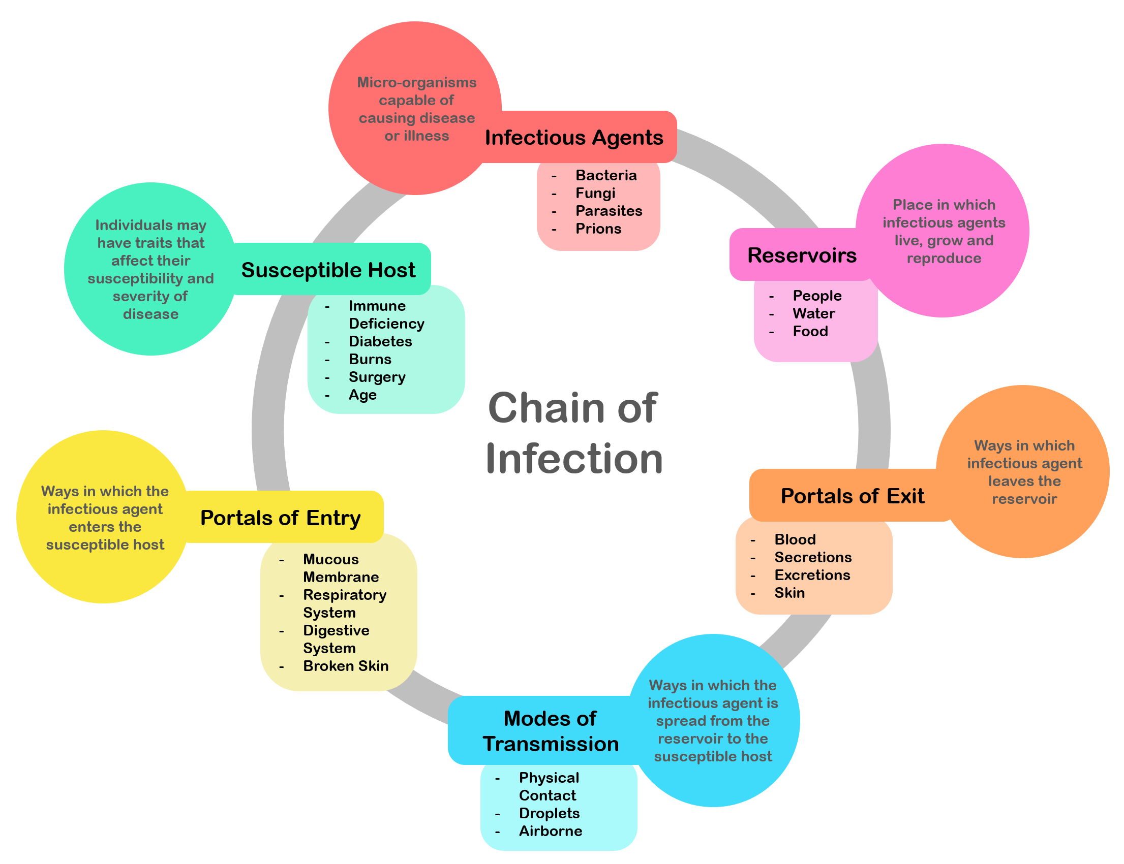
An infection is the invasion of tissue (biology), tissues by pathogens, their multiplication, and the reaction of host (biology), host tissues to the infectious agent and the toxins they produce. An infectious disease, also known as a transmissible disease or communicable disease, is an Disease#Terminology, illness resulting from an infection. Infections can be caused by a wide range of pathogens, most prominently pathogenic bacteria, bacteria and viruses. Hosts can fight infections using their immune systems. Mammalian hosts react to infections with an Innate immune system, innate response, often involving inflammation, followed by an Adaptive immune system, adaptive response. Treatment for infections depends on the type of pathogen involved. Common medications include: * Antibiotics for bacterial infections. * Antivirals for viral infections. * Antifungals for fungal infections. * Antiprotozoals for protozoan infections. * Antihelminthics for infections caused by parasi ... [...More Info...] [...Related Items...] OR: [Wikipedia] [Google] [Baidu] |


