|
Intracranial Dolichoectasias
The term dolichoectasia means elongation and distension. It is used to characterize arteries throughout the human body which have shown significant deterioration of their tunica intima (and occasionally the tunica media), weakening the vessel walls and causing the artery to elongate and distend. Signs and symptoms Vertebrobasillar dolichoectasia * Hemifacial spasm * Paresis * Trigeminal neuralgia Internal carotid dolichoectasia * Progressive visual field defect Cause Most commonly caused by hypertension, continued stress on the walls of the artery will degrade the vessel wall by damaging and loosening the collagen and elastin meshwork which comprises the intima. Similarly, hypercholesterolemia or hyperlipidemia can also provide sufficient trauma to the vessel wall resulting in dolichoectasia. As the arrangement of connective tissue is disturbed, the vessel wall is no longer able to hold its original conformation and begins to unravel due to the continued hypertension. High ... [...More Info...] [...Related Items...] OR: [Wikipedia] [Google] [Baidu] |
Vascular Surgery
Vascular surgery is a surgical subspecialty in which diseases of the vascular system, or arteries, veins and lymphatic circulation, are managed by medical therapy, minimally-invasive catheter procedures and surgical reconstruction. The specialty evolved from general and cardiac surgery and includes treatment of the body's other major and essential veins and arteries. Open surgery techniques, as well as endovascular techniques are used to treat vascular diseases. The vascular surgeon is trained in the diagnosis and management of diseases affecting all parts of the vascular system excluding the coronaries and intracranial vasculature. Vascular surgeons often assist other physicians to address traumatic vascular injury, hemorrhage control, and safe exposure of vascular structures. History Early leaders of the field included Russian surgeon Nikolai Korotkov, noted for developing early surgical techniques, American interventional radiologist Charles Theodore Dotter who is credite ... [...More Info...] [...Related Items...] OR: [Wikipedia] [Google] [Baidu] |
Tunica Intima
The tunica intima (New Latin "inner coat"), or intima for short, is the innermost tunica (layer) of an artery or vein. It is made up of one layer of endothelial cells and is supported by an internal elastic lamina. The endothelial cells are in direct contact with the blood flow. The three layers of a blood vessel are an inner layer (the tunica intima), a middle layer (the tunica media), and an outer layer (the tunica externa). In dissection, the inner coat (tunica intima) can be separated from the middle (tunica media) by a little maceration, or it may be stripped off in small pieces; but, because of its friability, it cannot be separated as a complete membrane. It is a fine, transparent, colorless structure which is highly elastic, and, after death, is commonly corrugated into longitudinal wrinkles. Structure The structure of the tunica intima depends on the blood vessel type. Elastic arteries – A single layer of Endothelial and a supporting layer of elastin-rich col ... [...More Info...] [...Related Items...] OR: [Wikipedia] [Google] [Baidu] |
Hemifacial Spasm
Hemifacial spasm (HFS) is a rare neuromuscular disease characterized by irregular, involuntary muscle contractions (spasms) on one side (hemi-) of the face (-facial). The facial muscles are controlled by the facial nerve (seventh cranial nerve), which originates at the brainstem and exits the skull below the ear where it separates into five main branches. This disease takes two forms: typical and atypical. In typical form, the twitching usually starts in the lower eyelid in orbicularis oculi muscle. As time progresses, it spreads to the whole lid, then to the orbicularis oris muscle around the lips, and buccinator muscle in the cheekbone area. The reverse process of twitching occurs in atypical hemifacial spasm; twitching starts in orbicularis oris muscle around the lips, and buccinator muscle in the cheekbone area in the lower face, then progresses up to the orbicularis oculi muscle in the eyelid as time progresses. The most common form is the typical form, and atypical form ... [...More Info...] [...Related Items...] OR: [Wikipedia] [Google] [Baidu] |
Paresis
In medicine, paresis () is a condition typified by a weakness of voluntary movement, or by partial loss of voluntary movement or by impaired movement. When used without qualifiers, it usually refers to the limbs, but it can also be used to describe the muscles of the eyes (ophthalmoparesis), the stomach (gastroparesis), and also the vocal cords (Vocal cord paresis). Neurologists use the term ''paresis'' to describe weakness, and ''plegia'' to describe paralysis in which all voluntary movement is lost. The term ''paresis'' comes from the grc, πάρεσις 'letting go' from παρίημι 'to let go, to let fall'. Types Limbs * Monoparesis – One leg or one arm * Paraparesis – Both legs * Hemiparesis – The loss of function to only one side of the body * Triparesis Three limbs. This can either mean both legs and one arm, both arms and a leg, or a combination of one arm, one leg, and face * Double Hemiparesis all four limbs are involved, but one side of the body is mo ... [...More Info...] [...Related Items...] OR: [Wikipedia] [Google] [Baidu] |
Trigeminal Neuralgia
Trigeminal neuralgia (TN or TGN), also called Fothergill disease, tic douloureux, or trifacial neuralgia is a long-term pain disorder that affects the trigeminal nerve, the nerve responsible for sensation in the face and motor functions such as biting and chewing. It is a form of neuropathic pain. There are two main types: typical and atypical trigeminal neuralgia. The typical form results in episodes of severe, sudden, shock-like pain in one side of the face that lasts for seconds to a few minutes. Groups of these episodes can occur over a few hours. The atypical form results in a constant burning pain that is less severe. Episodes may be triggered by any touch to the face. Both forms may occur in the same person. It is regarded as one of the most painful disorders known to medicine, and often results in depression. The exact cause is unknown, but believed to involve loss of the myelin of the trigeminal nerve. This might occur due to compression from a blood vessel as the ne ... [...More Info...] [...Related Items...] OR: [Wikipedia] [Google] [Baidu] |
Hypertension
Hypertension (HTN or HT), also known as high blood pressure (HBP), is a long-term medical condition in which the blood pressure in the arteries is persistently elevated. High blood pressure usually does not cause symptoms. Long-term high blood pressure, however, is a major risk factor for stroke, coronary artery disease, heart failure, atrial fibrillation, peripheral arterial disease, vision loss, chronic kidney disease, and dementia. Hypertension is a major cause of premature death worldwide. High blood pressure is classified as primary (essential) hypertension or secondary hypertension. About 90–95% of cases are primary, defined as high blood pressure due to nonspecific lifestyle and genetic factors. Lifestyle factors that increase the risk include excess salt in the diet, excess body weight, smoking, and alcohol use. The remaining 5–10% of cases are categorized as secondary high blood pressure, defined as high blood pressure due to an identifiable cause, such ... [...More Info...] [...Related Items...] OR: [Wikipedia] [Google] [Baidu] |
Vertebral Artery
The vertebral arteries are major arteries of the neck. Typically, the vertebral arteries originate from the subclavian arteries. Each vessel courses superiorly along each side of the neck, merging within the skull to form the single, midline basilar artery. As the supplying component of the ''vertebrobasilar vascular system'', the vertebral arteries supply blood to the upper spinal cord, brainstem, cerebellum, and posterior part of brain. Structure The vertebral arteries usually arise from the posterosuperior aspect of the central subclavian arteries on each side of the body, then enter deep to the transverse process at the level of the 6th cervical vertebrae (C6), or occasionally (in 7.5% of cases) at the level of C7. They then proceed superiorly, in the transverse foramen of each cervical vertebra. Once they have passed through the transverse foramen of C1 (also known as the atlas), the vertebral arteries travel across the posterior arch of C1 and through the suboccipi ... [...More Info...] [...Related Items...] OR: [Wikipedia] [Google] [Baidu] |
Basilar Artery
The basilar artery () is one of the arteries that supplies the brain with oxygen-rich blood. The two vertebral arteries and the basilar artery are known as the vertebral basilar system, which supplies blood to the posterior part of the circle of Willis and joins with blood supplied to the anterior part of the circle of Willis from the internal carotid arteries. Structure The basilar artery arises from the union of the two vertebral arteries at the junction between the medulla oblongata and the pons between the abducens nerves (CN VI). The diameter of the basilar artery range from 1.5 to 6.6 mm. It ascends superiorly in the basilar sulcus of the ventral pons and divides at the junction of the midbrain and pons into the posterior cerebral arteries. Its branches from caudal to rostral include: *anterior inferior cerebellar artery *labyrinthine artery (<15% of people, usually branches from the anterior inferior cerebellar artery) * |
Internal Carotid Artery
The internal carotid artery (Latin: arteria carotis interna) is an artery in the neck which supplies the anterior circulation of the brain. In human anatomy, the internal and external carotids arise from the common carotid arteries, where these bifurcate at cervical vertebrae C3 or C4. The internal carotid artery supplies the brain, including the eyes, while the external carotid nourishes other portions of the head, such as the face, scalp, skull, and meninges. Classification Terminologia Anatomica in 1998 subdivided the artery into four parts: "cervical", "petrous", "cavernous", and "cerebral". However, in clinical settings, the classification system of the internal carotid artery usually follows the 1996 recommendations by Bouthillier, describing seven anatomical segments of the internal carotid artery, each with a corresponding alphanumeric identifier—C1 cervical, C2 petrous, C3 lacerum, C4 cavernous, C5 clinoid, C6 ophthalmic, and C7 communicating. The Bouthillier nome ... [...More Info...] [...Related Items...] OR: [Wikipedia] [Google] [Baidu] |
Polycystic Kidney Disease
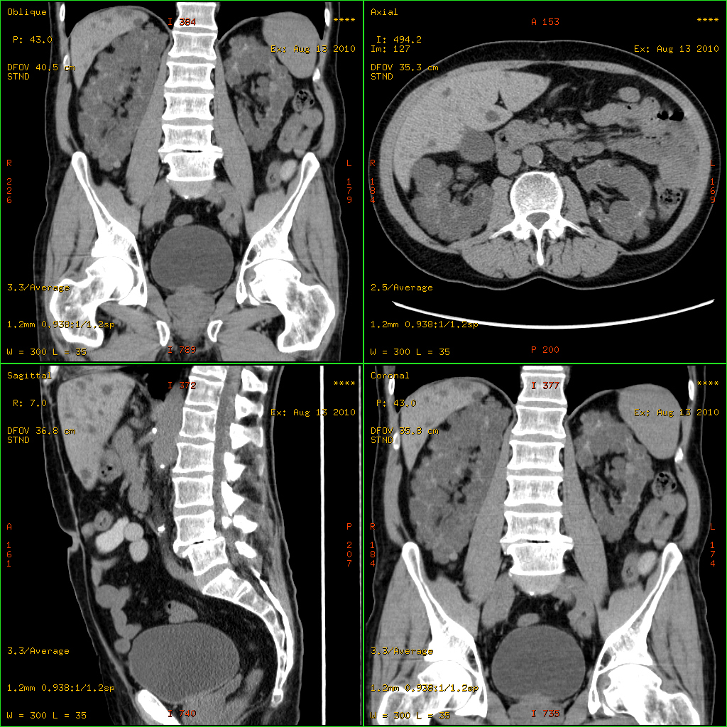
Polycystic kidney disease (PKD or PCKD, also known as polycystic kidney syndrome) is a genetic disorder in which the renal tubules become structurally abnormal, resulting in the development and growth of multiple cysts within the kidney. These cysts may begin to develop in utero, in infancy, in childhood, or in adulthood. Cysts are non-functioning tubules filled with fluid pumped into them, which range in size from microscopic to enormous, crushing adjacent normal tubules and eventually rendering them non-functional as well. PKD is caused by abnormal genes that produce a specific abnormal protein; this protein has an adverse effect on tubule development. PKD is a general term for two types, each having their own pathology and genetic cause: autosomal dominant polycystic kidney disease (ADPKD) and autosomal recessive polycystic kidney disease (ARPKD). The abnormal gene exists in all cells in the body; as a result, cysts may occur in the liver, seminal vesicles, and pancreas. Th ... [...More Info...] [...Related Items...] OR: [Wikipedia] [Google] [Baidu] |
Intracerebral Hemorrhage
Intracerebral hemorrhage (ICH), also known as cerebral bleed, intraparenchymal bleed, and hemorrhagic stroke, or haemorrhagic stroke, is a sudden bleeding into the tissues of the brain, into its ventricles, or into both. It is one kind of bleeding within the skull and one kind of stroke. Symptoms can include headache, one-sided weakness, vomiting, seizures, decreased level of consciousness, and neck stiffness. Often, symptoms get worse over time. Fever is also common. Causes include brain trauma, aneurysms, arteriovenous malformations, and brain tumors. The biggest risk factors for spontaneous bleeding are high blood pressure and amyloidosis. Other risk factors include alcoholism, low cholesterol, blood thinners, and cocaine use. Diagnosis is typically by CT scan. Other conditions that may present similarly include ischemic stroke. Treatment should typically be carried out in an intensive care unit. Guidelines recommend decreasing the blood pressure to a systolic of 14 ... [...More Info...] [...Related Items...] OR: [Wikipedia] [Google] [Baidu] |


