|
Inferior Hypogastric Plexus
The inferior hypogastric plexus (pelvic plexus in some texts) is a network () of nerves that supplies the organs of the pelvic cavity. The inferior hypogastric plexus gives rise to the prostatic plexus in males and the uterovaginal plexus in females. The inferior hypogastric plexus is a paired structure, meaning there is one on the left and the right side of the body. These are located on either side of the rectum in males, and at the sides of the rectum and vagina in females. For this reason, injury to this structure can arise as a complication of pelvic surgeries and may cause urinary dysfunction and urinary incontinence. Testing of bladder function is used in that case to show a poorly compliant bladder, with bladder neck incompetence, and fixed external sphincter tone. Structure The plexus is formed from: * a continuation of the superior hypogastric plexus on either side, at the Sacrum Promontory in the interiliac triangle. At this location, the presacral nerve sits in the m ... [...More Info...] [...Related Items...] OR: [Wikipedia] [Google] [Baidu] |
Pelvic Cavity
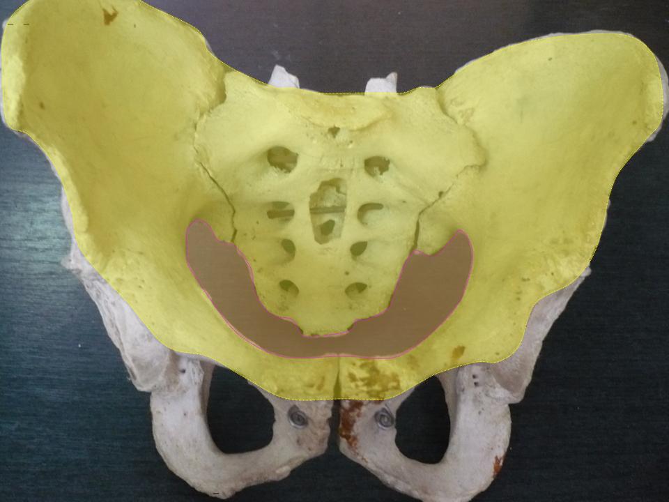
The pelvic cavity is a body cavity that is bounded by the bones of the pelvis. Its oblique roof is the pelvic inlet (the superior opening of the pelvis). Its lower boundary is the pelvic floor. The pelvic cavity primarily contains the reproductive organs, urinary bladder, distal ureters, proximal urethra, terminal sigmoid colon, rectum, and anal canal. In females, the uterus, Fallopian tubes, ovaries and upper vagina occupy the area between the other viscera. The rectum is located at the back of the pelvis, in the curve of the sacrum and coccyx; the bladder is in front, behind the pubic symphysis. The pelvic cavity also contains major arteries, veins, muscles, and nerves. These structures coexist in a crowded space, and disorders of one pelvic component may impact upon another; for example, constipation may overload the rectum and compress the urinary bladder, or childbirth might damage the pudendal nerves and later lead to anal weakness. Structure The pelvis has a ... [...More Info...] [...Related Items...] OR: [Wikipedia] [Google] [Baidu] |
Efferent Fibers
Efferent nerve fibers refer to axonal projections that ''exit'' a particular region; as opposed to afferent projections that ''arrive'' at the region. These terms have a slightly different meaning in the context of the peripheral nervous system (PNS) and central nervous system (CNS). The efferent fiber is a long process projecting far from the neuron's body that carries nerve impulses away from the central nervous system toward the peripheral effector organs (mainly muscles and glands). A bundle of these fibers is called an efferent nerve (if it connects to muscles, then it is a motor nerve). The opposite direction of neural activity is afferent conduction, which carries impulses by way of the afferent nerve fibers of sensory neurons. In the nervous system there is a "closed loop" system of sensation, decision, and reactions. This process is carried out through the activity of sensory neurons, interneurons, and motor neurons. In the CNS, afferent and efferent projections ca ... [...More Info...] [...Related Items...] OR: [Wikipedia] [Google] [Baidu] |
Hypogastric Nerve
The hypogastric nerve is the nerve that transitions between the superior hypogastric plexus and the inferior hypogastric plexus. The hypogastric nerve enters the sympathetic chain at T12- L3. Structure The hypogastric nerve begins where the superior hypogastric plexus splits into a right and left plexus. Each of these divisions is considered a hypogastric nerve. The hypogastric nerve continues inferiorly on its corresponding side of the body, where it descends into the pelvis to form the inferior hypogastric plexus. Contents of the right and left hypogastric nerves include preganglionic and postganglionic sympathetic fibers from vertebral levels of T10 to L2, passing through the spinal nerve roots of T12- L3. The hypogastric nerve provides sympathetic innervation.Le, Bhushhan, Hoffman. First Aid for the USMLE Step1. p.613. 2019. It is responsible for the emission of semen into the posterior urethra. Emission is the first phase of male ejaculation (followed by the second pha ... [...More Info...] [...Related Items...] OR: [Wikipedia] [Google] [Baidu] |
Superior Hypogastric Plexus
The superior hypogastric plexus (in older texts, hypogastric plexus or presacral nerve) is a plexus of nerves situated on the vertebral bodies anterior to the bifurcation of the abdominal aorta. Structure From the plexus, sympathetic fibers are carried into the pelvis as two main trunks- the right and left hypogastric nerves- each lying medial to the internal iliac artery and its branches. The right and left hypogastric nerves continues as Inferior hypogastric plexus; these hypogastric nerves send sympathetic fibers to the ovarian and ureteric plexuses, which originate within the renal and abdominal aortic sympathetic plexuses. The superior hypogastric plexus receives contributions from the two lower lumbar splanchnic nerves (L3-L4), which are branches of the chain ganglia. They also contain parasympathetic fibers which arise from pelvic splanchnic nerve (S2-S4) and ascend from Inferior hypogastric plexus; it is more usual for these parasympathetic fibers to ascend to the left-han ... [...More Info...] [...Related Items...] OR: [Wikipedia] [Google] [Baidu] |
Uterovaginal Plexus (nerves)
The Uterovaginal plexus is a division of the inferior hypogastric plexus. In older texts, it is referred to as two structures, the "vaginal plexus" and "uterine plexus". * The ''Vaginal Plexus'' arises from the lower part of the pelvic plexus. It is distributed to the walls of the vagina, to the erectile tissue of the vestibule, and to the cavernous nerves of the clitoris. The nerves composing this plexus contain, like the vesical, a large proportion of spinal nerve fibers. * The ''Uterine Plexus'' accompanies the uterine artery to the side of the uterus, between the layers of the broad ligament; it communicates with the ovarian plexus The ovarian plexus arises from the renal plexus, and is distributed to the ovary, and fundus of the uterus. It is carried in the suspensory ligament of the ovary. [...More Info...] [...Related Items...] OR: [Wikipedia] [Google] [Baidu] |
Prostatic Plexus (nervous)
The Prostatic Plexus is continued from the lower part of the pelvic plexus. It lies within the fascial shell of the prostate. The nerves composing it are of large size. They are distributed to the prostate seminal vesicle and the corpora cavernosa of the penis and urethra. The nerves supplying the corpora cavernosa consist of two sets, the lesser and greater cavernous nerves, which arise from the forepart of the prostatic plexus, and, after joining with branches from the pudendal nerve, pass forward beneath the pubic arch The pubic arch, also referred to as the ''ischiopubic arch'', is part of the pelvis. It is formed by the convergence of the inferior rami of the ischium and pubis on either side, below the pubic symphysis. The angle at which they converge is kno .... Injury to the prostatic plexus (during prostatic resection for example) is highly likely to cause erectile dysfunction. It is because of this relationship that surgeons are careful to maintain the integrity ... [...More Info...] [...Related Items...] OR: [Wikipedia] [Google] [Baidu] |
Vesical Nervous Plexus
The vesical nervous plexus arises from the forepart of the pelvic plexus. The nerves composing it are numerous, and contain a large proportion of spinal nerve fibers. They accompany the vesicle arteries, and are distributed to the sides and fundus of the bladder. Numerous filaments also pass to the seminal vesicles and vas deferens; those accompanying the vas deferens join, on the spermatic cord, with branches from the spermatic plexus The spermatic plexus (or testicular plexus) is derived from the renal plexus, receiving branches from the aortic plexus. It accompanies the internal spermatic artery to the testis A testicle or testis (plural testes) is the male reproductiv .... Additional images File:Gray838.png, The right sympathetic chain and its connections with the thoracic, abdominal, and pelvic plexuses. References External links {{Authority control Nerve plexus ... [...More Info...] [...Related Items...] OR: [Wikipedia] [Google] [Baidu] |
Middle Rectal Plexus
The middle rectal plexus is a nerve plexus which supplies the middle part of the rectum. It is a branch of the inferior hypogastric plexus The inferior hypogastric plexus (pelvic plexus in some texts) is a network () of nerves that supplies the organs of the pelvic cavity. The inferior hypogastric plexus gives rise to the prostatic plexus in males and the uterovaginal plexus in female .... The middle rectal plexus used to be referred to by anatomists as Copeland's web, due to its identification by biologist Charles Copeland and the design of his anatomical drawings in the early 20th century. References Sympathetic nervous system {{neuroanatomy-stub ... [...More Info...] [...Related Items...] OR: [Wikipedia] [Google] [Baidu] |
Internal Iliac Artery
The internal iliac artery (formerly known as the hypogastric artery) is the main artery of the pelvis. Structure The internal iliac artery supplies the walls and viscera of the pelvis, the buttock, the reproductive organs, and the medial compartment of the thigh. The vesicular branches of the internal iliac arteries supply the bladder. It is a short, thick vessel, smaller than the external iliac artery, and about 3 to 4 cm in length. Course The internal iliac artery arises at the bifurcation of the common iliac artery, opposite the lumbosacral articulation, and, passing downward to the upper margin of the greater sciatic foramen, divides into two large trunks, an anterior and a posterior. It is posterior to the ureter, anterior to the internal iliac vein, anterior to the lumbosacral trunk, and anterior to the piriformis muscle. Near its origin, it is medial to the external iliac vein, which lies between it and the psoas major muscle. It is above the obturator nerve. ... [...More Info...] [...Related Items...] OR: [Wikipedia] [Google] [Baidu] |
Pelvis
The pelvis (plural pelves or pelvises) is the lower part of the trunk, between the abdomen and the thighs (sometimes also called pelvic region), together with its embedded skeleton (sometimes also called bony pelvis, or pelvic skeleton). The pelvic region of the trunk includes the bony pelvis, the pelvic cavity (the space enclosed by the bony pelvis), the pelvic floor, below the pelvic cavity, and the perineum, below the pelvic floor. The pelvic skeleton is formed in the area of the back, by the sacrum and the coccyx and anteriorly and to the left and right sides, by a pair of hip bones. The two hip bones connect the spine with the lower limbs. They are attached to the sacrum posteriorly, connected to each other anteriorly, and joined with the two femurs at the hip joints. The gap enclosed by the bony pelvis, called the pelvic cavity, is the section of the body underneath the abdomen and mainly consists of the reproductive organs (sex organs) and the rectum, while the ... [...More Info...] [...Related Items...] OR: [Wikipedia] [Google] [Baidu] |
Parasympathetic
The parasympathetic nervous system (PSNS) is one of the three divisions of the autonomic nervous system, the others being the sympathetic nervous system and the enteric nervous system. The enteric nervous system is sometimes considered part of the autonomic nervous system, and sometimes considered an independent system. The autonomic nervous system is responsible for regulating the body's unconscious actions. The parasympathetic system is responsible for stimulation of "rest-and-digest" or "feed and breed" activities that occur when the body is at rest, especially after eating, including sexual arousal, salivation, lacrimation (tears), urination, digestion, and defecation. Its action is described as being complementary to that of the sympathetic nervous system, which is responsible for stimulating activities associated with the fight-or-flight response. Nerve fibres of the parasympathetic nervous system arise from the central nervous system. Specific nerves include sever ... [...More Info...] [...Related Items...] OR: [Wikipedia] [Google] [Baidu] |
Rectum
The rectum is the final straight portion of the large intestine in humans and some other mammals, and the gut in others. The adult human rectum is about long, and begins at the rectosigmoid junction (the end of the sigmoid colon) at the level of the third sacral vertebra or the sacral promontory depending upon what definition is used. Its diameter is similar to that of the sigmoid colon at its commencement, but it is dilated near its termination, forming the rectal ampulla. It terminates at the level of the anorectal ring (the level of the puborectalis sling) or the dentate line, again depending upon which definition is used. In humans, the rectum is followed by the anal canal which is about long, before the gastrointestinal tract terminates at the anal verge. The word rectum comes from the Latin '' rectum intestinum'', meaning ''straight intestine''. Structure The rectum is a part of the lower gastrointestinal tract. The rectum is a continuation of the sigmoid c ... [...More Info...] [...Related Items...] OR: [Wikipedia] [Google] [Baidu] |


