|
Involuntary Action
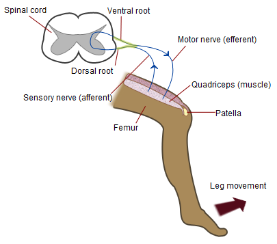
In biology, a reflex, or reflex action, is an involuntary, unplanned sequence or action and nearly instantaneous response to a stimulus. Reflexes are found with varying levels of complexity in organisms with a nervous system. A reflex occurs via neural pathways in the nervous system called reflex arcs. A stimulus initiates a neural signal, which is carried to a synapse. The signal is then transferred across the synapse to a motor neuron, which evokes a target response. These neural signals do not always travel to the brain, so many reflexes are an automatic response to a stimulus that does not receive or need conscious thought. Many reflexes are fine-tuned to increase organism survival and self-defense. This is observed in reflexes such as the startle reflex, which provides an automatic response to an unexpected stimulus, and the feline righting reflex, which reorients a cat's body when falling to ensure safe landing. The simplest type of reflex, a short-latency reflex, has ... [...More Info...] [...Related Items...] OR: [Wikipedia] [Google] [Baidu] |
Biology
Biology is the scientific study of life and living organisms. It is a broad natural science that encompasses a wide range of fields and unifying principles that explain the structure, function, growth, History of life, origin, evolution, and distribution of life. Central to biology are five fundamental themes: the cell (biology), cell as the basic unit of life, genes and heredity as the basis of inheritance, evolution as the driver of biological diversity, energy transformation for sustaining life processes, and the maintenance of internal stability (homeostasis). Biology examines life across multiple biological organisation, levels of organization, from molecules and cells to organisms, populations, and ecosystems. Subdisciplines include molecular biology, physiology, ecology, evolutionary biology, developmental biology, and systematics, among others. Each of these fields applies a range of methods to investigate biological phenomena, including scientific method, observation, ... [...More Info...] [...Related Items...] OR: [Wikipedia] [Google] [Baidu] |
Cervical Spinal Nerve 6
The cervical spinal nerve 6 (C6) is a spinal nerve of the cervical segment. It originates from the spinal column from above the cervical vertebra 6 (C6). The C6 nerve root shares a common branch from C5, and has a role in innervating many muscles of the rotator cuff and distal arm, including: * Subclavius *Supraspinatus * Infraspinatus *Biceps brachii * Brachialis *Deltoid * Teres minor *Brachioradialis *Serratus anterior *Subscapularis * Pectoralis major * Coracobrachialis * Teres major *Supinator * Extensor carpi radialis longus *Latissimus dorsi Damage to the C6 motor neuron, by way of impingement, ischemia, trauma, or degeneration of nerve tissue, can cause denervation Denervation is any loss of nerve supply regardless of the cause. If the nerves lost to denervation are part of neural communication to an organ system or for a specific tissue function, alterations to or compromise of physiological functioning ca ... of one or more of the associated muscles. Muscle atr ... [...More Info...] [...Related Items...] OR: [Wikipedia] [Google] [Baidu] |
H-reflex
The H-reflex (or Hoffmann's reflex) is a reflectory reaction of muscles after electrical stimulation of sensory fibers ( Ia afferents stemming from muscle spindles) in their innervating nerves (for example, those located behind the knee). The H-reflex test is performed using an electric stimulator, which gives usually a square-wave current of short duration and small amplitude (higher stimulations might involve alpha fibers, causing an F-wave, compromising the results), and an EMG set, to record the muscle response. That response is usually a clear wave, called H-wave, 28-35 ms after the stimulus, not to be confused with an F-wave. An M-wave, an early response, occurs 3-6 ms after the onset of stimulation. The H and F-waves are later responses. As the stimulus increases, the amplitude of the F-wave increases only slightly, and the H-wave decreases, and at supramaximal stimulus, the H-wave will disappear. The M-wave does the opposite of the H-wave. As the stimulus increases ... [...More Info...] [...Related Items...] OR: [Wikipedia] [Google] [Baidu] |
Sacral Spinal Nerve 2
The sacral spinal nerve 2 (S2) is a spinal nerve of the sacral segment. Nervous System -- Groups of Nerves It originates from the from below the 2nd body of the 
Muscles S2 supplies many muscles, either directly or through nerves originating from S2. They are not innervated with S2 as single origin, but partly by S2 and partly by other spinal nerves. They are ...[...More Info...] [...Related Items...] OR: [Wikipedia] [Google] [Baidu] |
Sacral Spinal Nerve 1
The sacral spinal nerve 1 (S1) is a spinal nerve of the sacral segment. Nervous System -- Groups of Nerves It originates from the from below the 1st body of the . 
Muscles S1 supplies many muscles, either directly or through nerves originating from S1. They are not innervated with S1 as single origin, but partly by S1 and partly by other spinal nerves. The mu ...[...More Info...] [...Related Items...] OR: [Wikipedia] [Google] [Baidu] |
Ankle Jerk Reflex
The ankle jerk reflex, also known as the Achilles reflex, occurs when the Achilles tendon is tapped while the foot is dorsiflexed. It is a type of stretch reflex that tests the function of the gastrocnemius muscle and the nerve that supplies it. A positive result would be the jerking of the foot towards its plantar surface. Being a deep tendon reflex, it is monosynaptic. It is also a stretch reflex. These are monosynaptic spinal segmental reflexes. When they are intact, integrity of the following is confirmed: cutaneous innervation, motor supply, and cortical input to the corresponding spinal segment. Root value This reflex is mediated by the S1 spinal segment of the spinal cord. Procedure and components Ankle of the patient is relaxed. It is helpful to support the ball of the foot at least somewhat to put some tension in the Achilles tendon, but don’t completely dorsiflex the ankle. A small strike is given on the Achilles tendon using a rubber hammer to elicit the resp ... [...More Info...] [...Related Items...] OR: [Wikipedia] [Google] [Baidu] |
Lumbar Spinal Nerve 4
The lumbar nerves are the five pairs of spinal nerves emerging from the lumbar vertebrae. They are divided into posterior and anterior divisions. Structure The lumbar nerves are five spinal nerves which arise from either side of the spinal cord below the thoracic spinal cord and above the sacral spinal cord. They arise from the spinal cord between each pair of lumbar spinal vertebrae and travel through the intervertebral foramina. The nerves then split into an anterior branch, which travels forward, and a posterior branch, which travels backwards and supplies the area of the back. Posterior divisions The middle divisions of the posterior branches run close to the articular processes of the vertebrae and end in the multifidus muscle. The outer branches supply the erector spinae muscles. The nerves give off branches to the skin. These pierce the aponeurosis of the greater trochanter. Anterior divisions The anterior divisions of the lumbar nerves () increase in size from ... [...More Info...] [...Related Items...] OR: [Wikipedia] [Google] [Baidu] |
Patellar Reflex
The patellar reflex, also called the knee reflex or knee-jerk, is a stretch reflex which tests the L2, L3, and L4 segments of the spinal cord. Many animals, most significantly humans, have been seen to have the patellar reflex, including dogs, cats, horses, and other mammalian species. Mechanism Striking of the patellar tendon with a reflex hammer just below the patella stretches the muscle spindle in the quadriceps muscle. This produces a signal which travels back to the spinal cord and synapses (without interneurons) at the level of L3 or L4 in the spinal cord, completely independent of higher centres. From there, an alpha motor neuron conducts an efferent impulse back to the quadriceps femoris muscle, triggering contraction. This contraction, coordinated with the relaxation of the antagonistic flexor hamstring muscle causes the leg to kick. There is a latency of around 18 ms between stretch of the patellar tendon and the beginning of contraction of the quadriceps femori ... [...More Info...] [...Related Items...] OR: [Wikipedia] [Google] [Baidu] |
Cervical Spinal Nerve 8
The cervical spinal nerve 8 (C8) is a spinal nerve of the cervical segment. It originates from the spinal column from below the cervical vertebra 7 (C7). Innervation The C8 nerve forms part of the radial and ulnar nerves via the brachial plexus, and therefore has motor and sensory function in the upper limb. Sensory The C8 nerve receives sensory afferents from the C8 dermatome. This consists of all the skin on the little finger, and continuing up slightly past the wrist on the palmar and dorsal aspects of the hand and forearm.Drake et al. Gray's Anatomy for Students. Second Edition (2010). Clinically, a test of the pad of the little finger is often used to assess C8 integrity.Aland et al. University of Queensland School of Medicine Clinical Skills Handbook 2010 Motor The C8 nerve contributes to the motor innervation of many of the muscles in the trunk and upper limb. Its primary function is the flexion of the fingers, and this is used as the clinical test for C8 inte ... [...More Info...] [...Related Items...] OR: [Wikipedia] [Google] [Baidu] |
Triceps Reflex
The triceps reflex, a deep tendon reflex, is a reflex that elicits involuntary contraction of the triceps brachii muscle. It is sensed and transmitted by the radial nerve. The reflex is tested as part of the neurological examination to assess the sensory and motor pathways within the C7 and C8 spinal nerves. Testing The test can be performed by tapping the triceps tendonA tendon is a strip or sheet of connective tissue that transmits the force generated by the contraction of muscle to the bone by attaching with it. Thus, in simple words, a tendon attaches a muscle to a bone with the sharp end of a reflex hammer while the forearm is hanging loose at a right angle to the arm. A sudden contraction of the triceps muscle causes extension,A straightening at the elbow joint) of the forearm and indicates a normal reflex. Reflex arc The arc involves the stretch receptors in the triceps tendon, from where the information travels along the radial nerve, through the C7/C8 nerve roo ... [...More Info...] [...Related Items...] OR: [Wikipedia] [Google] [Baidu] |


