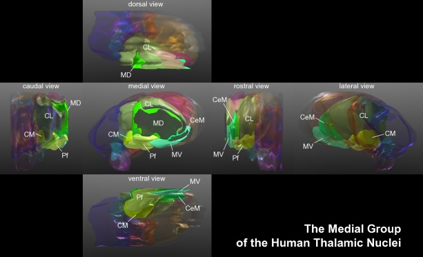
|
Intralaminar Nuclear Group
The intralaminar thalamic nuclei (ITN) are collections of neurons in the internal medullary lamina of the thalamus.Mancall, E., Brock, D. & Gray, H. (2011). Gray's clinical neuroanatomy the anatomic basis for clinical neuroscience. Philadelphia: Elsevier/Saunders. Anatomy Structure The ITN are generally divided in two groups as follows: * anterior (rostral) group ** central lateral nucleus ** central medial nucleus (''not'' referred to as "centromedial") ** paracentral nucleus * posterior (caudal) intralaminar group ** centromedian nucleus ** parafascicular nucleus Some sources also include a "central dorsal" nucleus. Afferents Midline intralaminar nuclei receive afferents from the brain stem, spinal cord, and cerebellum. Connections with the cerebral cortex and basal nuclei are reciprocal. Afferents from the spinothalamic tract as well as periaqueductal gray are part of a pathway involved in pain processing. Efferents The intralaminar nuclei project efferents to the h ... [...More Info...] [...Related Items...] OR: [Wikipedia] [Google] [Baidu] |
Thalamus
The thalamus (: thalami; from Greek language, Greek Wikt:θάλαμος, θάλαμος, "chamber") is a large mass of gray matter on the lateral wall of the third ventricle forming the wikt:dorsal, dorsal part of the diencephalon (a division of the forebrain). Nerve fibers project out of the thalamus to the cerebral cortex in all directions, known as the thalamocortical radiations, allowing hub (network science), hub-like exchanges of information. It has several functions, such as the relaying of sensory neuron, sensory and motor neuron, motor signals to the cerebral cortex and the regulation of consciousness, sleep, and alertness. Anatomically, the thalami are paramedian symmetrical structures (left and right), within the vertebrate brain, situated between the cerebral cortex and the midbrain. It forms during embryonic development as the main product of the diencephalon, as first recognized by the Swiss embryologist and anatomist Wilhelm His Sr. in 1893. Anatomy The thalami ar ... [...More Info...] [...Related Items...] OR: [Wikipedia] [Google] [Baidu] |
Neuron
A neuron (American English), neurone (British English), or nerve cell, is an membrane potential#Cell excitability, excitable cell (biology), cell that fires electric signals called action potentials across a neural network (biology), neural network in the nervous system. They are located in the nervous system and help to receive and conduct impulses. Neurons communicate with other cells via synapses, which are specialized connections that commonly use minute amounts of chemical neurotransmitters to pass the electric signal from the presynaptic neuron to the target cell through the synaptic gap. Neurons are the main components of nervous tissue in all Animalia, animals except sponges and placozoans. Plants and fungi do not have nerve cells. Molecular evidence suggests that the ability to generate electric signals first appeared in evolution some 700 to 800 million years ago, during the Tonian period. Predecessors of neurons were the peptidergic secretory cells. They eventually ga ... [...More Info...] [...Related Items...] OR: [Wikipedia] [Google] [Baidu] |
Medullary Laminae Of Thalamus
Medullary laminae of thalamus are layers of myelinated fibres that appear on cross sections of the thalamus. They also are commonly referred to as laminae medullares thalami or medullary layers of thalamus. The specific layers are: *Lamina medullaris lateralis (external medullary lamina) — separates ventral and lateral thalamus from the subthalamus and thalamic reticular nucleus *Lamina medullaris medialis (internal medullary lamina) — positioned between the dorsomedial and ventral nuclei of thalamus, encloses the intralaminar nuclei (centromedian nucleus In the anatomy of the brain, the centromedian nucleus, also known as the centrum medianum, (CM or Cm-Pf) is a nucleus in the posterior group of the intralaminar thalamic nuclei (ITN) in the thalamus. (This must not be confused with the central me ..., paracentral, and central lateral) External links * References Thalamus {{neuroanatomy-stub ... [...More Info...] [...Related Items...] OR: [Wikipedia] [Google] [Baidu] |
Thalamus
The thalamus (: thalami; from Greek language, Greek Wikt:θάλαμος, θάλαμος, "chamber") is a large mass of gray matter on the lateral wall of the third ventricle forming the wikt:dorsal, dorsal part of the diencephalon (a division of the forebrain). Nerve fibers project out of the thalamus to the cerebral cortex in all directions, known as the thalamocortical radiations, allowing hub (network science), hub-like exchanges of information. It has several functions, such as the relaying of sensory neuron, sensory and motor neuron, motor signals to the cerebral cortex and the regulation of consciousness, sleep, and alertness. Anatomically, the thalami are paramedian symmetrical structures (left and right), within the vertebrate brain, situated between the cerebral cortex and the midbrain. It forms during embryonic development as the main product of the diencephalon, as first recognized by the Swiss embryologist and anatomist Wilhelm His Sr. in 1893. Anatomy The thalami ar ... [...More Info...] [...Related Items...] OR: [Wikipedia] [Google] [Baidu] |
Central Lateral Nucleus
In the human brain, the central lateral nucleus is a part of the anterior intralaminar nucleus in the thalamus. The intralaminar nuclei project to many different regions of the brain, The thalamus The thalamus (: thalami; from Greek language, Greek Wikt:θάλαμος, θάλαμος, "chamber") is a large mass of gray matter on the lateral wall of the third ventricle forming the wikt:dorsal, dorsal part of the diencephalon (a division of ... acts generally as a relay point for the brain for other areas of the brain to link to. The central lateral nucleus acts in a vital role in consciousness. References Neuroanatomy Thalamic nuclei {{Anatomy-stub ... [...More Info...] [...Related Items...] OR: [Wikipedia] [Google] [Baidu] |
Centromedian Nucleus
In the anatomy of the brain, the centromedian nucleus, also known as the centrum medianum, (CM or Cm-Pf) is a nucleus in the posterior group of the intralaminar thalamic nuclei (ITN) in the thalamus. (This must not be confused with the central medial nucleus, which is in the anterior group of the ITN.) There are two centromedian nuclei arranged bilaterally. In humans, each centromedian nucleus contains about 2200 neurons per cubic millimetre and has a volume of about 310 cubic millimetres with 664,000 neurons in total. It measures less than 10 millimetres in every dimension. It belongs to the caudal intralaminar group of thalamic nuclei and is situated within the medial thalamus. It is bordered superiorly by the mediodorsal nucleus, medially by the parafascicular nucleus, and posteriorly by the pulvinar. Input and output It sends nerve fibres to the subthalamic nucleus and putamen. It receives nerve fibres from the cerebral cortex, vestibular nuclei, globus pallidus, superior col ... [...More Info...] [...Related Items...] OR: [Wikipedia] [Google] [Baidu] |
Spinothalamic Tract
The spinothalamic tract is a nerve tract in the anterolateral system in the spinal cord. This tract is an ascending sensory pathway to the thalamus. From the ventral posterolateral nucleus in the thalamus, sensory information is relayed upward to the somatosensory cortex of the postcentral gyrus. The spinothalamic tract consists of two adjacent pathways: anterior and lateral. The anterior spinothalamic tract carries information about crude touch. The lateral spinothalamic tract conveys pain and temperature. In the spinal cord, the spinothalamic tract has somatotopic organization. This is the segmental organization of its cervical, thoracic, lumbar, and sacral components, which is arranged from most medial to most lateral respectively. The pathway crosses over ( decussates) at the level of the spinal cord, rather than in the brainstem like the dorsal column-medial lemniscus pathway and lateral corticospinal tract. It is one of the three tracts which make up the anterola ... [...More Info...] [...Related Items...] OR: [Wikipedia] [Google] [Baidu] |
Periaqueductal Gray
The periaqueductal gray (PAG), also known as the central gray, is a brain region that plays a critical role in autonomic function, motivated behavior and behavioural responses to threatening stimuli. PAG is also the primary control center for descending pain modulation. It has enkephalin-producing cells that suppress pain. The periaqueductal gray is the gray matter located around the cerebral aqueduct within the tegmentum of the midbrain. It projects to the nucleus raphe magnus, and also contains descending autonomic tracts. The ascending pain and temperature fibers of the spinothalamic tract send information to the PAG via the spinomesencephalic pathway (so-named because the fibers originate in the spine and terminate in the PAG, in the mesencephalon or midbrain). This region has been used as the target for brain-stimulating implants in patients with chronic pain. Role in analgesia Stimulation of the periaqueductal gray matter of the midbrain activates enkephalin-r ... [...More Info...] [...Related Items...] OR: [Wikipedia] [Google] [Baidu] |
Progressive Supranuclear Palsy
Progressive supranuclear palsy (PSP) is a late-onset neurodegenerative disease involving the gradual deterioration and death of specific volumes of the brain, linked to 4-repeat tau pathology. The condition leads to symptoms including Balance disorder, loss of balance, Hypokinesia, slowing of movement, Ophthalmoparesis, difficulty moving the eyes, and cognitive impairment. PSP may be mistaken for other types of neurodegeneration such as Parkinson's disease, frontotemporal dementia and Alzheimer's disease. It is the second most common tauopathy behind Alzheimer's disease. The cause of the condition is uncertain, but involves the accumulation of tau protein within the brain. Medications such as L-DOPA, levodopa and amantadine may be useful in some cases. PSP was first officially described by Richardson, Steele, and Olszewski in 1963 as a form of progressive parkinsonism. However, the earliest known case presenting clinical features consistent with PSP, along with pathological co ... [...More Info...] [...Related Items...] OR: [Wikipedia] [Google] [Baidu] |
Parkinson's Disease
Parkinson's disease (PD), or simply Parkinson's, is a neurodegenerative disease primarily of the central nervous system, affecting both motor system, motor and non-motor systems. Symptoms typically develop gradually and non-motor issues become more prevalent as the disease progresses. The motor symptoms are collectively called parkinsonism and include tremors, bradykinesia, spasticity, rigidity as well as postural instability (i.e., difficulty maintaining balance). Non-motor symptoms develop later in the disease and include behavior change (individual), behavioral changes or mental disorder, neuropsychiatric problems such as sleep abnormalities, psychosis, anosmia, and mood swings. Most Parkinson's disease cases are idiopathic disease, idiopathic, though contributing factors have been identified. Pathophysiology involves progressive nerve cell death, degeneration of nerve cells in the substantia nigra, a midbrain region that provides dopamine to the basal ganglia, a system invo ... [...More Info...] [...Related Items...] OR: [Wikipedia] [Google] [Baidu] |
Traumatic Brain Injury
A traumatic brain injury (TBI), also known as an intracranial injury, is an injury to the brain caused by an external force. TBI can be classified based on severity ranging from mild traumatic brain injury (mTBI/concussion) to severe traumatic brain injury. TBI can also be characterized based on mechanism (closed head injury, closed or penetrating head injury) or other features (e.g., occurring in a specific location or over a widespread area). Head injury is a broader category that may involve damage to other structures such as the scalp and skull. TBI can result in physical, cognitive, social, emotional and behavioral symptoms, and outcomes can range from complete recovery to permanent disability or death. Causes include Falling (accident), falls, vehicle collisions, and violence. Brain trauma occurs as a consequence of a sudden acceleration or deceleration of the brain within the skull or by a complex combination of both movement and sudden impact. In addition to the damage ... [...More Info...] [...Related Items...] OR: [Wikipedia] [Google] [Baidu] |
Closed-head Injury
Closed-head injury is a type of traumatic brain injury in which the skull and dura mater remain intact. Closed-head injuries are the leading cause of death in children under 4 years old and the most common cause of physical disability and cognitive impairment in young people. Overall, closed-head injuries and other forms of mild traumatic brain injury account for about 75% of the estimated 1.7 million brain injuries that occur annually in the United States. Brain injuries such as closed-head injuries may result in lifelong physical, cognitive, or psychological impairment and, thus, are of utmost concern with regards to public health. Symptoms If symptoms of a head injury are seen after an accident, medical care is necessary to diagnose and treat the injury. Without medical attention, injuries can progress and cause further brain damage, disability, or death. Common symptoms Because the brain swelling that produces these symptoms is often a slow process, these symptoms may not surfa ... [...More Info...] [...Related Items...] OR: [Wikipedia] [Google] [Baidu] |


