|
Gyrification
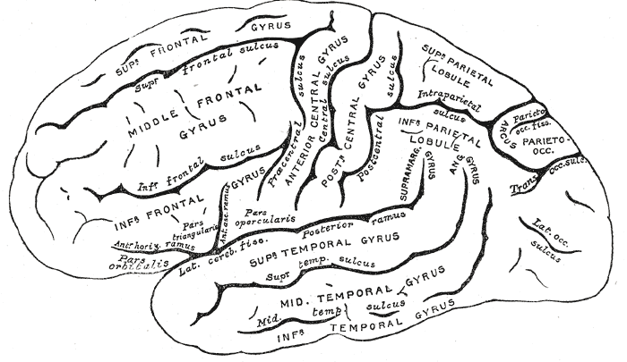
Gyrification is the process of forming the characteristic folds of the cerebral cortex. The peak of such a fold is called a ''gyrus'' (pl. ''gyri''), and its trough is called a ''Sulcus (neuroanatomy), sulcus'' (pl. ''sulci''). The neurons of the cerebral cortex reside in a thin layer of gray matter, only 2–4 mm thick, at the surface of the brain. Much of the interior volume is occupied by white matter, which consists of long axonal projections to and from the cortical neurons residing near the surface. Gyrification allows a larger cortical surface area, and hence greater cognitive functionality to fit inside a smaller cranium. In most mammals, gyrification begins during prenatal development, fetal development. Primates, cetaceans, and ungulates have extensive cortical gyri, with a few species exceptions, while small rodents such as the rat, and mouse have none. Gyrification in some animals, for example the ferret, continues well into postnatal life. Human brain devel ... [...More Info...] [...Related Items...] OR: [Wikipedia] [Google] [Baidu] |
Cerebral Cortex
The cerebral cortex, also known as the cerebral mantle, is the outer layer of neural tissue of the cerebrum of the brain in humans and other mammals. It is the largest site of Neuron, neural integration in the central nervous system, and plays a key role in attention, perception, awareness, thought, memory, language, and consciousness. The six-layered neocortex makes up approximately 90% of the Cortex (anatomy), cortex, with the allocortex making up the remainder. The cortex is divided into left and right parts by the longitudinal fissure, which separates the two cerebral hemispheres that are joined beneath the cortex by the corpus callosum and other commissural fibers. In most mammals, apart from small mammals that have small brains, the cerebral cortex is folded, providing a greater surface area in the confined volume of the neurocranium, cranium. Apart from minimising brain and cranial volume, gyrification, cortical folding is crucial for the Neural circuit, brain circuitry ... [...More Info...] [...Related Items...] OR: [Wikipedia] [Google] [Baidu] |
Gyrus
In neuroanatomy, a gyrus (: gyri) is a ridge on the cerebral cortex. It is generally surrounded by one or more sulci (depressions or furrows; : sulcus). Gyri and sulci create the folded appearance of the brain in humans and other mammals. Structure The gyri are part of a system of folds and ridges that create a larger surface area for the human brain and other mammalian brains. Because the brain is confined to the skull, brain size is limited. Ridges and depressions create folds allowing a larger cortical surface area, and greater cognitive function, to exist in the confines of a smaller cranium. Development The human brain undergoes gyrification during fetal and neonatal development. In embryonic development, all mammalian brains begin as smooth structures derived from the neural tube. A cerebral cortex without surface convolutions is lissencephalic, meaning 'smooth-brained'. As development continues, gyri and sulci begin to take shape on the fetal brain, with deepe ... [...More Info...] [...Related Items...] OR: [Wikipedia] [Google] [Baidu] |
Sulcus (neuroanatomy)
In neuroanatomy, a sulcus (Latin: "furrow"; : sulci) is a shallow Sulcus (morphology), depression or groove in the cerebral cortex. One or more sulci surround a gyrus (pl. gyri), a ridge on the surface of the cortex, creating the characteristic folded appearance of the brain in humans and most other mammals. The larger sulci are also called Sulcus (morphology)#Brain, fissures. The cortex develops in the fetal stage of corticogenesis, preceding the cortical folding stage known as gyrification. The large fissures and main sulci are the first to develop. Mammals that have a folded cortex are known as ''gyrencephalic'', and the small-brained mammals that have a smooth cortex, such as rats and mice are termed lissencephaly, lissencephalic. Structure Sulci, the grooves, and gyri, the folds or ridges, make up the gyrification, folded surface of the cerebral cortex. Larger or deeper sulci are also often termed fissures. The folded cortex creates a larger surface area for the brain in h ... [...More Info...] [...Related Items...] OR: [Wikipedia] [Google] [Baidu] |
Working Memory
Working memory is a cognitive system with a limited capacity that can Memory, hold information temporarily. It is important for reasoning and the guidance of decision-making and behavior. Working memory is often used synonymously with short-term memory, but some theorists consider the two forms of memory distinct, assuming that working memory allows for the manipulation of stored information, whereas short-term memory only refers to the short-term storage of information. Working memory is a theoretical concept central to cognitive psychology, neuropsychology, and neuroscience. History The term "working memory" was coined by George Armitage Miller, Miller, Eugene Galanter, Galanter, and Karl H. Pribram, Pribram, and was used in the 1960s in the context of Computational theory of mind, theories that likened the mind to a computer. In 1968, Atkinson–Shiffrin memory model, Atkinson and Shiffrin used the term to describe their "short-term store". The term short-term store was the na ... [...More Info...] [...Related Items...] OR: [Wikipedia] [Google] [Baidu] |
Central Sulcus
In neuroanatomy, the central sulcus (also central fissure, fissure of Rolando, or Rolandic fissure, after Luigi Rolando) is a sulcus, or groove, in the cerebral cortex in the brains of vertebrates. It is sometimes confused with the longitudinal fissure. The central sulcus is a prominent landmark of the brain, separating the parietal lobe from the frontal lobe and the primary motor cortex from the primary somatosensory cortex. Evolution of the central sulcus The evolution of the central sulcus is theorized to have occurred in mammals when the complete dissociation of the original somatosensory cortex from its mirror duplicate developed in placental mammals such as primates, though the development did not stop there as time progressed the distinction between the two cortices grew. Evolution in primates The central sulcus is more prominent in apes as a result of fine-tuning of the motor system in apes. Hominins (bipedal apes) continued this trend through increased use of the ... [...More Info...] [...Related Items...] OR: [Wikipedia] [Google] [Baidu] |
Precentral Gyrus
The precentral gyrus is a prominent gyrus on the surface of the posterior frontal lobe of the brain. It is the site of the primary motor cortex that in humans is cytoarchitecturally defined as Brodmann area 4. Structure The precentral gyrus lies in front of the postcentral gyrus - mostly on the lateral (convex) side of each cerebral hemisphere - from which it is separated by the central sulcus. Its anterior border is represented by the precentral sulcus, while inferiorly it borders to the lateral sulcus (Sylvian fissure). Medially, it is contiguous with the paracentral lobule. The internal pyramidal layer (layer V) of the precentral cortex contains giant (70-100 micrometers) pyramidal neurons called Betz cells, which send long axons to the contralateral motor nuclei of the cranial nerves and to the lower motor neurons in the ventral horn of the spinal cord. These axons form the corticospinal tract. The Betz cells along with their long axons are referred to as upper motor n ... [...More Info...] [...Related Items...] OR: [Wikipedia] [Google] [Baidu] |
Postcentral Gyrus
In neuroanatomy, the postcentral gyrus is a prominent gyrus in the lateral parietal lobe of the human brain. It is the location of the primary somatosensory cortex, the main sensory receptive area for the sense of touch. Like other sensory areas, there is a map of sensory space in this location, called the '' sensory homunculus''. The primary somatosensory cortex was initially defined from surface stimulation studies of Wilder Penfield, and parallel surface potential studies of Bard, Woolsey, and Marshall. Although initially defined to be roughly the same as Brodmann areas 3, 1, and 2, more recent work by Kaas has suggested that for homogeny with other sensory fields only area 3 should be referred to as "primary somatosensory cortex", as it receives the bulk of the thalamocortical projections from the sensory input fields. Structure The lateral postcentral gyrus is bounded by: * medial longitudinal fissure medially (to the middle) * central sulcus rostrally (in front) ... [...More Info...] [...Related Items...] OR: [Wikipedia] [Google] [Baidu] |
Gestation
Gestation is the period of development during the carrying of an embryo, and later fetus, inside viviparous animals (the embryo develops within the parent). It is typical for mammals, but also occurs for some non-mammals. Mammals during pregnancy can have one or more gestations at the same time, for example in a multiple birth. The time interval of a gestation is called the '' gestation period''. In obstetrics, '' gestational age'' refers to the time since the onset of the last menses, which on average is fertilization age plus two weeks. Mammals In mammals, pregnancy begins when a zygote (fertilized ovum) implants in the female's uterus and ends once the fetus leaves the uterus during labor or an abortion (whether induced or spontaneous). Humans In humans, pregnancy can be defined clinically, biochemically or biologically. Clinically, pregnancy starts from first day of the mother's last period. Biochemically, pregnancy starts when a woman's human chorionic gonado ... [...More Info...] [...Related Items...] OR: [Wikipedia] [Google] [Baidu] |
Action Potential
An action potential (also known as a nerve impulse or "spike" when in a neuron) is a series of quick changes in voltage across a cell membrane. An action potential occurs when the membrane potential of a specific Cell (biology), cell rapidly rises and falls. This depolarization then causes adjacent locations to similarly depolarize. Action potentials occur in several types of Membrane potential#Cell excitability, excitable cells, which include animal cells like neurons and myocyte, muscle cells, as well as some plant cells. Certain endocrine cells such as pancreatic beta cells, and certain cells of the anterior pituitary gland are also excitable cells. In neurons, action potentials play a central role in cell–cell interaction, cell–cell communication by providing for—or with regard to saltatory conduction, assisting—the propagation of signals along the neuron's axon toward axon terminal, synaptic boutons situated at the ends of an axon; these signals can then connect wit ... [...More Info...] [...Related Items...] OR: [Wikipedia] [Google] [Baidu] |
Childbirth
Childbirth, also known as labour, parturition and delivery, is the completion of pregnancy, where one or more Fetus, fetuses exits the Womb, internal environment of the mother via vaginal delivery or caesarean section and becomes a newborn to the world. In 2019, there were about 140.11 million human births globally. In Developed country, developed countries, most deliveries occur in hospitals, while in Developing country, developing countries most are home births. The most common childbirth method worldwide is vaginal delivery. It involves four stages of labour: the cervical effacement, shortening and Cervical dilation, opening of the cervix during the first stage, descent and birth of the baby during the second, the delivery of the placenta during the third, and the recovery of the mother and infant during the fourth stage, which is referred to as the Postpartum period, postpartum. The first stage is characterised by abdominal cramping or also back pain in the case of B ... [...More Info...] [...Related Items...] OR: [Wikipedia] [Google] [Baidu] |
Lateral Sulcus
The lateral sulcus (or lateral fissure, also called Sylvian fissure, after Franciscus Sylvius) is the most prominent sulcus (neuroanatomy), sulcus of each cerebral hemisphere in the human brain. The lateral sulcus (neuroanatomy), sulcus is a deep fissure (anatomy), fissure in each hemisphere that separates the frontal lobe, frontal and parietal lobes from the temporal lobe. The insular cortex lies deep within the lateral sulcus. Anatomy The lateral sulcus divides both the frontal lobe and parietal lobe above from the temporal lobe below. It is in both Cerebral hemisphere, hemispheres of the brain. The lateral Sulcus (neuroanatomy), sulcus is one of the earliest-developing sulci of the human brain, appearing around the fourteenth week of gestational age. The insular cortex lies deep within the lateral sulcus. The lateral sulcus has a number of side branches. Two of the most prominent and most regularly found are the ascending (also called vertical) ramus and the horizontal ramus ... [...More Info...] [...Related Items...] OR: [Wikipedia] [Google] [Baidu] |


