|
First Cervical Vertebra
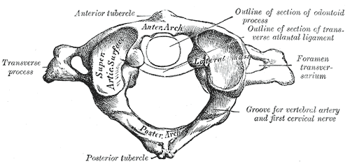
In anatomy, the atlas (C1) is the most superior (first) cervical vertebra of the spine and is located in the neck. The bone is named for Atlas of Greek mythology, just as Atlas bore the weight of the heavens, the first cervical vertebra supports the head. However, the term atlas was first used by the ancient Romans for the seventh cervical vertebra (C7) due to its suitability for supporting burdens. In Greek mythology, Atlas was condemned to bear the weight of the heavens as punishment for rebelling against Zeus. Ancient depictions of Atlas show the globe of the heavens resting at the base of his neck, on C7. Sometime around 1522, anatomists decided to call the first cervical vertebra the atlas. Scholars believe that by switching the designation atlas from the seventh to the first cervical vertebra Renaissance anatomists were commenting that the point of man's burden had shifted from his shoulders to his head—that man's true burden was not a physical load, but rather, his mi ... [...More Info...] [...Related Items...] OR: [Wikipedia] [Google] [Baidu] |
Anatomy
Anatomy () is the branch of morphology concerned with the study of the internal structure of organisms and their parts. Anatomy is a branch of natural science that deals with the structural organization of living things. It is an old science, having its beginnings in prehistoric times. Anatomy is inherently tied to developmental biology, embryology, comparative anatomy, evolutionary biology, and phylogeny, as these are the processes by which anatomy is generated, both over immediate and long-term timescales. Anatomy and physiology, which study the structure and function of organisms and their parts respectively, make a natural pair of related disciplines, and are often studied together. Human anatomy is one of the essential basic sciences that are applied in medicine, and is often studied alongside physiology. Anatomy is a complex and dynamic field that is constantly evolving as discoveries are made. In recent years, there has been a significant increase in the use of ... [...More Info...] [...Related Items...] OR: [Wikipedia] [Google] [Baidu] |
Warren Harmon Lewis
Warren Harmon Lewis (June 17, 1870 – July 3, 1964) was an American embryologist and cell biologist. He became professor of physiological anatomy at the Johns Hopkins University School of Medicine in 1913 and from 1919 to 1940, he worked along with his wife Margaret Reed Lewis at the Carnegie Institute of Washington. Life and work Lewis was born in Suffield to John Lewis and Adelaide Eunice Harmon. The family moved to Chicago where his father practiced law. Lewis went to Oak Park public schools and also attended the Chicago Manual Training School. He was interested in botany as a boy and collected plants. He joined the University of Michigan in 1890 where he studied zoology as also French, German and mathematics. He also took an interest in sports and music. In 1894 he obtained a microscope and was influenced by Jacob Reighard to study medicine. In 1896 he joined Johns Hopkins University in the school of medicine. He spent summers at the Marine Biological Laboratory at Woo ... [...More Info...] [...Related Items...] OR: [Wikipedia] [Google] [Baidu] |
Ligamentum Nuchae
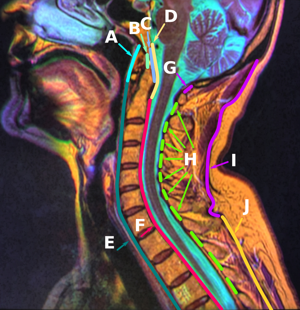
The nuchal ligament is a ligament at the back of the neck that is continuous with the supraspinous ligament. Structure The nuchal ligament extends from the external occipital protuberance on the skull and median nuchal line to the spinous process of the seventh cervical vertebra in the lower part of the neck. From the anterior border of the nuchal ligament, a fibrous lamina is given off. This is attached to the posterior tubercle of the atlas, and to the spinous processes of the cervical vertebrae, and forms a septum between the muscles on either side of the neck. The trapezius and splenius capitis muscle attach to the nuchal ligament. Function It is a tendon-like structure that has developed independently in humans and other animals well adapted for running. In some four-legged animals, particularly ungulates and canids, the nuchal ligament serves to sustain the weight of the head. Clinical significance In Chiari malformation treatment, decompression and duraplasty ... [...More Info...] [...Related Items...] OR: [Wikipedia] [Google] [Baidu] |
Recti Capitis Posteriores Minores
The rectus capitis posterior minor (or rectus capitis posticus minor) is a muscle in the upper back part of the neck. It is one of the suboccipital muscles. Its inferior attachment is at the posterior arch of atlas; its superior attachment is onto the occipital bone at and below the inferior nuchal line. The muscle is innervated by the suboccipital nerve (the posterior ramus of first cervical spinal nerve). The muscle acts as a weak extensor of the head. Anatomy The rectus capitis posterior major muscle is one of the suboccipital muscles. The muscle extends vertically superior-ward from its inferiro attachment to its superior attachment. The muscle becomes broader superiorly. Attachments The inferior attachment is (by a narrow tendon) onto the posterior tubercle of the posterior arch of atlas. Its superior attachment is onto the medial portion of the inferior nuchal line and the external surface of the occipital bone inferior to it (between this line superiorly and ... [...More Info...] [...Related Items...] OR: [Wikipedia] [Google] [Baidu] |
Rudiment (biology)
Vestigiality is the retention, during the process of evolution, of genetically determined structures or attributes that have lost some or all of the ancestral function in a given species. Assessment of the vestigiality must generally rely on comparison with homologous features in related species. The emergence of vestigiality occurs by normal evolutionary processes, typically by loss of function of a feature that is no longer subject to positive selection pressures when it loses its value in a changing environment. The feature may be selected against more urgently when its function becomes definitively harmful, but if the lack of the feature provides no advantage, and its presence provides no disadvantage, the feature may not be phased out by natural selection and persist across species. Examples of vestigial structures (also called degenerate, atrophied, or rudimentary organs) are the loss of functional wings in island-dwelling birds; the human vomeronasal organ; and the h ... [...More Info...] [...Related Items...] OR: [Wikipedia] [Google] [Baidu] |
Occipital Bone
The occipital bone () is a neurocranium, cranial dermal bone and the main bone of the occiput (back and lower part of the skull). It is trapezoidal in shape and curved on itself like a shallow dish. The occipital bone lies over the occipital lobes of the cerebrum. At the base of the skull in the occipital bone, there is a large oval opening called the foramen magnum, which allows the passage of the spinal cord. Like the other cranial bones, it is classed as a flat bone. Due to its many attachments and features, the occipital bone is described in terms of separate parts. From its front to the back is the basilar part of occipital bone, basilar part, also called the basioccipital, at the sides of the foramen magnum are the lateral parts of occipital bone, lateral parts, also called the exoccipitals, and the back is named as the squamous part of occipital bone, squamous part. The basilar part is a thick, somewhat quadrilateral piece in front of the foramen magnum and directed toward ... [...More Info...] [...Related Items...] OR: [Wikipedia] [Google] [Baidu] |
Anterior Atlantoaxial Ligament
The anterior atlantoaxial ligament is a strong membrane, fixed above the lower border of the anterior arch of the atlas; below, to the front of the body of the axis. It is strengthened in the middle line by a rounded cord, which connects the tubercle on the anterior arch of the atlas to the body of the axis. It is a continuation upward of the anterior longitudinal ligament. Structure Anatomical relations The anterior atlantoaxial ligament is situated anterior to the longus capitis muscle The longus capitis muscle (Latin for ''long muscle of the head'', alternatively rectus capitis anticus major) is broad and thick above, narrow below, and arises by four tendinous slips, from the anterior tubercles of the transverse processes of t .... See also * Atlanto-axial joint References External links Description at spineuniverse.com Ligaments of the head and neck Bones of the vertebral column {{Portal bar, Anatomy ... [...More Info...] [...Related Items...] OR: [Wikipedia] [Google] [Baidu] |
Anterior Atlantooccipital Membrane
The anterior atlantooccipital membrane (anterior atlantooccipital ligament) is a broad, dense membrane extending between the anterior margin of the foramen magnum (superiorly), and (the superior margin of) the anterior arch of atlas (inferiorly). The membrane helps limit excessive movement at the atlanto-occipital joints. Anatomy Structure It is composed of broad, densely woven fibers; especially towards the midline where the membrane is continuous medially with the anterior longitudinal ligament. It is innervated by the cervical spinal nerve 1. Relations Medially, it is continuous with the anterior longitudinal ligament. Laterally, it is blends with either articular capsule In anatomy, a joint capsule or articular capsule is an envelope surrounding a synovial joint. [...More Info...] [...Related Items...] OR: [Wikipedia] [Google] [Baidu] |
Odontoid Process
In anatomy, the axis (from Latin ''axis'', "axle") is the second cervical vertebra (C2) of the spine, immediately inferior to the atlas, upon which the head rests. The spinal cord passes through the axis. The defining feature of the axis is its strong bony protrusion known as the dens, which rises from the superior aspect of the bone. Structure The body is deeper in front or in the back and is prolonged downward anteriorly to overlap the upper and front part of the third vertebra. It presents a median longitudinal ridge in front, separating two lateral depressions for the attachment of the longus colli muscles. Dens The dens, also called the odontoid process, or the peg, is the most pronounced projecting feature of the axis. The dens exhibits a slight constriction where it joins the main body of the vertebra. The condition where the dens is separated from the body of the axis is called ''os odontoideum'' and may cause nerve and circulation compression syndrome. On its ante ... [...More Info...] [...Related Items...] OR: [Wikipedia] [Google] [Baidu] |
Anterior Longitudinal Ligament
The anterior longitudinal ligament is a ligament that extends across the anterior/ventral aspect of the vertebral bodies and intervertebral discs the spine. It may be partially cut to treat certain abnormal curvatures in the vertebral column, such as kyphosis. Anatomy The anterior longitudinal ligament extends superoinferiorly between the basiocciput of the skull and the anterior tubercle of the atlas (cervical vertebra C1) superiorly, and the superior part of the sacrum inferiorly; inferiorly, it ends at the sacral promontory. It broadens inferiorly. Inferiorly, it becomes continuous with the anterior sacrococcygeal ligament. Superiorly, between the skull and atlas, the ligament is continuous laterally with the anterior atlantooccipital membrane. The ligament is thick and slightly more narrow over the vertebral bodies and thinner but slightly wider over the intervertebral discs. It tends to be narrower and thicker around thoracic vertebrae, and wider and thinner around ... [...More Info...] [...Related Items...] OR: [Wikipedia] [Google] [Baidu] |
Muscle
Muscle is a soft tissue, one of the four basic types of animal tissue. There are three types of muscle tissue in vertebrates: skeletal muscle, cardiac muscle, and smooth muscle. Muscle tissue gives skeletal muscles the ability to muscle contraction, contract. Muscle tissue contains special Muscle contraction, contractile proteins called actin and myosin which interact to cause movement. Among many other muscle proteins, present are two regulatory proteins, troponin and tropomyosin. Muscle is formed during embryonic development, in a process known as myogenesis. Skeletal muscle tissue is striated consisting of elongated, multinucleate muscle cells called muscle fibers, and is responsible for movements of the body. Other tissues in skeletal muscle include tendons and perimysium. Smooth and cardiac muscle contract involuntarily, without conscious intervention. These muscle types may be activated both through the interaction of the central nervous system as well as by innervation ... [...More Info...] [...Related Items...] OR: [Wikipedia] [Google] [Baidu] |


