|
Femur Fracture
A femoral fracture is a bone fracture that involves the femur. They are typically sustained in high-impact trauma, such as Traffic collision, car crashes, due to the large amount of force needed to break the bone. Fractures of the diaphysis, or middle of the femur, are managed differently from those at the head, neck, and trochanter; those are conventionally called hip fractures (because they involve the hip joint region). Thus, mentions of femoral fracture in medicine usually refer implicitly to femoral fractures at the shaft or distally. Signs and symptoms Fractures are commonly obvious, since femoral fractures are often caused by high energy trauma. Signs of fracture include swelling, deformity, and shortening of the leg. Extensive soft-tissue injury, bleeding, and shock are common. The most common symptom is severe pain, which prevents movement of the leg. Diagnosis Physical exam Femoral shaft fractures occur during extensive trauma, and they can act as distracting ... [...More Info...] [...Related Items...] OR: [Wikipedia] [Google] [Baidu] |
Bone Fracture
A bone fracture (abbreviated FRX or Fx, Fx, or #) is a medical condition in which there is a partial or complete break in the continuity of any bone in the body. In more severe cases, the bone may be broken into several fragments, known as a ''comminuted fracture''. An open fracture (or compound fracture) is a bone fracture where the broken bone breaks through the skin. A bone fracture may be the result of high force Impact force, impact or Stress fracture, stress, or a minimal trauma injury as a result of certain medical conditions that weaken the bones, such as osteoporosis, osteopenia, bone cancer, or osteogenesis imperfecta, where the fracture is then properly termed a pathologic fracture. Most bone fractures require urgent medical attention to prevent further injury. Signs and symptoms Although bone tissue contains no nociceptors, pain receptors, a bone fracture is painful for several reasons: * Breaking in the continuity of the periosteum, with or without similar disconti ... [...More Info...] [...Related Items...] OR: [Wikipedia] [Google] [Baidu] |
Winquist And Hansen Classification
The Winquist and Hansen classification is a system of categorizing femoral shaft fractures based upon the degree of comminution Comminution is the reduction of solid materials from one average particle size to a smaller average particle size, by crushing, grinding, cutting, vibrating, or other processes. Comminution is related to pulverization and grinding. All use m .... Classification References Bone fractures Orthopedic classifications Injuries of hip and thigh Eponyms in medicine {{Orthopedics-stub ... [...More Info...] [...Related Items...] OR: [Wikipedia] [Google] [Baidu] |
Osteoporosis
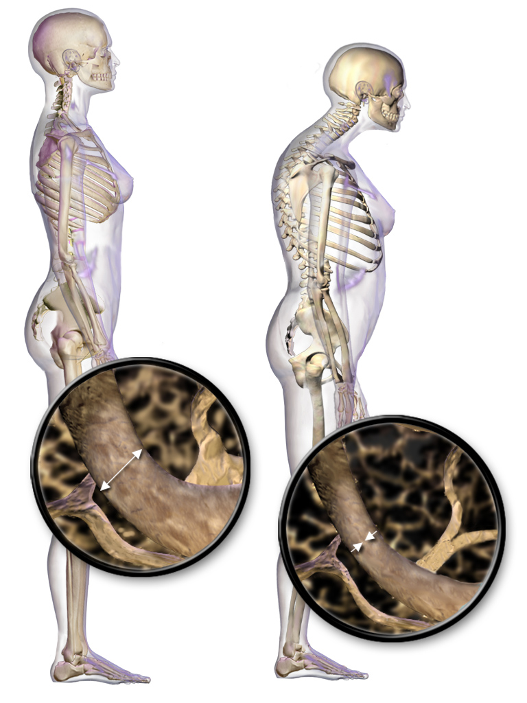
Osteoporosis is a systemic skeletal disorder characterized by low bone mass, micro-architectural deterioration of bone tissue leading to more porous bone, and consequent increase in Bone fracture, fracture risk. It is the most common reason for a broken bone among the Old age, elderly. Bones that commonly break include the vertebrae in the Vertebral column, spine, the bones of the forearm, the wrist, and the hip. Until a broken bone occurs there are typically no symptoms. Bones may weaken to such a degree that a break may occur with minor stress or spontaneously. After the broken bone heals, some people may have chronic pain and a decreased ability to carry out normal activities. Osteoporosis may be due to lower-than-normal peak bone mass, maximum bone mass and greater-than-normal bone loss. Bone loss increases after menopause in women due to lower levels of estrogen, and after andropause in older men due to lower levels of testosterone. Osteoporosis may also occur due to a ... [...More Info...] [...Related Items...] OR: [Wikipedia] [Google] [Baidu] |
Acute Respiratory Distress Syndrome
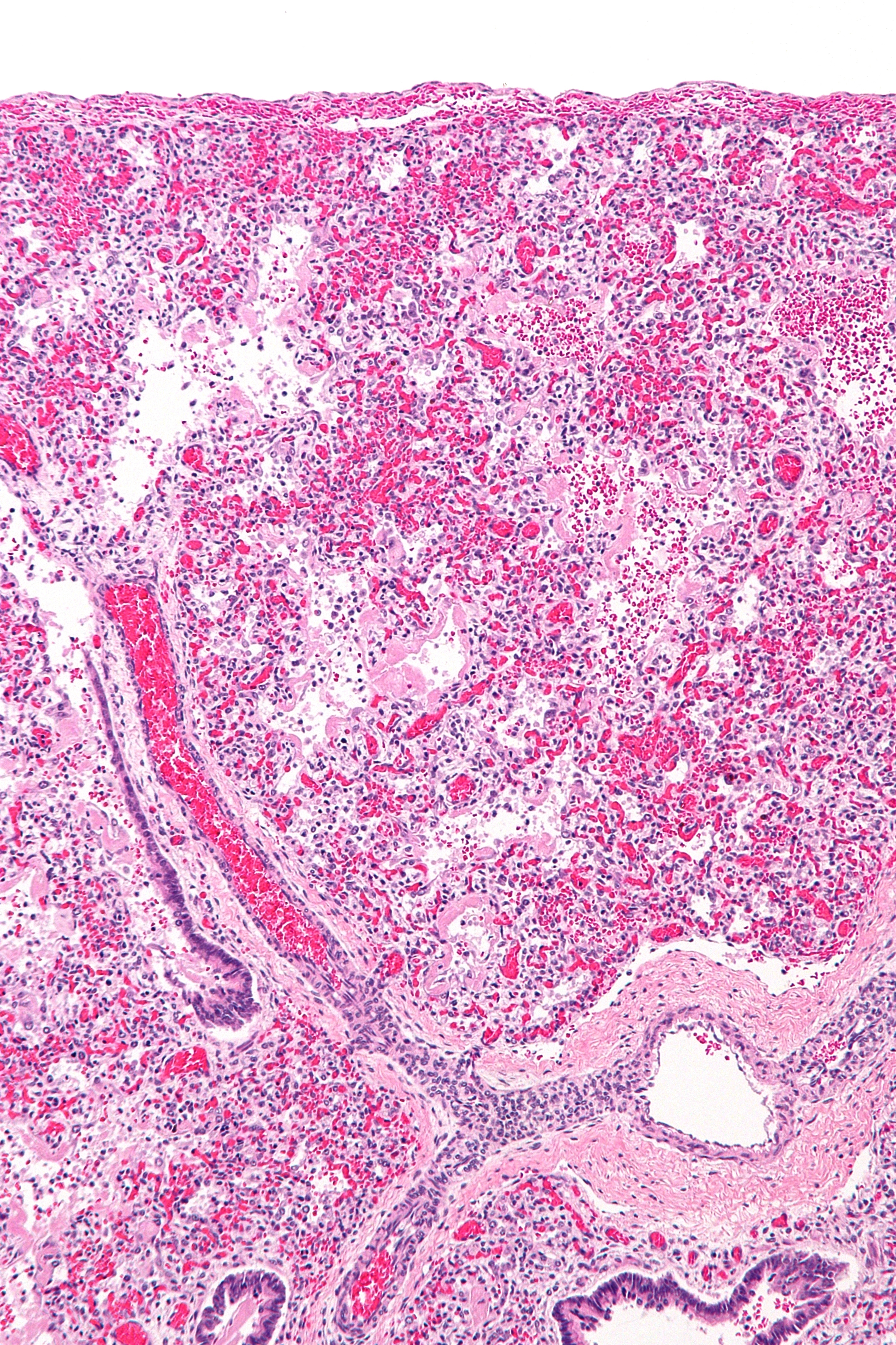
Acute respiratory distress syndrome (ARDS) is a type of respiratory failure characterized by rapid onset of widespread inflammation in the lungs. Symptoms include shortness of breath (dyspnea), rapid breathing (tachypnea), and bluish skin coloration (cyanosis). For those who survive, a decreased quality of life is common. Causes may include sepsis, pancreatitis, trauma, pneumonia, and aspiration. The underlying mechanism involves diffuse injury to cells which form the barrier of the microscopic air sacs of the lungs, surfactant dysfunction, activation of the immune system, and dysfunction of the body's regulation of blood clotting. In effect, ARDS impairs the lungs' ability to exchange oxygen and carbon dioxide. Adult diagnosis is based on a PaO2/FiO2 ratio (ratio of partial pressure arterial oxygen and fraction of inspired oxygen) of less than 300 mm Hg despite a positive end-expiratory pressure (PEEP) of more than 5 cm H2O. Cardiogenic pulmonary edema, ... [...More Info...] [...Related Items...] OR: [Wikipedia] [Google] [Baidu] |
Fat Embolism
Fat embolism syndrome occurs when adipose tissue, fat enters the blood stream (fat embolism) and results in symptoms. Symptoms generally begin within a day. This may include a petechia, petechial rash, decreased level of consciousness, and shortness of breath. Other symptoms may include fever and decreased urine output. The risk of death is about 10%. Fat embolism most commonly occurs as a result of bone fracture, fractures of bones such as the femur or pelvis. Other potential causes include pancreatitis, orthopedic surgery, bone marrow transplant, and liposuction. The underlying mechanism involves widespread inflammation. Diagnosis is based on symptoms. Treatment is mostly supportive care. This may involve oxygen therapy, intravenous fluids, albumin, and mechanical ventilation. While small amounts of fat commonly occur in the blood after a bone fracture, fat embolism syndrome is rare. The condition was first diagnosed in 1862 by Zenker. Signs and symptoms The symptoms of fat ... [...More Info...] [...Related Items...] OR: [Wikipedia] [Google] [Baidu] |
External Fixation
External fixation is a surgical treatment wherein Kirschner wire, Kirschner pins and wires are inserted and affixed into bone and then exit the body to be attached to an external apparatus composed of rings and threaded rods — the Ilizarov apparatus, the Taylor Spatial Frame, and the Octopod External Fixator — which immobilises the damaged limb to facilitate healing. As an alternative to internal fixation, wherein bone-stabilising mechanical components are surgically emplaced in the body of the patient, external fixation is used to stabilize bone tissues and soft tissues at a distance from the site of the injury. History In Classical Greece, the physician Hippocrates described an external fixation apparatus composed of leather rings connected with four wooden rods from a Cornel tree to splint the fracture of a tibia, tibia bone. In 1840, Jean-Francois Malgaigne described a spike driven into the tibia and held by straps to immobilise a fractured tibia. In 1843 he used a claw ... [...More Info...] [...Related Items...] OR: [Wikipedia] [Google] [Baidu] |
Comorbidity
In medicine, comorbidity refers to the simultaneous presence of two or more medical conditions in a patient; often co-occurring (that is, concomitant or concurrent) with a primary condition. It originates from the Latin term (meaning "sickness") prefixed with ("together") and suffixed with ''-ity'' (to indicate a state or condition). Comorbidity includes all additional ailments a patient may experience alongside their primary diagnosis, which can be either physiological or psychological in nature. In the context of mental health, comorbidity frequently refers to the concurrent existence of mental disorders, for example, the co-occurrence of depressive and anxiety disorders. The concept of multimorbidity is related to comorbidity but is different in its definition and approach, focusing on the presence of multiple diseases or conditions in a patient without the need to specify one as primary. Definition The term "comorbid" has three definitions: # to indicate a medical con ... [...More Info...] [...Related Items...] OR: [Wikipedia] [Google] [Baidu] |
Pelvis
The pelvis (: pelves or pelvises) is the lower part of an Anatomy, anatomical Trunk (anatomy), trunk, between the human abdomen, abdomen and the thighs (sometimes also called pelvic region), together with its embedded skeleton (sometimes also called bony pelvis or pelvic skeleton). The pelvic region of the trunk includes the bony pelvis, the pelvic cavity (the space enclosed by the bony pelvis), the pelvic floor, below the pelvic cavity, and the perineum, below the pelvic floor. The pelvic skeleton is formed in the area of the back, by the sacrum and the coccyx and anteriorly and to the left and right sides, by a pair of hip bones. The two hip bones connect the spine with the lower limbs. They are attached to the sacrum posteriorly, connected to each other anteriorly, and joined with the two femurs at the hip joints. The gap enclosed by the bony pelvis, called the pelvic cavity, is the section of the body underneath the abdomen and mainly consists of the reproductive organs and ... [...More Info...] [...Related Items...] OR: [Wikipedia] [Google] [Baidu] |
Traction Splint
A traction splint most commonly refers to a splinting device that uses straps attaching over the pelvis or hip as an anchor, a metal rod(s) to mimic normal bone stability and limb length, and a mechanical device to apply traction (used in an attempt to reduce pain, realign the limb, and minimize vascular and neurological complication) to the limb. The use of traction splints to treat complete long bone fractures of the femur is common in prehospital care. Evidence to support their usage, however, is poor. A dynamic traction splint has also been developed for intra-articular fractures of the phalanges of the hand. Medical uses Traction splints are most commonly used for fractures of the femur (or upper leg bone). For these fractures they may reduce pain and decrease the amount of bleeding which occurs into the soft tissues of the leg. Some state that they are appropriate for middle tibia fractures which are displaced or bent. Others state they should not be used for lower ... [...More Info...] [...Related Items...] OR: [Wikipedia] [Google] [Baidu] |
Comminution
Comminution is the reduction of solid materials from one average particle size to a smaller average particle size, by crushing, grinding, cutting, vibrating, or other processes. Comminution is related to pulverization and grinding. All use mechanical devices, and many types of mills have been invented. Concomitant with size reduction, comminution increases the surface area of the solid. For example, a pulverizer mill is used to pulverize coal for combustion in the steam-generating furnaces of coal power plants. A cement mill produces finely ground ingredients for portland cement. A hammer mill is used on farms for grinding grain and chaff for animal feed. A demolition pulverizer is an attachment for an excavator to break up large pieces of concrete. Comminution is important in mineral processing, where rocks are broken into small particles to help liberate the ore from gangue. Comminution or grinding is also important in ceramics, electronics, and battery research. Mech ... [...More Info...] [...Related Items...] OR: [Wikipedia] [Google] [Baidu] |
Femoral Shaft
In human anatomy, the body of femur (or shaft of femur) is the almost cylindrical, long part of the femur. It is a little broader above than in the center, broadest and somewhat flattened from before backward below. It is slightly arched, so as to be convex in front, and concave behind, where it is strengthened by a prominent longitudinal ridge, the linea aspera. It presents for examination three borders, separating three surfaces. Of the borders, one, the linea aspera, is posterior, one is medial, and the other, lateral. Borders The borders of the femur are the linea aspera, a medial border, and a lateral border. Linea aspera border The linea aspera is a prominent longitudinal ridge or crest, on the middle third of the bone, presenting a medial and a lateral lip, and a narrow rough, intermediate line. Above, the linea aspera is prolonged by three ridges. The lateral ridge termed the gluteal tuberosity is very rough, and runs almost vertically upward to the base of the greate ... [...More Info...] [...Related Items...] OR: [Wikipedia] [Google] [Baidu] |
Femur
The femur (; : femurs or femora ), or thigh bone is the only long bone, bone in the thigh — the region of the lower limb between the hip and the knee. In many quadrupeds, four-legged animals the femur is the upper bone of the hindleg. The Femoral head, top of the femur fits into a socket in the pelvis called the hip joint, and the bottom of the femur connects to the shinbone (tibia) and kneecap (patella) to form the knee. In humans the femur is the largest and thickest bone in the body. Structure The femur is the only bone in the upper Human leg, leg. The two femurs converge Anatomical terms of location, medially toward the knees, where they articulate with the Anatomical terms of location, proximal ends of the tibiae. The angle at which the femora converge is an important factor in determining the femoral-tibial angle. In females, thicker pelvic bones cause the femora to converge more than in males. In the condition genu valgum, ''genu valgum'' (knock knee), the femurs conve ... [...More Info...] [...Related Items...] OR: [Wikipedia] [Google] [Baidu] |


