|
Echoencephalography
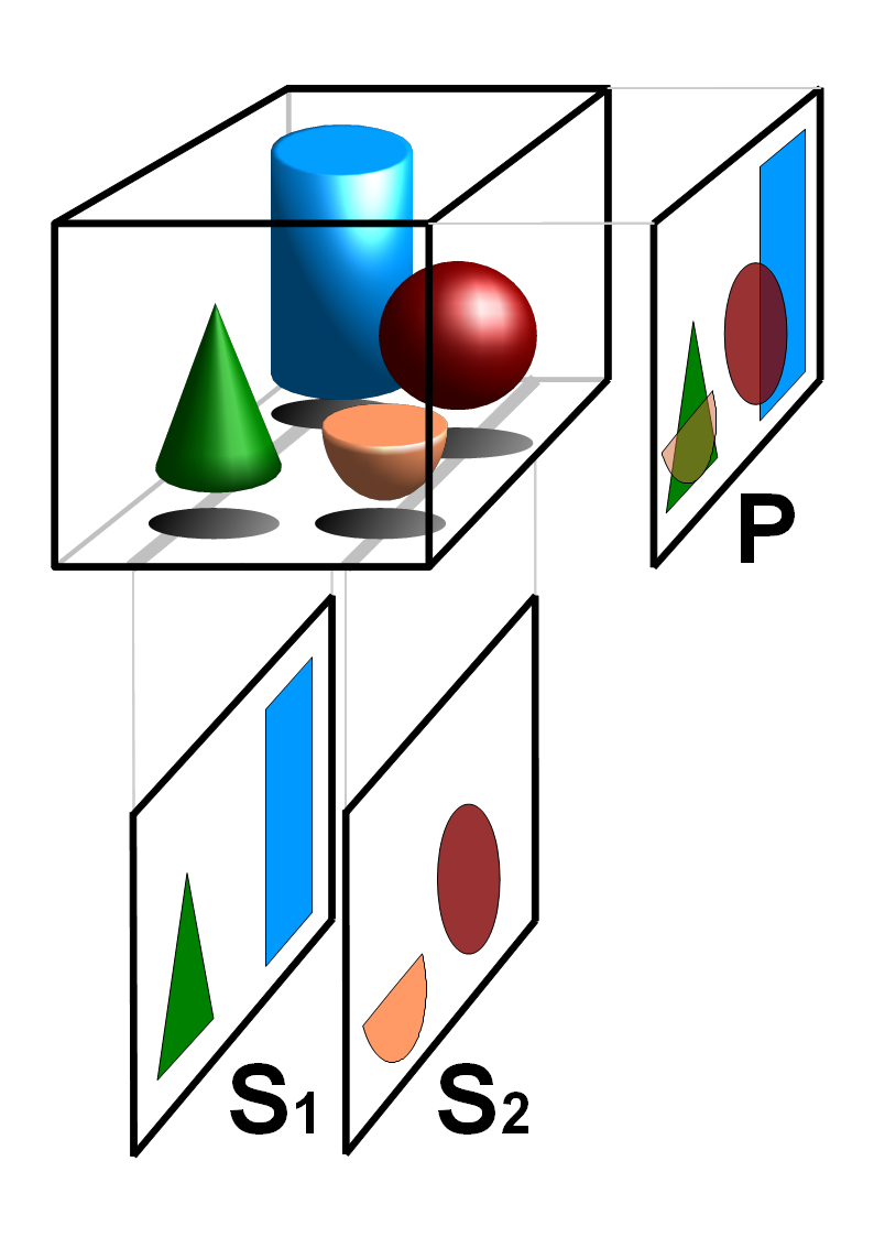
Echoencephalography is the detailing of interfaces in the brain by means of ultrasonic waves. See also * Electroencephalography (EEG) *Magnetoencephalography (MEG) *Tomography * Medical ultrasonography * Echocardiography, magnetocardiography Magnetocardiography (MCG) is a technique to measure the magnetic fields produced by electrical currents in the heart using extremely sensitive devices such as the superconducting quantum interference device (SQUID). If the magnetic field is mea ... (MCG), and electrocardiography (ECG or EKG), for diagnosing heart problems References Medical ultrasonography {{med-imaging-stub ... [...More Info...] [...Related Items...] OR: [Wikipedia] [Google] [Baidu] |
Brain
A brain is an organ that serves as the center of the nervous system in all vertebrate and most invertebrate animals. It is located in the head, usually close to the sensory organs for senses such as vision. It is the most complex organ in a vertebrate's body. In a human, the cerebral cortex contains approximately 14–16 billion neurons, and the estimated number of neurons in the cerebellum is 55–70 billion. Each neuron is connected by synapses to several thousand other neurons. These neurons typically communicate with one another by means of long fibers called axons, which carry trains of signal pulses called action potentials to distant parts of the brain or body targeting specific recipient cells. Physiologically, brains exert centralized control over a body's other organs. They act on the rest of the body both by generating patterns of muscle activity and by driving the secretion of chemicals called hormones. This centralized control allows rapid and coordinated responses ... [...More Info...] [...Related Items...] OR: [Wikipedia] [Google] [Baidu] |
Electroencephalography
Electroencephalography (EEG) is a method to record an electrogram of the spontaneous electrical activity of the brain. The biosignals detected by EEG have been shown to represent the postsynaptic potentials of pyramidal neurons in the neocortex and allocortex. It is typically non-invasive, with the EEG electrodes placed along the scalp (commonly called "scalp EEG") using the International 10-20 system, or variations of it. Electrocorticography, involving surgical placement of electrodes, is sometimes called "intracranial EEG". Clinical interpretation of EEG recordings is most often performed by visual inspection of the tracing or quantitative EEG analysis. Voltage fluctuations measured by the EEG bioamplifier and electrodes allow the evaluation of normal brain activity. As the electrical activity monitored by EEG originates in neurons in the underlying brain tissue, the recordings made by the electrodes on the surface of the scalp vary in accordance with their orientation ... [...More Info...] [...Related Items...] OR: [Wikipedia] [Google] [Baidu] |
Magnetoencephalography
Magnetoencephalography (MEG) is a functional neuroimaging technique for mapping brain activity by recording magnetic fields produced by electrical currents occurring naturally in the brain, using very sensitive magnetometers. Arrays of SQUIDs (superconducting quantum interference devices) are currently the most common magnetometer, while the SERF (spin exchange relaxation-free) magnetometer is being investigated for future machines. Applications of MEG include basic research into perceptual and cognitive brain processes, localizing regions affected by pathology before surgical removal, determining the function of various parts of the brain, and neurofeedback. This can be applied in a clinical setting to find locations of abnormalities as well as in an experimental setting to simply measure brain activity. History MEG signals were first measured by University of Illinois physicist David Cohen in 1968, before the availability of the SQUID, using a copper induction coil as the ... [...More Info...] [...Related Items...] OR: [Wikipedia] [Google] [Baidu] |
Tomography
Tomography is imaging by sections or sectioning that uses any kind of penetrating wave. The method is used in radiology, archaeology, biology, atmospheric science, geophysics, oceanography, plasma physics, materials science, astrophysics, quantum information, and other areas of science. The word ''tomography'' is derived from Ancient Greek τόμος ''tomos'', "slice, section" and γράφω ''graphō'', "to write" or, in this context as well, "to describe." A device used in tomography is called a tomograph, while the image produced is a tomogram. In many cases, the production of these images is based on the mathematical procedure tomographic reconstruction, such as X-ray computed tomography technically being produced from multiple projectional radiographs. Many different reconstruction algorithms exist. Most algorithms fall into one of two categories: filtered back projection (FBP) and iterative reconstruction (IR). These procedures give inexact results: they repres ... [...More Info...] [...Related Items...] OR: [Wikipedia] [Google] [Baidu] |
Medical Ultrasonography
Medical ultrasound includes diagnostic techniques (mainly imaging techniques) using ultrasound, as well as therapeutic applications of ultrasound. In diagnosis, it is used to create an image of internal body structures such as tendons, muscles, joints, blood vessels, and internal organs, to measure some characteristics (e.g. distances and velocities) or to generate an informative audible sound. Its aim is usually to find a source of disease or to exclude pathology. The usage of ultrasound to produce visual images for medicine is called medical ultrasonography or simply sonography. The practice of examining pregnant women using ultrasound is called obstetric ultrasonography, and was an early development of clinical ultrasonography. Ultrasound is composed of sound waves with frequencies which are significantly higher than the range of human hearing (>20,000 Hz). Ultrasonic images, also known as sonograms, are created by sending pulses of ultrasound into tissue usin ... [...More Info...] [...Related Items...] OR: [Wikipedia] [Google] [Baidu] |
Echocardiography
An echocardiography, echocardiogram, cardiac echo or simply an echo, is an ultrasound of the heart. It is a type of medical imaging of the heart, using standard ultrasound or Doppler ultrasound. Echocardiography has become routinely used in the diagnosis, management, and follow-up of patients with any suspected or known heart diseases. It is one of the most widely used diagnostic imaging modalities in cardiology. It can provide a wealth of helpful information, including the size and shape of the heart (internal chamber size quantification), pumping capacity, location and extent of any tissue damage, and assessment of valves. An echocardiogram can also give physicians other estimates of heart function, such as a calculation of the cardiac output, ejection fraction, and diastolic function (how well the heart relaxes). Echocardiography is an important tool in assessing wall motion abnormality in patients with suspected cardiac disease. It is a tool which helps in reaching an ... [...More Info...] [...Related Items...] OR: [Wikipedia] [Google] [Baidu] |
Magnetocardiography
Magnetocardiography (MCG) is a technique to measure the magnetic fields produced by electrical currents in the heart using extremely sensitive devices such as the superconducting quantum interference device (SQUID). If the magnetic field is measured using a multichannel device, a map of the magnetic field is obtained over the chest; from such a map, using mathematical algorithms that take into account the conductivity structure of the torso, it is possible to locate the source of the activity. For example, sources of abnormal rhythms or arrhythmia may be located using MCG. History The first MCG measurements were made by Baule and McFee using two large coils placed over the chest, connected in opposition to cancel out the relatively large magnetic background. Heart signals were indeed seen, but were very noisy. The next development was by David Cohen, who used a magnetically shielded room to reduce the background, and a smaller coil with better electronics; the heart signals ... [...More Info...] [...Related Items...] OR: [Wikipedia] [Google] [Baidu] |
Electrocardiography
Electrocardiography is the process of producing an electrocardiogram (ECG or EKG), a recording of the heart's electrical activity. It is an electrogram of the heart which is a graph of voltage versus time of the electrical activity of the heart using electrodes placed on the skin. These electrodes detect the small electrical changes that are a consequence of cardiac muscle depolarization followed by repolarization during each cardiac cycle (heartbeat). Changes in the normal ECG pattern occur in numerous cardiac abnormalities, including cardiac rhythm disturbances (such as atrial fibrillation and ventricular tachycardia), inadequate coronary artery blood flow (such as myocardial ischemia and myocardial infarction), and electrolyte disturbances (such as hypokalemia and hyperkalemia). Traditionally, "ECG" usually means a 12-lead ECG taken while lying down as discussed below. However, other devices can record the electrical activity of the heart such as a Holter monitor but al ... [...More Info...] [...Related Items...] OR: [Wikipedia] [Google] [Baidu] |


