|
Epithalamus
The epithalamus (: epithalami) is a posterior (dorsal) segment of the diencephalon. The epithalamus includes the habenular nuclei, the stria medullaris, the anterior and posterior paraventricular nuclei, the posterior commissure, and the pineal gland. Functions The function of the epithalamus is to connect the limbic system to other parts of the brain. The epithalamus also serves as a connecting point for the dorsal diencephalic conduction system, which is responsible for carrying information from the limbic forebrain to limbic midbrain structures. Some functions of its components include the secretion of melatonin from the pineal gland (circadian rhythms), regulation of motor pathways and emotions, and how energy is conserved in the body. A study has shown that the lateral habenula, in the epithalamus, produces spontaneous theta oscillatory activity that was correlated with theta oscillation in the hippocampus. The same study also found that the increase in theta waves in ... [...More Info...] [...Related Items...] OR: [Wikipedia] [Google] [Baidu] |
Pineal Gland
The pineal gland (also known as the pineal body or epiphysis cerebri) is a small endocrine gland in the brain of most vertebrates. It produces melatonin, a serotonin-derived hormone, which modulates sleep, sleep patterns following the diurnal cycles. The shape of the gland resembles a pine cone, which gives it its name. The pineal gland is located in the epithalamus, near the center of the brain, between the two cerebral hemisphere, hemispheres, tucked in a groove where the two halves of the thalamus join. It is one of the neuroendocrinology, neuroendocrine Circumventricular organs, secretory circumventricular organs in which capillaries are mostly Vascular permeability, permeable to solutes in the blood. The pineal gland is present in almost all vertebrates, but is absent in Protochordata, protochordates in which there is a simple pineal homologue. The hagfish, archaic vertebrates, lack a pineal gland. In some species of amphibians and reptiles, the gland is linked to a light-s ... [...More Info...] [...Related Items...] OR: [Wikipedia] [Google] [Baidu] |
Diencephalon
In the human brain, the diencephalon (or interbrain) is a division of the forebrain (embryonic ''prosencephalon''). It is situated between the telencephalon and the midbrain (embryonic ''mesencephalon''). The diencephalon has also been known as the tweenbrain in older literature. It consists of structures that are on either side of the third ventricle, including the thalamus, the hypothalamus, the epithalamus and the subthalamus. The diencephalon is one of the main brain vesicle, vesicles of the brain formed during human embryonic development, embryonic development. During the third week of development a neural tube is created from the ectoderm, one of the three primary germ layers, and forms three main vesicles: the prosencephalon, the Midbrain, mesencephalon and the Hindbrain, rhombencephalon. The prosencephalon gradually divides into the telencephalon (the cerebrum) and the diencephalon. Structure The diencephalon consists of the following structures: * Thalamus * Hypothalamus ... [...More Info...] [...Related Items...] OR: [Wikipedia] [Google] [Baidu] |
Lateral Habenula
The habenula (diminutive of Latin meaning rein) is a small bilateral neuronal structure in the brain of vertebrates, that has also been called a microstructure since it is no bigger than a pea. The naming as little rein describes its elongated shape in the epithalamus, where it borders the third ventricle, and lies in front of the pineal gland. Although it is a microstructure each habenular nucleus is divided into two distinct regions of nuclei, a medial habenula (MHb), and a lateral habenula (LHb) both having different neuronal populations, inputs, and outputs. The medial habenula can be subdivided into five subnuclei, the lateral habenula into four subnuclei. Research has shown morphological complexity in the MHb and LHb. Different inputs to the MHb are discriminated between the different subnuclei. In the two regions of nuclei there is a difference in gene expression giving different functions to each. The habenula is a conserved structure across vertebrates. In mammals it ... [...More Info...] [...Related Items...] OR: [Wikipedia] [Google] [Baidu] |
Stria Medullaris Of Thalamus
The stria medullaris (SM), (Latin, furrow and pith or marrow) is a part of the epithalamus and forms a bilateral white matter tract of the initial segment of the dorsal diencephalic conduction system (DDCS). It contains afferent fibers from the septal nuclei, lateral preoptico- hypothalamic region, and anterior thalamic nuclei to the habenula. It forms a horizontal ridge on the medial surface of the thalamus on the border between dorsal and medial surfaces of the thalamus. The SM, in conjunction with the habenula and the habenular commissure, forms the habenular trigone. It is considered to be the primary afferent of the DDCS. History Vesalius showed it in his 1543 De Humani Corporis Fabrica Libri Septem. It first received its present name from Wenzel and Wenzel in 1812. The SM can be referred to as the stria medullaris thalami or habenular stria. Historically it has also been called the ''columna medullaris'', ''markiger Streisen'' and ''rené''. It was once thought to be ... [...More Info...] [...Related Items...] OR: [Wikipedia] [Google] [Baidu] |
Paraventricular Nucleus Of Hypothalamus
The paraventricular nucleus (PVN) is a nucleus in the hypothalamus, located next to the third ventricle. Many of its neurons project to the posterior pituitary where they secrete oxytocin, and a smaller amount of vasopressin. Other secretions are corticotropin-releasing hormone (CRH) and thyrotropin-releasing hormone (TRH). CRH and TRH are secreted into the hypophyseal portal system, and target different neurons in the anterior pituitary. Dysfunctions of the PVN can cause hypersomnia in mice. In humans, the dysfunction of the PVN and the other nuclei around it can lead to drowsiness for up to 20 hours per day. The PVN is thought to mediate many diverse functions through different hormones, including osmoregulation, appetite, wakefulness, and the response of the body to stress. Location The paraventricular nucleus lies adjacent to the third ventricle. It lies within the periventricular zone and is not to be confused with the periventricular nucleus, which occupies a ... [...More Info...] [...Related Items...] OR: [Wikipedia] [Google] [Baidu] |
Basal Ganglia
The basal ganglia (BG) or basal nuclei are a group of subcortical Nucleus (neuroanatomy), nuclei found in the brains of vertebrates. In humans and other primates, differences exist, primarily in the division of the globus pallidus into external and internal regions, and in the division of the striatum. Positioned at the base of the forebrain and the top of the midbrain, they have strong connections with the cerebral cortex, thalamus, brainstem and other brain areas. The basal ganglia are associated with a variety of functions, including regulating voluntary motor control, motor movements, procedural memory, procedural learning, habituation, habit formation, conditional learning, eye movements, cognition, and emotion. The main functional components of the basal ganglia include the striatum, consisting of both the dorsal striatum (caudate nucleus and putamen) and the ventral striatum (nucleus accumbens and olfactory tubercle), the globus pallidus, the ventral pallidum, the substa ... [...More Info...] [...Related Items...] OR: [Wikipedia] [Google] [Baidu] |
Neuroscience Information Framework
The Neuroscience Information Framework is a repository of global neuroscience web resources, including experimental, clinical, and translational neuroscience databases, knowledge bases, atlases, and genetic/ genomic resources and provides many authoritative links throughout the neuroscience portal of Wikipedia. Description The Neuroscience Information Framework (NIF) is an initiative of the NIH Blueprint for Neuroscience Research, which was established in 2004 by the National Institutes of Health. Development of the NIF started in 2008, when the University of California, San Diego School of Medicine obtained an NIH contract to create and maintain "a dynamic inventory of web-based neurosciences data, resources, and tools that scientists and students can access via any computer connected to the Internet". The project is headed by Maryann Martone, co-director of the National Center for Microscopy and Imaging Research (NCMIR), part of the multi-disciplinary Center for Research in Bi ... [...More Info...] [...Related Items...] OR: [Wikipedia] [Google] [Baidu] |
Suprachiasmatic Nucleus
The suprachiasmatic nucleus or nuclei (SCN) is a small region of the brain in the hypothalamus, situated directly above the optic chiasm. It is responsible for regulating sleep cycles in animals. Reception of light inputs from photosensitive retinal ganglion cells allow it to coordinate the subordinate cellular clocks of the body and entrain to the environment. The neuronal and hormonal activities it generates regulate many different body functions in an approximately 24-hour cycle. The SCN also interacts with many other regions of the brain. It contains several cell types, neurotransmitters and peptides, including vasopressin and vasoactive intestinal peptide. Disruptions or damage to the SCN has been associated with different mood disorders and sleep disorders, suggesting the significance of the SCN in regulating circadian timing. Neuroanatomy The SCN is situated in the anterior part of the hypothalamus immediately dorsal, or ''superior'' (hence supra) to the optic c ... [...More Info...] [...Related Items...] OR: [Wikipedia] [Google] [Baidu] |
Periventricular White Matter Lesions
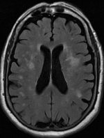
Leukoaraiosis is a particular abnormal change in appearance of white matter near the lateral ventricles. It is often seen in aged individuals, but sometimes in young adults. On MRI, leukoaraiosis changes appear as white matter hyperintensities (WMHs) in T2 FLAIR images. On CT scans, leukoaraiosis appears as hypodense periventricular white-matter lesions. The term "leukoaraiosis" was coined in 1986 by Hachinski, Potter, and Merskey as a descriptive term for rarefaction ("araiosis") of the white matter, showing up as decreased density on CT and increased signal intensity on T2/FLAIR sequences (white matter hyperintensities) performed as part of MRI brain scans. These white matter changes are also commonly referred to as periventricular white matter disease, or white matter hyperintensities (WMH), due to their bright white appearance on T2 MRI scans. Many patients can have leukoaraiosis without any associated clinical abnormality. However, underlying vascular mechanisms are susp ... [...More Info...] [...Related Items...] OR: [Wikipedia] [Google] [Baidu] |
Calcification
Calcification is the accumulation of calcium salts in a body tissue. It normally occurs in the formation of bone, but calcium can be deposited abnormally in soft tissue,Miller, J. D. Cardiovascular calcification: Orbicular origins. ''Nature Materials'' 12, 476-478 (2013). causing it to harden. Calcifications may be classified on whether there is mineral balance or not, and the location of the calcification. Calcification may also refer to the processes of normal mineral deposition in biological systems, such as the formation of stromatolites or mollusc shells (see Biomineralization). Signs and symptoms Calcification can manifest itself in many ways in the body depending on the location. In the pulpal structure of a tooth, calcification often presents asymptomatically, and is diagnosed as an incidental finding during radiographic interpretation. Individual teeth with calcified pulp will typically respond negatively to vitality testing; teeth with calcified pulp often lack ... [...More Info...] [...Related Items...] OR: [Wikipedia] [Google] [Baidu] |


