|
Distal Radius Fracture
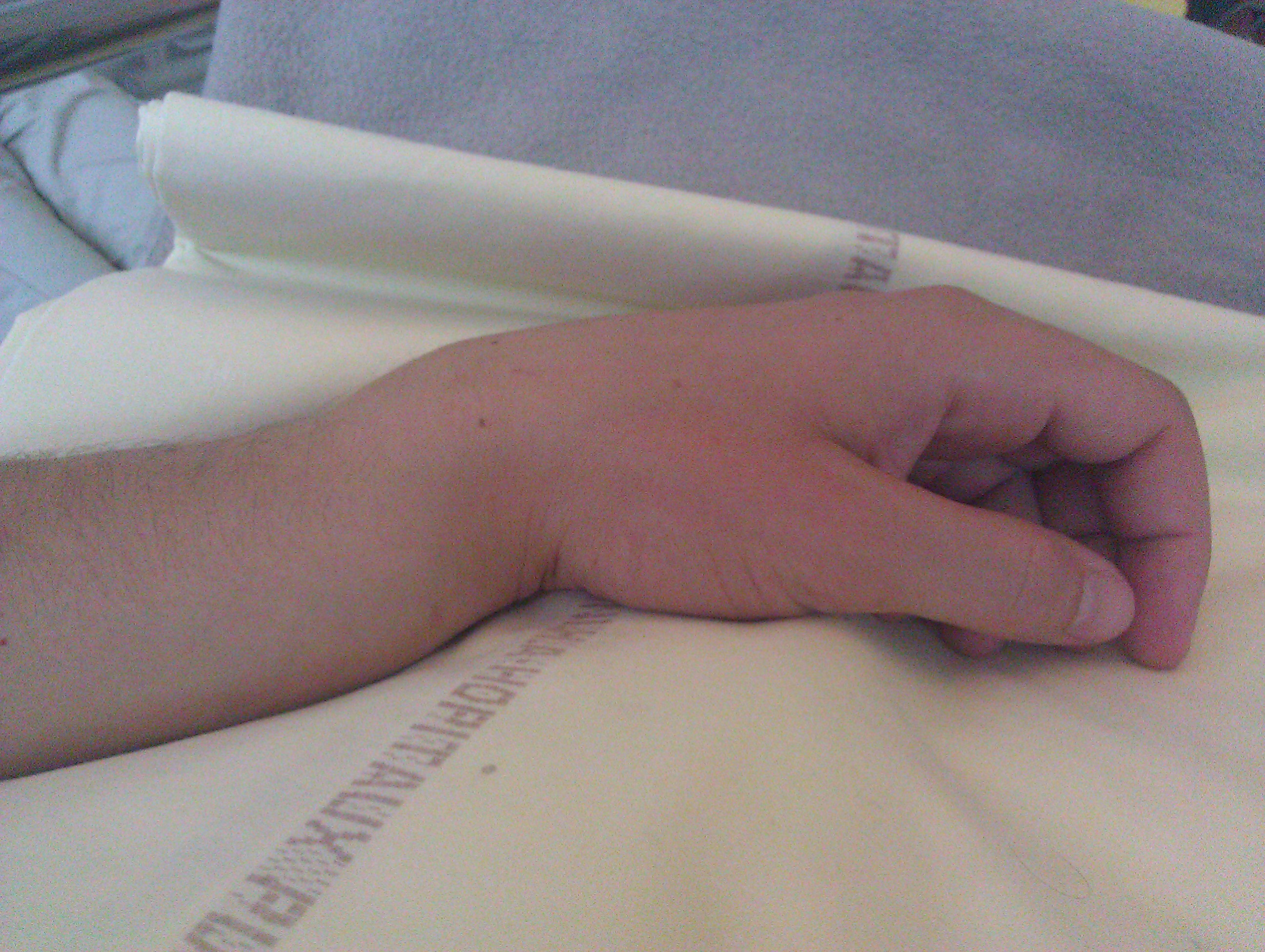
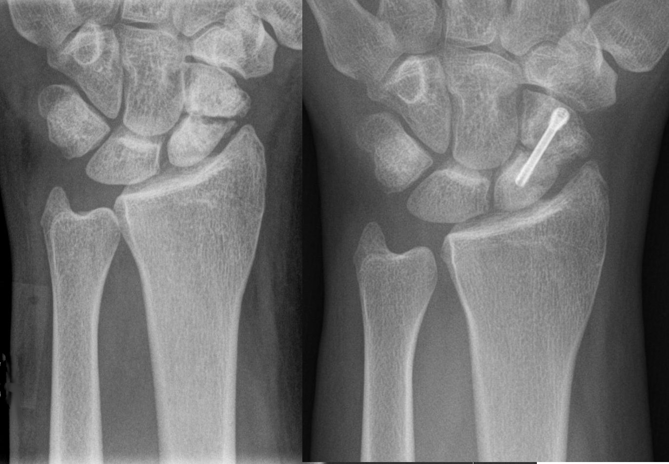
A distal radius fracture, also known as wrist fracture, is a fracture (bone), break of the part of the radius (bone), radius bone which is close to the wrist. Symptoms include pain, bruising, and rapid-onset swelling. The ulna bone may also be broken. In younger people, these fractures typically occur during sports or a motor vehicle collision. In older people, the most common cause is falling on an outstretched hand. Specific types include Colles' fracture, Colles, Smith's fracture, Smith, Barton's fracture, Barton, and Chauffeur's fractures. The diagnosis is generally suspected based on symptoms and confirmed with radiography, X-rays. Treatment is with orthopedic cast, casting for six weeks or surgery. Surgery is generally indicated if the joint surface is broken and does not line up, the radius is overly short, or the joint surface of the radius is tilted more than 10% backwards. Among those who are cast, repeated X-rays are recommended within three weeks to verify that a go ... [...More Info...] [...Related Items...] OR: [Wikipedia] [Google] [Baidu] |
Colles Fracture
A Colles' fracture is a type of distal radius fracture, fracture of the distal forearm in which the broken end of the Radius (bone), radius is bent dorsal (anatomy), backwards. Symptoms may include pain, Edema, swelling, deformity, and bruising. Complications may include damage to the median nerve. It typically occurs as a result of a fall on an outstretched hand. Risk factors include osteoporosis. The diagnosis may be confirmed via radiography, X-rays. The tip of the ulna may also be broken. Treatment may include orthopedic cast, casting or surgery. Reduction (orthopedic surgery), Surgical reduction and casting is possible in the majority of cases in people over the age of 50. Pain management can be achieved during the reduction with procedural sedation and analgesia or a hematoma block. A year or two may be required for healing to occur. About 15% of people have a Colles' fracture at some point in their life. They occur more commonly in young adults and older people than in c ... [...More Info...] [...Related Items...] OR: [Wikipedia] [Google] [Baidu] |
Median Nerve
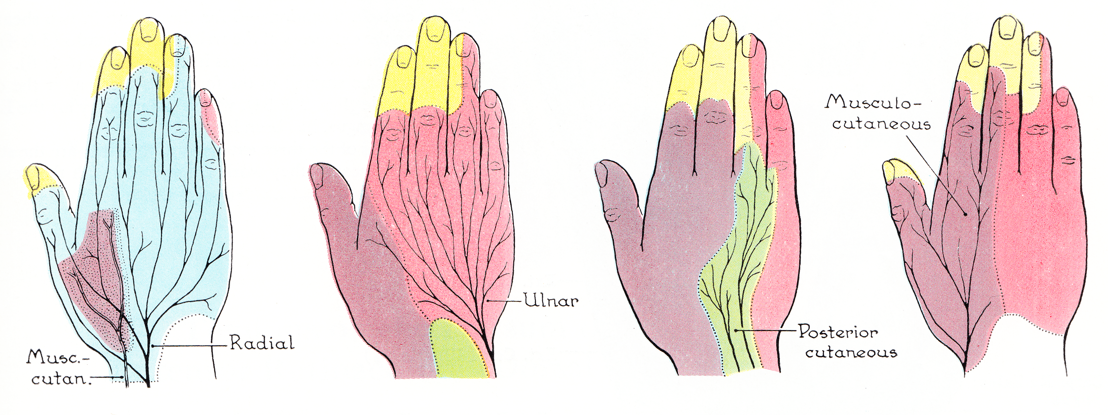
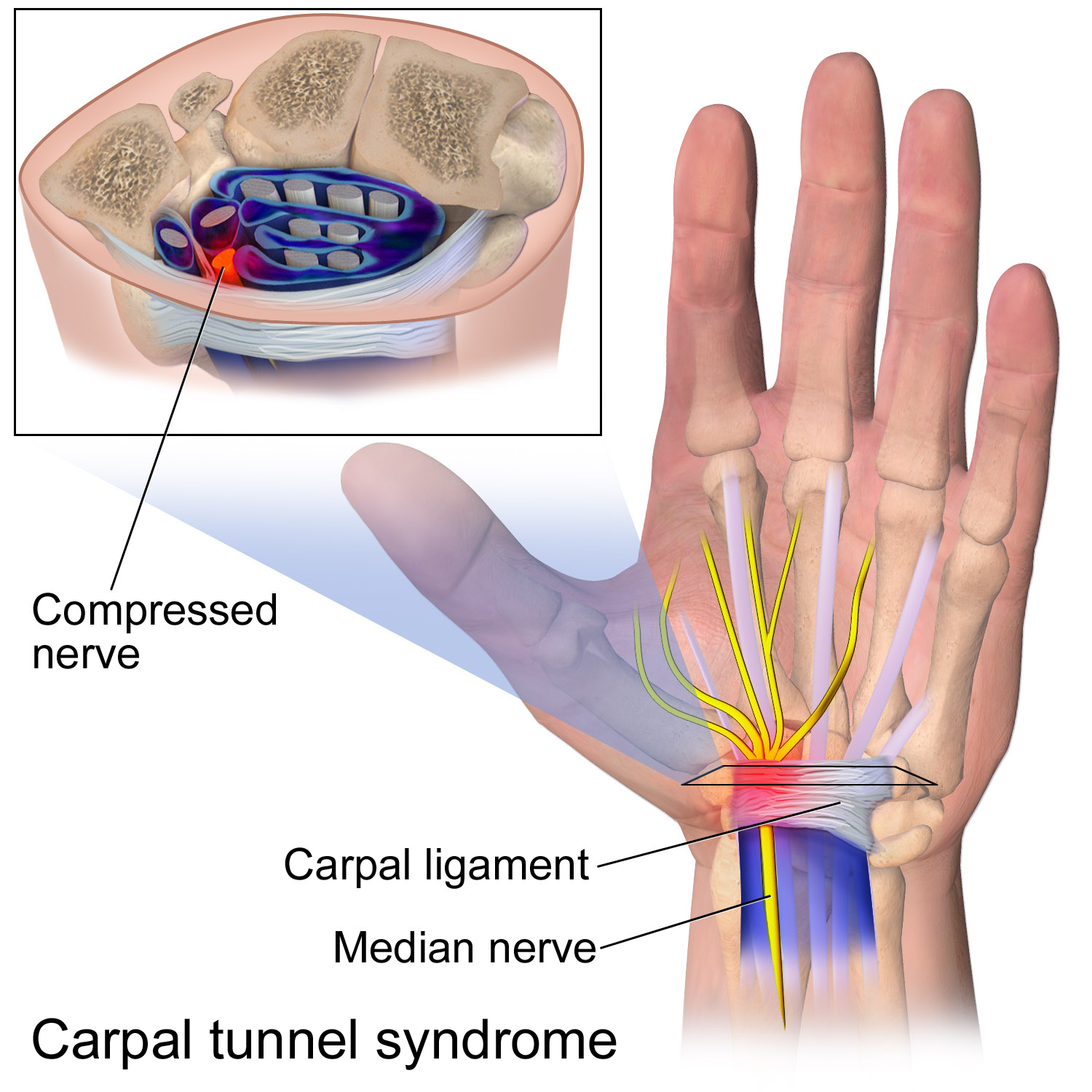
The median nerve is a nerve in humans and other animals in the upper limb. It is one of the five main nerves originating from the brachial plexus. The median nerve originates from the lateral and medial cords of the brachial plexus, and has contributions from ventral roots of C6-C7 (lateral cord) and C8 and T1 (medial cord). The median nerve is the only nerve that passes through the carpal tunnel. Carpal tunnel syndrome is the disability that results from the median nerve being pressed in the carpal tunnel. Structure The median nerve arises from the branches from lateral and medial cords of the brachial plexus, courses through the anterior part of arm, forearm, and hand, and terminates by supplying the muscles of the hand. Arm After receiving inputs from both the lateral and medial cords of the brachial plexus, the median nerve enters the arm from the axilla at the inferior margin of the teres major muscle. It then passes vertically down and courses lateral to the brac ... [...More Info...] [...Related Items...] OR: [Wikipedia] [Google] [Baidu] |
Extensor Pollicis Longus Muscle
In human anatomy, the extensor pollicis longus muscle (EPL) is a skeletal muscle located dorsally on the forearm. It is much larger than the extensor pollicis brevis, the origin of which it partly covers and acts to stretch the thumb together with this muscle. Structure The extensor pollicis longus arises from the dorsal surface of the ulna and from the interosseous membrane, next to the origins of abductor pollicis longus and extensor pollicis brevis. Passing through the third tendon compartment, lying in a narrow, oblique groove on the back of the lower end of the radius,''Gray's Anatomy'' 1918, see infobox it crosses the wrist close to the dorsal midline before turning towards the thumb using Lister's tubercle on the distal end of the radius as a pulley. It obliquely crosses the tendons of the extensores carpi radialis longus and brevis, and is separated from the extensor pollicis brevis by a triangular interval, the anatomical snuff box in which the radial artery is foun ... [...More Info...] [...Related Items...] OR: [Wikipedia] [Google] [Baidu] |
Forearm
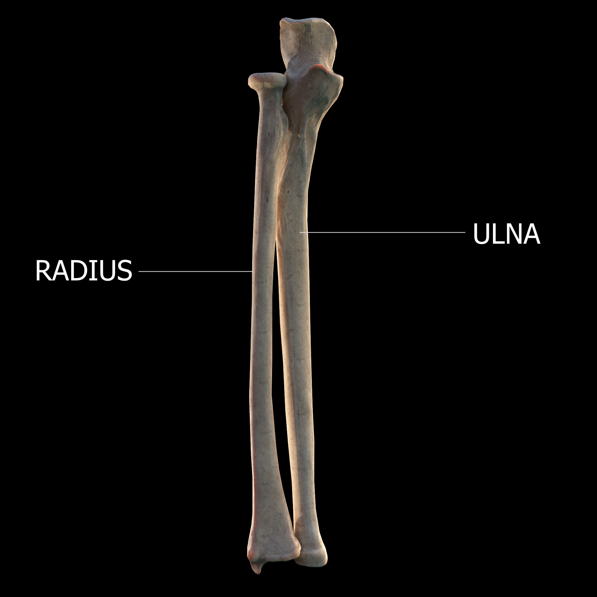
The forearm is the region of the upper limb between the elbow and the wrist. The term forearm is used in anatomy to distinguish it from the arm, a word which is used to describe the entire appendage of the upper limb, but which in anatomy, technically, means only the region of the upper arm, whereas the lower "arm" is called the forearm. It is homologous to the region of the leg that lies between the knee and the ankle joints, the crus. The forearm contains two long bones, the radius and the ulna, forming the two radioulnar joints. The interosseous membrane connects these bones. Ultimately, the forearm is covered by skin, the anterior surface usually being less hairy than the posterior surface. The forearm contains many muscles, including the flexors and extensors of the wrist, flexors and extensors of the digits, a flexor of the elbow ( brachioradialis), and pronators and supinators that turn the hand to face down or upwards, respectively. In cross-section, the forearm can ... [...More Info...] [...Related Items...] OR: [Wikipedia] [Google] [Baidu] |
Ulnar Styloid Process
The styloid process of the ulna is a bony prominence found at distal end of the ulna in the forearm. Structure The styloid process of the ulna projects from the medial and back part of the ulna. It descends a little lower than the head. The head is separated from the styloid process by a depression for the attachment of the apex of the triangular articular disk, and behind, by a shallow groove for the tendon of the extensor carpi ulnaris muscle. The styloid process of the ulna varies from 2 to 6 mm in length. Function The rounded end of the styloid process of the ulna connects to the ulnar collateral ligament of the wrist. The radioulnar ligaments also attaches to the base of the styloid process of the ulna. Clinical significance Fractures of the styloid process of the ulna seldom require treatment when they occur in association with a distal radius fracture. The major exception is when the joint between these bones, the distal radioulnar joint (or DRUJ), is unstable ... [...More Info...] [...Related Items...] OR: [Wikipedia] [Google] [Baidu] |
Osteoarthritis
Osteoarthritis is a type of degenerative joint disease that results from breakdown of articular cartilage, joint cartilage and underlying bone. A form of arthritis, it is believed to be the fourth leading cause of disability in the world, affecting 1 in 7 adults in the United States alone. The most common symptoms are joint pain and Joint stiffness, stiffness. Usually the symptoms progress slowly over years. Other symptoms may include joint effusion, joint swelling, decreased range of motion, and, when the back is affected, weakness or numbness of the arms and legs. The most commonly involved joints are the two near the ends of the fingers and the joint at the base of the thumbs, the knee and hip joints, and the joints of the neck and lower back. The symptoms can interfere with work and normal daily activities. Unlike some other types of arthritis, only the joints, not internal organs, are affected. Possible causes include previous joint injury, abnormal joint or limb development ... [...More Info...] [...Related Items...] OR: [Wikipedia] [Google] [Baidu] |
Malunion
A malunion is when a fractured bone does not heal properly. Some ways that it shows is by having the bone being twisted, shorter, or bent. Malunions can occur by having the bones improperly aligned when immobilized, having the cast taken off too early, or never seeking medical treatment after the break. Malunions are painful and commonly produce swelling around the area, possible immobilization, and deterioration of the bone and tissue. Signs and symptoms Malunions are presented by excessive swelling, twisting, bending, and possibly shortening of the bone. Patients may have trouble placing weight on or near the malunion. Diagnosis An X-ray is essential for the proper diagnosis of a malunion. The doctor will look into the patient’s history and the treatment process for the bone fracture. Oftentimes a CT scan and probably an MRI are also used in diagnosis. MRI are used to check of cartilage and ligament issues that developed due to the malunion and misalignment. CT scans ... [...More Info...] [...Related Items...] OR: [Wikipedia] [Google] [Baidu] |
Nonunion
Nonunion is permanent failure of healing following a broken bone unless intervention (such as surgery) is performed. A fracture with nonunion generally forms a structural resemblance to a fibrous joint, and is therefore often called a "false joint" or pseudoarthrosis (from Greek '' pseudo-'', meaning false, , meaning joint, and '' -osis'', meaning abnormal condition). The diagnosis is generally made when there is no healing between two sets of medical imaging, such as X-ray or CT scan. This is generally after 6–8 months.Page 542 in: Nonunion is a serious complication of a fracture and may occur when the fracture moves too much, has a poor |
Compartment Syndrome
Compartment syndrome is a serious medical condition in which increased pressure within a Fascial compartment, body compartment compromises blood flow and tissue function, potentially leading to permanent damage if not promptly treated. There are two types: Acute (medicine), acute and Chronic condition, chronic. Acute compartment syndrome can lead to a loss of the affected limb due to tissue death. Symptoms of acute compartment syndrome (ACS) include severe pain, decreased blood flow, decreased movement, numbness, and a pale limb. It is most often due to Injury, physical trauma, like a bone fracture (up to 75% of cases) or a crush injury. It can also occur after Reperfusion injury, blood flow returns following a period of poor circulation. Diagnosis is Clinical diagnosis, clinical, based on symptoms, not a specific test. However, it may be supported by measuring the pressure inside the Fascial compartment, compartment. It is classically described by pain out of proportion to the in ... [...More Info...] [...Related Items...] OR: [Wikipedia] [Google] [Baidu] |
Carpal Tunnel Syndrome
Carpal tunnel syndrome (CTS) is a nerve compression syndrome associated with the collected signs and symptoms of Pathophysiology of nerve entrapment#Compression, compression of the median nerve at the carpal tunnel in the wrist. Carpal tunnel syndrome usually has no known cause, but there are environmental and medical risk factors associated with the condition.> CTS can affect both wrists. Other conditions can cause CTS such as wrist fracture or rheumatoid arthritis. After fracture, the resulting swelling, bleeding, and deformity compress the median nerve. With rheumatoid arthritis, the enlarged synovial membrane, synovial lining of the tendons causes compression. The main symptoms are numbness and Paresthesia, tingling of the thumb, index finger, middle finger, and the thumb side of the ring finger, as well as pain in the hand and fingers. Symptoms are typically most troublesome at night. Many people sleep with their wrists bent, and the ensuing symptoms may lead to awake ... [...More Info...] [...Related Items...] OR: [Wikipedia] [Google] [Baidu] |
Open Fracture
An open fracture, also called a compound fracture, is a type of bone fracture (broken bone) that has an open wound in the skin near the fractured bone. The skin wound is usually caused by the bone breaking through the surface of the skin. An open fracture can be life threatening or limb-threatening (person may be at risk of losing a limb) due to the risk of a deep infection and/or bleeding. Open fractures are often caused by high energy trauma such as road traffic accidents and are associated with a high degree of damage to the bone and nearby soft tissue. Other potential complications include nerve damage or impaired bone healing, including malunion or nonunion. The severity of open fractures can vary. For diagnosing and classifying open fractures, Gustilo-Anderson open fracture classification is the most commonly used method. This classification system can also be used to guide treatment, and to predict clinical outcomes. Advanced trauma life support is the first line of action in ... [...More Info...] [...Related Items...] OR: [Wikipedia] [Google] [Baidu] |
Radius (bone)
The radius or radial bone (: radii or radiuses) is one of the two large bones of the forearm, the other being the ulna. It extends from the Anatomical terms of location, lateral side of the Elbow-joint, elbow to the thumb side of the wrist and runs parallel to the ulna. The ulna is longer than the radius, but the radius is thicker. The radius is a long bone, Prism (geometry), prism-shaped and slightly curved longitudinally. The radius is part of two joint (anatomy), joints: the elbow and the wrist. At the elbow, it joins with the capitulum of the humerus, and in a separate region, with the ulna at the radial notch. At the wrist, the radius forms a joint with the ulna bone. The corresponding bone in the human leg, lower leg is the tibia. Structure The long narrow medullary cavity is enclosed in a strong wall of compact bone. It is thickest along the interosseous border and thinnest at the extremities, same over the cup-shaped articular surface (fovea) of the head. The tra ... [...More Info...] [...Related Items...] OR: [Wikipedia] [Google] [Baidu] |

