|
Crus Of Clitoris
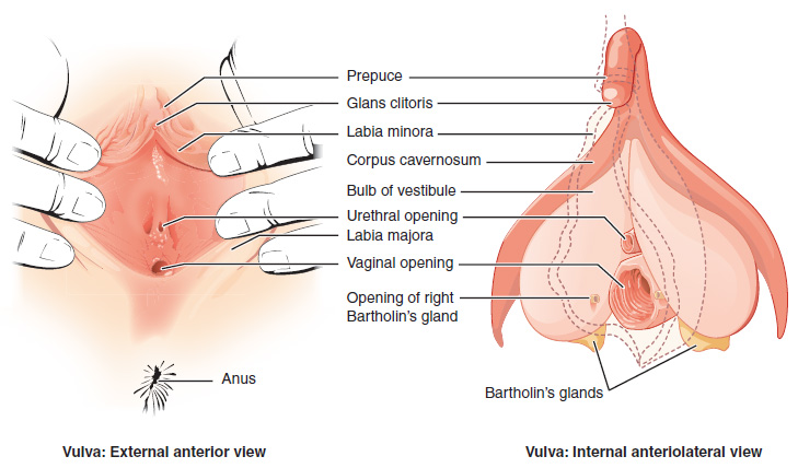
The clitoral crura (singular: clitoral crux) are two erectile tissue structures, which together form a V-shape. ''Crus'' is a Latin word that means "leg". Each "leg" of the ''V'' converges on the clitoral body. At each divergent point is a corpus cavernosum of clitoris. The crura are attached to the pubic arch, and are adjacent to the vestibular bulbs. The crura flank the urethra, urethral sponge, and vagina and extend back toward the pubis. Each clitoral crus connects to the rami of the pubis and the ischium. During sexual arousal, the crura become engorged with blood, as does all of the erectile tissue of the clitoris. The clitoral crura are each covered by an ischiocavernosus muscle. See also * Crus of penis For their anterior three-fourths the corpora cavernosa penis lie in intimate apposition with one another, but behind they diverge in the form of two tapering processes, known as the crura, which are firmly connected to the ischial rami. Traced f ... Referen ... [...More Info...] [...Related Items...] OR: [Wikipedia] [Google] [Baidu] |
Vulva
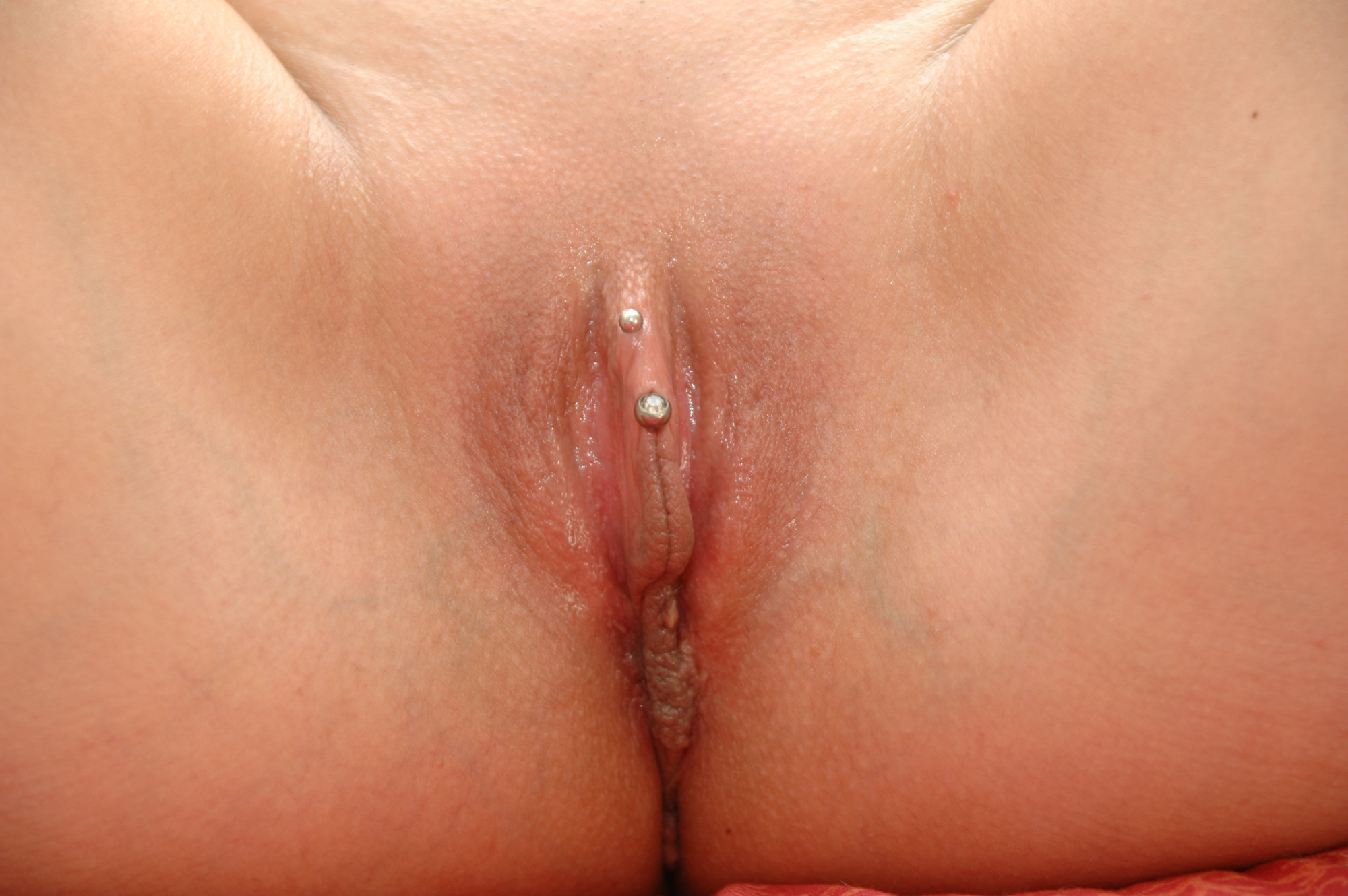
The vulva (plural: vulvas or vulvae; derived from Latin for wrapper or covering) consists of the external female sex organs. The vulva includes the mons pubis (or mons veneris), labia majora, labia minora, clitoris, vestibular bulbs, vulval vestibule, urinary meatus, the vaginal opening, hymen, and Bartholin's and Skene's vestibular glands. The urinary meatus is also included as it opens into the vulval vestibule. Other features of the vulva include the pudendal cleft, sebaceous glands, the urogenital triangle (anterior part of the perineum), and pubic hair. The vulva includes the entrance to the vagina, which leads to the uterus, and provides a double layer of protection for this by the folds of the outer and inner labia. Pelvic floor muscles support the structures of the vulva. Other muscles of the urogenital triangle also give support. Blood supply to the vulva comes from the three pudendal arteries. The internal pudendal veins give drainage. Afferent lymp ... [...More Info...] [...Related Items...] OR: [Wikipedia] [Google] [Baidu] |
Urethra
The urethra (from Greek οὐρήθρα – ''ourḗthrā'') is a tube that connects the urinary bladder to the urinary meatus for the removal of urine from the body of both females and males. In human females and other primates, the urethra connects to the urinary meatus above the vagina, whereas in marsupials, the female's urethra empties into the urogenital sinus. Females use their urethra only for urinating, but males use their urethra for both urination and ejaculation. The external urethral sphincter is a striated muscle that allows voluntary control over urination. The internal sphincter, formed by the involuntary smooth muscles lining the bladder neck and urethra, receives its nerve supply by the sympathetic division of the autonomic nervous system. The internal sphincter is present both in males and females. Structure The urethra is a fibrous and muscular tube which connects the urinary bladder to the external urethral meatus. Its length differs between ... [...More Info...] [...Related Items...] OR: [Wikipedia] [Google] [Baidu] |
Ischiocavernosus Muscle
The ischiocavernosus muscle (erectores penis ''or'' erector clitoridis in older texts) is a muscle just below the surface of the perineum, present in both men and women. Structure It arises by tendinous and fleshy fibers from the inner surface of the tuberosity of the ischium, behind the crus penis; and from the inferior pubic rami and ischium on either side of the crus. From these points fleshy fibers succeed, and end in an aponeurosis which is inserted into the sides and under surface of the crus penis. Function In females, the ischiocavernosus muscle assists with clitoral erection. In males, it helps to stabilize the erect penis An erection (clinically: penile erection or penile tumescence) is a physiological phenomenon in which the penis becomes firm, engorged, and enlarged. Penile erection is the result of a complex interaction of psychological, neural, vascular, a ... by compressing the crus penis and retarding the return of blood through the veins. Additional im ... [...More Info...] [...Related Items...] OR: [Wikipedia] [Google] [Baidu] |
Sexual Arousal
Sexual arousal (also known as sexual excitement) describes the physiological and psychological responses in preparation for sexual intercourse or when exposed to sexual stimuli. A number of physiological responses occur in the body and mind as preparation for sexual intercourse, and continue during intercourse. Male arousal will lead to an erection, and in female arousal the body's response is engorged sexual tissues such as nipples, vulva, clitoris, vaginal walls, and vaginal lubrication. Mental stimuli and physical stimuli such as touch, and the internal fluctuation of hormones, can influence sexual arousal. Sexual arousal has several stages and may not lead to any actual sexual activity beyond a mental arousal and the physiological changes that accompany it. Given sufficient sexual stimulation, sexual arousal reaches its climax during an orgasm. It may also be pursued for its own sake, even in the absence of an orgasm. Erotic stimuli Depending on the situation, a p ... [...More Info...] [...Related Items...] OR: [Wikipedia] [Google] [Baidu] |
Ischium
The ischium () forms the lower and back region of the hip bone (''os coxae''). Situated below the ilium and behind the pubis, it is one of three regions whose fusion creates the coxal bone. The superior portion of this region forms approximately one-third of the . Structure The i ...[...More Info...] [...Related Items...] OR: [Wikipedia] [Google] [Baidu] |
Pubis (bone)
In vertebrates, the pubic region ( la, pubis) is the most forward-facing ( ventral and anterior) of the three main regions making up the coxal bone. The left and right pubic regions are each made up of three sections, a superior ramus, inferior ramus, and a body. Structure The pubic region is made up of a ''body'', ''superior ramus'', and ''inferior ramus'' (). The left and right coxal bones join at the pubic symphysis. It is covered by a layer of fat, which is covered by the mons pubis. The pubis is the lower limit of the suprapubic region. In the female, the pubic region is anterior to the urethral sponge. Body The body forms the wide, strong, middle and flat part of the pubic region. The bodies of the left and right pubic regions join at the pubic symphysis. The rough upper edge is the pubic crest, ending laterally in the pubic tubercle. This tubercle, found roughly 3 cm from the pubic symphysis, is a distinctive feature on the lower part of the abdominal wal ... [...More Info...] [...Related Items...] OR: [Wikipedia] [Google] [Baidu] |
Vagina
In mammals, the vagina is the elastic, muscular part of the female genital tract. In humans, it extends from the vestibule to the cervix. The outer vaginal opening is normally partly covered by a thin layer of mucosal tissue called the hymen. At the deep end, the cervix (neck of the uterus) bulges into the vagina. The vagina allows for sexual intercourse and birth. It also channels menstrual flow, which occurs in humans and closely related primates as part of the menstrual cycle. Although research on the vagina is especially lacking for different animals, its location, structure and size are documented as varying among species. Female mammals usually have two external openings in the vulva; these are the urethral opening for the urinary tract and the vaginal opening for the genital tract. This is different from male mammals, who usually have a single urethral opening for both urination and reproduction. The vaginal opening is much larger than the nearby urethral openi ... [...More Info...] [...Related Items...] OR: [Wikipedia] [Google] [Baidu] |
Urethral Sponge
The urethral sponge is a spongy cushion of tissue, found in the lower genital area of females, that sits against both the pubic bone and vaginal wall, and surrounds the urethra. Functions The urethral sponge is composed of erectile tissue; during arousal, it becomes swollen with blood, compressing the urethra, helping, along with the pubococcygeus muscle, to prevent urination during sexual activity. Female ejaculation Additionally, the urethral sponge contains the Skene's glands, which may be involved in female ejaculation. Sexual stimulation The urethral sponge encompasses sensitive nerve endings, and can be stimulated through the front wall of the vagina. Some women experience intense pleasure from stimulation of the urethral sponge and others find the sensation irritating. The urethral sponge surrounds the clitoral nerve, and since the two are so closely interconnected, stimulation of the clitoris may stimulate the nerve endings of the urethral sponge and vice versa. So ... [...More Info...] [...Related Items...] OR: [Wikipedia] [Google] [Baidu] |
Vestibular Bulbs
In female anatomy, the vestibular bulbs, bulbs of the vestibule or clitoral bulbs are two elongated masses of erectile tissue typically described as being situated on either side of the vaginal opening. They are united to each other in front by a narrow median band. Some research indicates that they do not surround the vaginal opening, and are more closely related to the clitoris than to the vestibule. Structure Research indicates that the vestibular bulbs are more closely related to the clitoris than to the vestibule because of the similarity of the trabecular and erectile tissue within the clitoris and bulbs, and the absence of trabecular tissue in other genital organs, with the erectile tissue's trabecular nature allowing engorgement and expansion during sexual arousal. Ginger et al. state that although a number of texts report that they surround the vaginal opening, this does not appear to be the case and tunica albuginea does not envelop the erectile tissue of the bulb. ... [...More Info...] [...Related Items...] OR: [Wikipedia] [Google] [Baidu] |
Clitoral Hood
In the female human body, the clitoral hood (also called preputium clitoridis and clitoral prepuce) is a fold of skin that surrounds and protects the glans of the clitoris; it also covers the external shaft of the clitoris, develops as part of the labia minora and is homologous with the foreskin (also called the ''prepuce'') in the male reproductive system. The clitoral hood is composed of muccocutaneous tissues; these tissues are between the mucous membrane and the skin, and they may have immunological importance because they may be a point of entry of mucosal vaccines. The clitoral hood is also important not only in the protection of the clitoral glans, but also in pleasure, as its tissue forms part of the erogenous zones of the vulva. Development and variation The clitoral hood is formed during the fetal stage by the cellular lamella. The cellular lamella grows down on the dorsal side of the clitoris and is eventually fused with the clitoris. The clitoral hood is formed fr ... [...More Info...] [...Related Items...] OR: [Wikipedia] [Google] [Baidu] |
Pubic Arch
The pubic arch, also referred to as the ''ischiopubic arch'', is part of the pelvis. It is formed by the convergence of the inferior rami of the ischium and pubis on either side, below the pubic symphysis. The angle at which they converge is known as the subpubic angle. Function The pubic arch is one of three notches (the one in front) that separate the eminences of the lower circumference of the true pelvis. Clinical significance Subpubic angle The subpubic angle (or pubic angle) is the angle in the human body as the apex of the pubic arch, formed by the convergence of the inferior rami of the ischium and pubis on either side. The subpubic angle is important in forensic anthropology, in determining the sex of someone from skeletal remains. A subpubic angle of 50-82 degrees indicates a male; an angle of 90 degrees indicates a female.Anthony J. Bertino. Forensic Science - Fundamentals and Investigations. South-Western Cengage Learning, 2000. . Page 368 Other sources operate ... [...More Info...] [...Related Items...] OR: [Wikipedia] [Google] [Baidu] |


