|
Coronary Arteries
The coronary arteries are the arterial blood vessels of coronary circulation, which transport oxygenated blood to the heart muscle. The heart requires a continuous supply of oxygen to function and survive, much like any other tissue or organ of the body. The coronary arteries wrap around the entire heart. The two main branches are the left coronary artery and right coronary artery. The arteries can additionally be categorized based on the area of the heart for which they provide circulation. These categories are called ''epicardial'' (above the epicardium, or the outermost tissue of the heart) and ''microvascular'' (close to the endocardium, or the innermost tissue of the heart). Reduced function of the coronary arteries can lead to decreased flow of oxygen and nutrients to the heart. Not only does this affect supply to the heart muscle itself, but it also can affect the ability of the heart to pump blood throughout the body. Therefore, any disorder or disease of the coronary ... [...More Info...] [...Related Items...] OR: [Wikipedia] [Google] [Baidu] |
Arteries
An artery (plural arteries) () is a blood vessel in humans and most animals that takes blood away from the heart to one or more parts of the body (tissues, lungs, brain etc.). Most arteries carry oxygenated blood; the two exceptions are the pulmonary and the umbilical arteries, which carry deoxygenated blood to the organs that oxygenate it (lungs and placenta, respectively). The effective arterial blood volume is that extracellular fluid which fills the arterial system. The arteries are part of the circulatory system, that is responsible for the delivery of oxygen and nutrients to all cells, as well as the removal of carbon dioxide and waste products, the maintenance of optimum blood pH, and the circulation of proteins and cells of the immune system. Arteries contrast with veins, which carry blood back towards the heart. Structure The anatomy of arteries can be separated into gross anatomy, at the macroscopic level, and microanatomy, which must be studied with a micro ... [...More Info...] [...Related Items...] OR: [Wikipedia] [Google] [Baidu] |
Right Coronary Artery
In the blood supply of the heart, the right coronary artery (RCA) is an artery originating above the right cusp of the aortic valve, at the right aortic sinus in the heart. It travels down the right coronary sulcus, towards the crux of the heart. It supplies the right side of the heart, and the interventricular septum. Structure The right coronary artery originates above the right aortic sinus above the aortic valve. It passes through the right coronary sulcus (right atrioventricular groove), towards the crux of the heart. It gives off many branches, including the posterior interventricular artery, the right marginal artery, the conus artery, and the sinoatrial nodal artery. Segments * Proximal: starting at RCA origin, spanning half the distance to the acute margin * Middle: from proximal segment to the acute margin * Distal: from middle segment to origination point of the posterior interventricular artery, where the posterior interventricular sulcus meets the atri ... [...More Info...] [...Related Items...] OR: [Wikipedia] [Google] [Baidu] |
Atherosclerosis
Atherosclerosis is a pattern of the disease arteriosclerosis in which the wall of the artery develops abnormalities, called lesions. These lesions may lead to narrowing due to the buildup of atheromatous plaque. At onset there are usually no symptoms, but if they develop, symptoms generally begin around middle age. When severe, it can result in coronary artery disease, stroke, peripheral artery disease, or kidney problems, depending on which arteries are affected. The exact cause is not known and is proposed to be multifactorial. Risk factors include abnormal cholesterol levels, elevated levels of inflammatory markers, high blood pressure, diabetes, smoking, obesity, family history, genetic, and an unhealthy diet. Plaque is made up of fat, cholesterol, calcium, and other substances found in the blood. The narrowing of arteries limits the flow of oxygen-rich blood to parts of the body. Diagnosis is based upon a physical exam, electrocardiogram, and exercise stress test, amo ... [...More Info...] [...Related Items...] OR: [Wikipedia] [Google] [Baidu] |
Atherosclerosis 2011
Atherosclerosis is a pattern of the disease arteriosclerosis in which the wall of the artery develops abnormalities, called lesions. These lesions may lead to narrowing due to the buildup of atheromatous plaque. At onset there are usually no symptoms, but if they develop, symptoms generally begin around middle age. When severe, it can result in coronary artery disease, stroke, peripheral artery disease, or kidney problems, depending on which arteries are affected. The exact cause is not known and is proposed to be multifactorial. Risk factors include abnormal cholesterol levels, elevated levels of inflammatory markers, high blood pressure, diabetes, smoking, obesity, family history, genetic, and an unhealthy diet. Plaque is made up of fat, cholesterol, calcium, and other substances found in the blood. The narrowing of arteries limits the flow of oxygen-rich blood to parts of the body. Diagnosis is based upon a physical exam, electrocardiogram, and exercise stress test, amo ... [...More Info...] [...Related Items...] OR: [Wikipedia] [Google] [Baidu] |
Right Marginal Branch Of Right Coronary Artery
The right marginal branch of right coronary artery (or right marginal artery) is the largest marginal branch of the right coronary artery. It follows the acute margin of the heart. It supplies blood to both surfaces of the right ventricle. Structure The right marginal branch is the largest branch to split off from the right coronary artery. It often anastomoses with the nearby parallel posterior interventricular artery, which itself is usually a continuation of the right coronary artery. Variation The right marginal branch may reach the distal part of the posterior interventricular sulcus. Function The right marginal branch primarily supplies the right ventricle A ventricle is one of two large chambers toward the bottom of the heart that collect and expel blood towards the peripheral beds within the body and lungs. The blood pumped by a ventricle is supplied by an atrium, an adjacent chamber in the uppe .... Additional images File:Human heart with coronary arter ... [...More Info...] [...Related Items...] OR: [Wikipedia] [Google] [Baidu] |
Crux Cordis
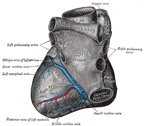
The crux cordis or crux of the heart (from Latin "crux" meaning "cross") is the area on the lower back side of the heart where the coronary sulcus (the groove separating the atria from the ventricles) and the posterior interventricular sulcus The posterior interventricular sulcus or posterior longitudinal sulcus is one of the two grooves that separates the ventricles of the heart and is on the diaphragmatic surface of the heart near the right margin. The other groove is the anterior i ... (the groove separating the left from the right ventricle) meet. It is important surgically because the atrioventricular nodal artery, a small but vital vessel, passes in proximity to the crux of the heart. It is the anastomotic point of right and left coronary artery. References Cardiac anatomy {{circulatory-stub ... [...More Info...] [...Related Items...] OR: [Wikipedia] [Google] [Baidu] |
Coronary Sulcus
The coronary sulcus (also called coronary groove, auriculoventricular groove, atrioventricular groove, AV groove) is a groove on the surface of the heart at the base of right auricle that separates the atria from the ventricles. The structure contains the trunks of the nutrient vessels of the heart, and is deficient in front, where it is crossed by the root of the pulmonary trunk. On the posterior surface of the heart, the coronary sulcus contains the coronary sinus. Structure In relation to the rib cage, the coronary sulcus spans from the medial side of the 3rd left costal cartilage, to the middle of the right 6th costal cartilage. Epicardial fat tends to be concentrated along the coronary sulcus. There are two coronary sulci in the heart, including left and right coronary sulci. Left coronary sulcus The left coronary sulcus originates posterior to the pulmonary trunk, and travels inferiorly separating the left atrium and left ventricle. The location of the left coronar ... [...More Info...] [...Related Items...] OR: [Wikipedia] [Google] [Baidu] |
Elsevier
Elsevier () is a Dutch academic publishing company specializing in scientific, technical, and medical content. Its products include journals such as '' The Lancet'', ''Cell'', the ScienceDirect collection of electronic journals, '' Trends'', the '' Current Opinion'' series, the online citation database Scopus, the SciVal tool for measuring research performance, the ClinicalKey search engine for clinicians, and the ClinicalPath evidence-based cancer care service. Elsevier's products and services also include digital tools for data management, instruction, research analytics and assessment. Elsevier is part of the RELX Group (known until 2015 as Reed Elsevier), a publicly traded company. According to RELX reports, in 2021 Elsevier published more than 600,000 articles annually in over 2,700 journals; as of 2018 its archives contained over 17 million documents and 40,000 e-books, with over one billion annual downloads. Researchers have criticized Elsevier for its high profit m ... [...More Info...] [...Related Items...] OR: [Wikipedia] [Google] [Baidu] |
Posterior Descending Artery
In the coronary circulation, the posterior interventricular artery (PIV, PIA, or PIVA), most often called the posterior descending artery (PDA), is an artery running in the posterior interventricular sulcus to the apex of the heart where it meets with the anterior interventricular artery or also known as Left Anterior Descending artery. It supplies the posterior third of the interventricular septum. The remaining anterior two-thirds is supplied by the anterior interventricular artery which is a septal branch of the left anterior descending artery, which is a branch of left coronary artery. It is typically a branch of the right coronary artery (70%, known as right dominance). Alternately, the PIV can be a branch of the circumflex coronary artery The circumflex branch of left coronary artery, or left circumflex artery or circumflex artery, is a branch of the left coronary artery. Description The left circumflex artery follows the left part of the coronary sulcus, running f ... [...More Info...] [...Related Items...] OR: [Wikipedia] [Google] [Baidu] |
Ventricle (heart)
A ventricle is one of two large chambers toward the bottom of the heart that collect and expel blood towards the peripheral beds within the body and lungs. The blood pumped by a ventricle is supplied by an atrium, an adjacent chamber in the upper heart that is smaller than a ventricle. Interventricular means between the ventricles (for example the interventricular septum), while intraventricular means within one ventricle (for example an intraventricular block). In a four-chambered heart, such as that in humans, there are two ventricles that operate in a double circulatory system: the right ventricle pumps blood into the pulmonary circulation to the lungs, and the left ventricle pumps blood into the systemic circulation through the aorta. Structure Ventricles have thicker walls than atria and generate higher blood pressures. The physiological load on the ventricles requiring pumping of blood throughout the body and lungs is much greater than the pressure generated by the atr ... [...More Info...] [...Related Items...] OR: [Wikipedia] [Google] [Baidu] |
Interventricular Septum
The interventricular septum (IVS, or ventricular septum, or during development septum inferius) is the stout wall separating the ventricles, the lower chambers of the heart, from one another. The ventricular septum is directed obliquely backward to the right and curved with the convexity toward the right ventricle; its margins correspond with the anterior and posterior interventricular sulci. The lower part of the septum, which is the major part, is thick and muscular, and its much smaller upper part is thin and membraneous. During each cardiac cycle the interventricular septum contracts by shortening longitudinally and becoming thicker. Structure The interventricular septum is the stout wall separating the ventricles, the lower chambers of the heart, from one another. The ventricular septum is directed obliquely backward to the right and curved with the convexity toward the right ventricle; its margins correspond with the anterior and posterior longitudinal sulci. Th ... [...More Info...] [...Related Items...] OR: [Wikipedia] [Google] [Baidu] |
Circumflex Branch Of Left Coronary Artery
The circumflex branch of left coronary artery, or left circumflex artery or circumflex artery, is a branch of the left coronary artery. Description The left circumflex artery follows the left part of the coronary sulcus, running first to the left and then to the right, reaching nearly as far as the posterior longitudinal sulcus. There have been multiple anomalies described, for example the left circumflex having an aberrant course from the right coronary artery. Branches The circumflex artery curves to the left around the heart within the coronary sulcus, giving rise to one or more left marginal arteries (also called obtuse marginal branches) as it curves toward the posterior surface of the heart. It helps form the posterior left ''ventricular branch'' or posterolateral artery. The circumflex artery ends at the point where it joins to form to the posterior interventricular artery in 15% of all cases, which lies in the posterior interventricular sulcus. In the other 85% of ... [...More Info...] [...Related Items...] OR: [Wikipedia] [Google] [Baidu] |


