|
Cerebellar Vermis
The cerebellar vermis (from Latin ''vermis,'' "worm") is located in the medial, cortico-nuclear zone of the cerebellum, which is in the posterior fossa of the cranium. The primary fissure in the vermis curves ventrolaterally to the superior surface of the cerebellum, dividing it into anterior and posterior lobes. Functionally, the vermis is associated with bodily posture and locomotion. The vermis is included within the spinocerebellum and receives somatic sensory input from the head and proximal body parts via ascending spinal pathways. The cerebellum develops in a rostro-caudal manner, with rostral regions in the midline giving rise to the vermis, and caudal regions developing into the cerebellar hemispheres. By 4 months of prenatal development, the vermis becomes fully foliated, while development of the hemispheres lags by 30–60 days. Postnatally, proliferation and organization of the cellular components of the cerebellum continues, with completion of the foliatio ... [...More Info...] [...Related Items...] OR: [Wikipedia] [Google] [Baidu] |
Cerebellum
The cerebellum (Latin for "little brain") is a major feature of the hindbrain of all vertebrates. Although usually smaller than the cerebrum, in some animals such as the mormyrid fishes it may be as large as or even larger. In humans, the cerebellum plays an important role in motor control. It may also be involved in some cognitive functions such as attention and language as well as emotional control such as regulating fear and pleasure responses, but its movement-related functions are the most solidly established. The human cerebellum does not initiate movement, but contributes to coordination, precision, and accurate timing: it receives input from sensory systems of the spinal cord and from other parts of the brain, and integrates these inputs to fine-tune motor activity. Cerebellar damage produces disorders in fine movement, equilibrium, posture, and motor learning in humans. Anatomically, the human cerebellum has the appearance of a separate structure attached to the ... [...More Info...] [...Related Items...] OR: [Wikipedia] [Google] [Baidu] |
Caudal (anatomical Term)
Standard anatomical terms of location are used to unambiguously describe the anatomy of animals, including humans. The terms, typically derived from Latin or Greek roots, describe something in its standard anatomical position. This position provides a definition of what is at the front ("anterior"), behind ("posterior") and so on. As part of defining and describing terms, the body is described through the use of anatomical planes and anatomical axes. The meaning of terms that are used can change depending on whether an organism is bipedal or quadrupedal. Additionally, for some animals such as invertebrates, some terms may not have any meaning at all; for example, an animal that is radially symmetrical will have no anterior surface, but can still have a description that a part is close to the middle ("proximal") or further from the middle ("distal"). International organisations have determined vocabularies that are often used as standard vocabularies for subdisciplines of ana ... [...More Info...] [...Related Items...] OR: [Wikipedia] [Google] [Baidu] |
Cerebellar Hypoplasia
Cerebellar hypoplasia is characterized by reduced cerebellar volume, even though cerebellar shape is (near) normal. It consists of a heterogeneous group of disorders of cerebellar maldevelopment presenting as early-onset non–progressive congenital ataxia, hypotonia and motor learning disability. Various causes have been incriminated, including hereditary, metabolic, toxic and viral agents. It was first reported by French neurologist Octave Crouzon in 1929. In 1940, an unclaimed body came for dissection in London Hospital and was discovered to have no cerebellum. This unique case was appropriately named "human brain without a cerebellum" and was used every year in the Department of Anatomy at Cambridge University in a neuroscience course for medical students. Cerebellar hypoplasia can sometimes present alongside hypoplasia of the corpus callosum or pons. It can also be associated with hydrocephalus or an enlarged fourth ventricle; this is called Dandy–Walker malform ... [...More Info...] [...Related Items...] OR: [Wikipedia] [Google] [Baidu] |
Pontocerebellar Hypoplasia
Pontocerebellar hypoplasia (PCH) is a heterogeneous group of rare neurodegenerative disorders caused by genetic mutations and characterised by progressive atrophy of various parts of the brain such as the cerebellum or brainstem (particularly the pons). Where known, these disorders are inherited in an autosomal recessive fashion. There is no known cure for PCH. Signs and symptoms There are different signs and symptoms for different forms of pontocerebellar hypoplasia, at least six of which have been described by researchers. All forms involve abnormal development of the brain, leading to slow development, movement problems, and intellectual impairment. The following values seem to be aberrant in children with CASK gene defects: lactate, pyruvate, 2-ketoglutaric acid, adipic acid, and suberic acid which seems to support the thesis that CASK affects mitochondrial function. Causes Pontocerebellar hypoplasia is caused by mutations in genes including VRK1 (PCH1); TSEN2, TSE ... [...More Info...] [...Related Items...] OR: [Wikipedia] [Google] [Baidu] |
Rhombencephalosynapsis
Rhombencephalosynapsis is a rare genetic brain abnormality of malformation of the cerebellum. The cerebellar vermis is either absent or only partially formed, and fusion is seen in varying degree between the cerebellar hemispheres, fusion of the middle cerebellar peduncles, and fusion of the dentate nuclei. Findings range from mild truncal ataxia, to severe cerebral palsy. Rhombencephalosynapsis is a constantly found feature of Gomez-Lopez-Hernandez syndrome. One case of which has shown a co-occurrence with autism-spectrum disorder. Presentation Clinical indications range from mild truncal ataxia with unaffected cognitive abilities, to severe cerebral palsy and intellectual disability. Genetics An association with mutations in the ''MN1'' gene has been reported in cases of atypical rhomboencephalosynapsis. Pathology Rhombencephalosynapsis is a rare brain disorder of malformation of the cerebellum that may be detected on ultrasound of the fetus. The vermis is either abse ... [...More Info...] [...Related Items...] OR: [Wikipedia] [Google] [Baidu] |
Dandy–Walker Malformation
Dandy–Walker malformation (DWM), also known as Dandy–Walker syndrome (DWS), is a rare congenital brain malformation in which the part joining the two hemispheres of the cerebellum (the cerebellar vermis) does not fully form, and the fourth ventricle and space behind the cerebellum (the posterior fossa) are enlarged with cerebrospinal fluid. Most of those affected develop hydrocephalus within the first year of life, which can present as increasing head size, vomiting, excessive sleepiness, irritability, downward deviation of the eyes and seizures. Other, less common symptoms are generally associated with comorbid genetic conditions and can include congenital heart defects, eye abnormalities, intellectual disability, congenital tumours, other brain defects such as agenesis of the corpus callosum, skeletal abnormalities, an occipital encephalocele or underdeveloped genitalia or kidneys. It is sometimes discovered in adolescents or adults due to mental health problems. DWM ... [...More Info...] [...Related Items...] OR: [Wikipedia] [Google] [Baidu] |
Phenotypes
In genetics, the phenotype () is the set of observable characteristics or phenotypic trait, traits of an organism. The term covers the organism's morphology (biology), morphology or physical form and structure, its Developmental biology, developmental processes, its biochemical and physiological properties, its behavior, and the products of behavior. An organism's phenotype results from two basic factors: the Gene expression, expression of an organism's genetic code, or its genotype, and the influence of environmental factors. Both factors may interact, further affecting phenotype. When two or more clearly different phenotypes exist in the same population of a species, the species is called Polymorphism (biology), polymorphic. A well-documented example of polymorphism is Labrador Retriever coat colour genetics, Labrador Retriever coloring; while the coat color depends on many genes, it is clearly seen in the environment as yellow, black, and brown. Richard Dawkins in 1978 a ... [...More Info...] [...Related Items...] OR: [Wikipedia] [Google] [Baidu] |
Prenatal Ultrasound
Obstetric ultrasonography, or prenatal ultrasound, is the use of medical ultrasonography in pregnancy, in which sound waves are used to create real-time visual images of the developing embryo or fetus in the uterus (womb). The procedure is a standard part of prenatal care in many countries, as it can provide a variety of information about the health of the mother, the timing and progress of the pregnancy, and the health and development of the embryo or fetus. The International Society of Ultrasound in Obstetrics and Gynecology (ISUOG) recommends that pregnant women have routine obstetric ultrasounds between 18 weeks' and 22 weeks' gestational age (the anatomy scan) in order to confirm pregnancy dating, to measure the fetus so that growth abnormalities can be recognized quickly later in pregnancy, and to assess for congenital malformations and multiple pregnancies (twins, etc). Additionally, the ISUOG recommends that pregnant patients who desire genetic testing have obstetric ul ... [...More Info...] [...Related Items...] OR: [Wikipedia] [Google] [Baidu] |
Infant Development
Child development involves the biological, psychological and emotional changes that occur in human beings between birth and the conclusion of adolescence. Childhood is divided into 3 stages of life which include early childhood, middle childhood, and late childhood (preadolescence). Early childhood typically ranges from infancy to the age of 6 years old. During this period, development is significant, as many of life's milestones happen during this time period such as first words, learning to crawl, and learning to walk. There is speculation that middle childhood/preadolescence or ages 6–12 are the most crucial years of a child's life. Adolescence is the stage of life that typically starts around the major onset of puberty, with markers such as menarche and spermarche, typically occurring at 12–13 years of age. It has been defined as ages 10 to 19 by the World Health Organization. In the course of development, the individual human progresses from dependency to increasing auton ... [...More Info...] [...Related Items...] OR: [Wikipedia] [Google] [Baidu] |
Arbor Vitae (anatomy)
The arbor vitae ( Latin for " tree of life") is the cerebellar white matter, so called for its branched, tree-like appearance. In some ways it more resembles a fern and is present in both cerebellar hemispheres. It brings sensory and motor information to and from the cerebellum. The arbor vitae is located deep in the cerebellum. Situated within the arbor vitae are the deep cerebellar nuclei; the dentate, globose, emboliform and the fastigial nuclei. These four different structures lead to the efferent projections of the cerebellum. Related Godfrey Blount's 1899 book ''Arbor Vitae'' was ‘a book on the nature and development of imaginative design for the use of teachers and craftsmen’.Blount, Arbor Vitae, 1899 Additional Images File:Arbor vitae by Sanjoy Sanyal.webm, Dissection video (1 min 14 s). Describing the arbor vitae. File:Midline sagittal view of the brainstem and cerebellum - Arbor vitae.png, Midsagittal section of the brainstem. File:Slide2qq.JPG, Midsagittal se ... [...More Info...] [...Related Items...] OR: [Wikipedia] [Google] [Baidu] |
Cell Migration
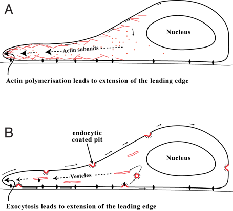
Cell migration is a central process in the development and maintenance of multicellular organisms. Tissue formation during embryonic development, wound healing and immune responses all require the orchestrated movement of cells in particular directions to specific locations. Cells often migrate in response to specific external signals, including chemical signals and mechanical signals. Errors during this process have serious consequences, including intellectual disability, vascular disease, tumor formation and metastasis. An understanding of the mechanism by which cells migrate may lead to the development of novel therapeutic strategies for controlling, for example, invasive tumour cells. Due to the highly viscous environment (low Reynolds number), cells need to continuously produce forces in order to move. Cells achieve active movement by very different mechanisms. Many less complex prokaryotic organisms (and sperm cells) use flagella or cilia to propel themselves. Eukaryot ... [...More Info...] [...Related Items...] OR: [Wikipedia] [Google] [Baidu] |
Cell Proliferation
Cell proliferation is the process by which ''a cell grows and divides to produce two daughter cells''. Cell proliferation leads to an exponential increase in cell number and is therefore a rapid mechanism of tissue growth. Cell proliferation requires both cell growth and cell division to occur at the same time, such that the average size of cells remains constant in the population. Cell division can occur without cell growth, producing many progressively smaller cells (as in cleavage of the zygote), while cell growth can occur without cell division to produce a single larger cell (as in growth of neurons). Thus, cell proliferation is not synonymous with either cell growth or cell division, despite the fact that these terms are sometimes used interchangeably. Stem cells undergo cell proliferation to produce proliferating "transit amplifying" daughter cells that later differentiate to construct tissues during normal development and tissue growth, during tissue regeneration ... [...More Info...] [...Related Items...] OR: [Wikipedia] [Google] [Baidu] |


