|
Colcemid
Demecolcine (INN; also known as colcemid) is a drug used in chemotherapy. It is closely related to the natural alkaloid colchicine with the replacement of the acetyl group on the amino moiety with methyl, but it is less toxic. It depolymerises microtubules and limits microtubule formation (inactivates spindle fibre formation), thus arresting cells in metaphase and allowing cell harvest and karyotyping to be performed. During cell division, demecolcine inhibits mitosis at metaphase by inhibiting spindle formation. Medically, demecolcine has been used to improve the results of cancer radiotherapy by synchronising tumour cells at metaphase, the radiosensitive stage of the cell cycle. In animal cloning procedures, demecolcine makes an ovum eject its nucleus, creating space for insertion of a new nucleus. Mechanism of action Demecolcine is a microtubule-depolymerizing drug like vinblastine. It acts by two distinct mechanisms. At very low concentration it binds to microtubule plus en ... [...More Info...] [...Related Items...] OR: [Wikipedia] [Google] [Baidu] |
|
|
Vinblastine
Vinblastine, sold under the brand name Velban among others, is a chemotherapy medication, typically used with other medications, to treat a number of types of cancer. This includes Hodgkin's lymphoma, non-small-cell lung cancer, bladder cancer, brain cancer, melanoma, and testicular cancer. It is given by injection into a vein. Most people experience some side effects. Commonly it causes a change in sensation, constipation, weakness, loss of appetite, and headaches. Severe side effects include low blood cell counts and shortness of breath. It should not be given to people who have a current bacterial infection. Use during pregnancy will likely harm the baby. Vinblastine works by blocking cell division. Vinblastine was isolated in 1958. An example of a natural herbal remedy that has since been developed into a conventional medicine, vinblastine was originally obtained from the Madagascar periwinkle. It is on the World Health Organization's List of Essential Medicines. ... [...More Info...] [...Related Items...] OR: [Wikipedia] [Google] [Baidu] |
|
 |
Microtubule Inhibitors
Microtubules are polymers of tubulin that form part of the cytoskeleton and provide structure and shape to eukaryotic cells. Microtubules can be as long as 50 micrometres, as wide as 23 to 27 nm and have an inner diameter between 11 and 15 nm. They are formed by the polymerization of a dimer of two globular proteins, alpha and beta tubulin into protofilaments that can then associate laterally to form a hollow tube, the microtubule. The most common form of a microtubule consists of 13 protofilaments in the tubular arrangement. Microtubules play an important role in a number of cellular processes. They are involved in maintaining the structure of the cell and, together with microfilaments and intermediate filaments, they form the cytoskeleton. They also make up the internal structure of cilia and flagella. They provide platforms for intracellular transport and are involved in a variety of cellular processes, including the movement of secretory vesicles, organelles, ... [...More Info...] [...Related Items...] OR: [Wikipedia] [Google] [Baidu] |
|
Pyrogallol Ethers
Pyrogallol is an organic compound with the formula C6H3(OH)3. It is a water-soluble, white solid although samples are typically brownish because of its sensitivity toward oxygen. It is one of three isomers of benzenetriols. Production and reactions It is produced in the manner first reported by Scheele in 1786: heating gallic acid to induce decarboxylation. Gallic acid is also obtained from tannin. Many alternative routes have been devised. One preparation involves treating ''para''-chlorophenoldisulfonic acid with potassium hydroxide, a variant on the time-honored route to phenols from sulfonic acids. Polyhydroxybenzenes are relatively electron-rich. One manifestation is the easy C-acetylation of pyrogallol. Uses It was once used in hair dyeing, dyeing of suturing materials. It also has antiseptic properties. In alkaline solution, pyrogallol undergoes deprotonation. Such solutions absorb oxygen from the air, turning brown. This conversion can be used to determine t ... [...More Info...] [...Related Items...] OR: [Wikipedia] [Google] [Baidu] |
|
|
Micronucleus
A micronucleus is a small nucleus that forms whenever a chromosome or a fragment of a chromosome is not incorporated into one of the daughter nuclei during cell division. It usually is a sign of genotoxic events and chromosomal instability. Micronuclei are commonly seen in cancerous cells and may indicate genomic damage events that can increase the risk of developmental or degenerative diseases. Micronuclei form during anaphase from lagging acentric chromosomes or chromatid fragments caused by incorrectly repaired or unrepaired DNA breaks or by nondisjunction of chromosomes. This improper segregation of chromosomes may result from hypomethylation of repeat sequences present in pericentromeric DNA, irregularities in kinetochore proteins or their assembly, a dysfunctional spindle apparatus, or flawed anaphase checkpoint genes. Micronuclei can contribute to genome instability by promoting a catastrophic mutational event called chromothripsis. Many micronucleus assays have been dev ... [...More Info...] [...Related Items...] OR: [Wikipedia] [Google] [Baidu] |
|
 |
DNA Fragmentation
DNA fragmentation is the separation or breaking of DNA strands into pieces. It can be done intentionally by laboratory personnel or by cells, or can occur spontaneously. Spontaneous or accidental DNA fragmentation is fragmentation that gradually accumulates in a cell. It can be measured by e.g. the comet assay or by the TUNEL assay. Its main units of measurement is the DNA Fragmentation Index (DFI). A DFI of 20% or more significantly reduces the success rates after ICSI. DNA fragmentation was first documented by Williamson in 1970 when he observed discrete oligomeric fragments occurring during cell death in primary neonatal liver cultures. He described the cytoplasmic DNA isolated from mouse liver cells after culture as characterized by DNA fragments with a molecular weight consisting of multiples of 135 kDa. This finding was consistent with the hypothesis that these DNA fragments were a specific degradation product of nuclear DNA. Intentional DNA fragmentation is often neces ... [...More Info...] [...Related Items...] OR: [Wikipedia] [Google] [Baidu] |
 |
Aneuploidy
Aneuploidy is the presence of an abnormal number of chromosomes in a cell (biology), cell, for example a human somatic (biology), somatic cell having 45 or 47 chromosomes instead of the usual 46. It does not include a difference of one or more ploidy#Haploid and monoploid, complete sets of chromosomes. A cell with any number of complete chromosome sets is called a ''ploidy#Euploid, euploid'' cell. An extra or missing chromosome is a common cause of some genetic disorders. Some cancer cells also have abnormal numbers of chromosomes. About 68% of human solid tumors are aneuploid. Aneuploidy originates during cell division when the chromosomes do not separate properly between the two cells (nondisjunction). Most cases of aneuploidy in the autosomes result in miscarriage, and the most common extra autosomal chromosomes among live births are Down syndrome, 21, Edwards syndrome, 18 and Patau syndrome, 13. Chromosome abnormality, Chromosome abnormalities are detected in 1 of 160 live huma ... [...More Info...] [...Related Items...] OR: [Wikipedia] [Google] [Baidu] |
|
Mitosis
Mitosis () is a part of the cell cycle in eukaryote, eukaryotic cells in which replicated chromosomes are separated into two new Cell nucleus, nuclei. Cell division by mitosis is an equational division which gives rise to genetically identical cells in which the total number of chromosomes is maintained. Mitosis is preceded by the S phase of interphase (during which DNA replication occurs) and is followed by telophase and cytokinesis, which divide the cytoplasm, organelles, and cell membrane of one cell into two new cell (biology), cells containing roughly equal shares of these cellular components. The different stages of mitosis altogether define the mitotic phase (M phase) of a cell cycle—the cell division, division of the mother cell into two daughter cells genetically identical to each other. The process of mitosis is divided into stages corresponding to the completion of one set of activities and the start of the next. These stages are preprophase (specific to plant ce ... [...More Info...] [...Related Items...] OR: [Wikipedia] [Google] [Baidu] |
|
 |
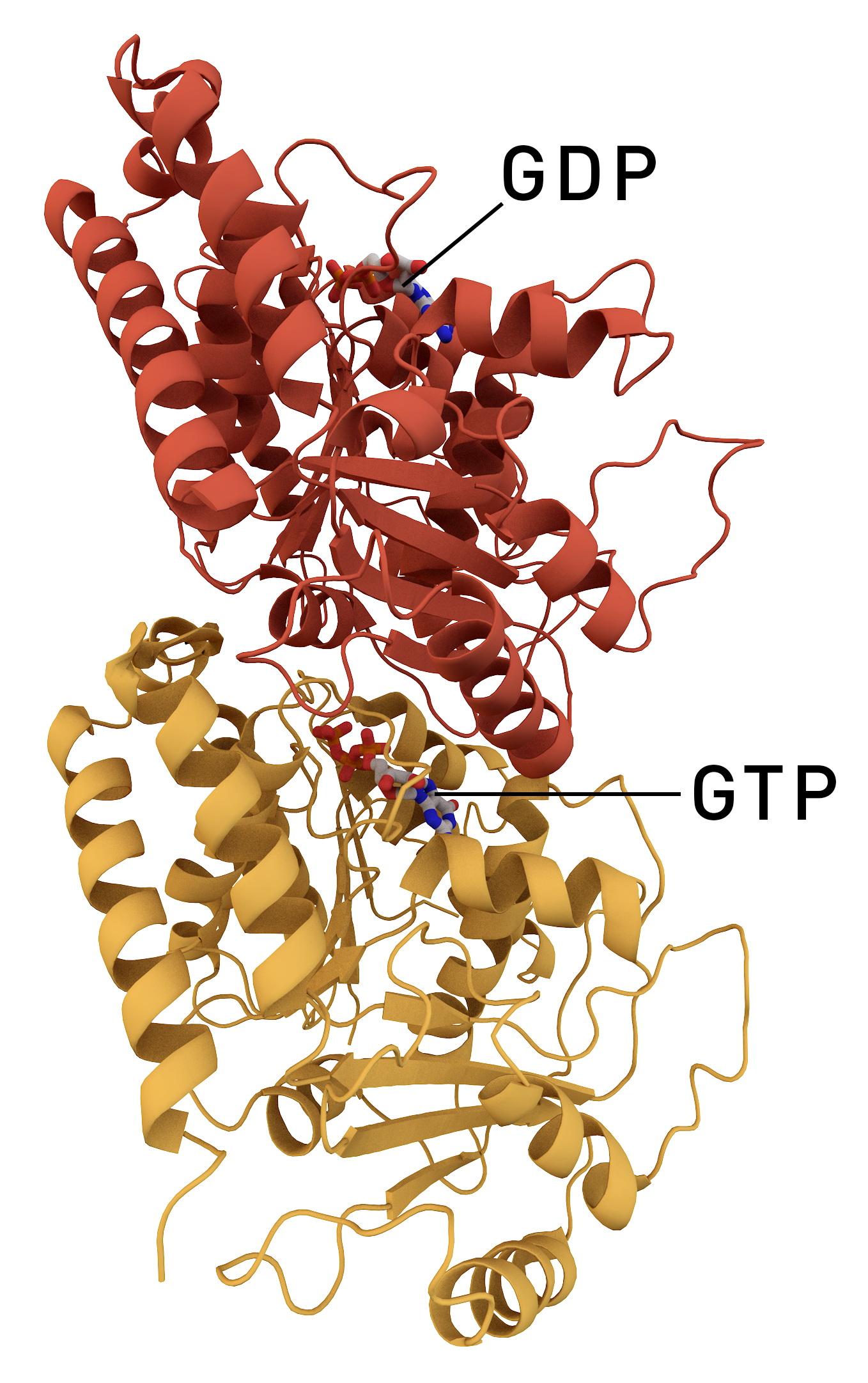
Microtubule
Microtubules are polymers of tubulin that form part of the cytoskeleton and provide structure and shape to eukaryotic cells. Microtubules can be as long as 50 micrometres, as wide as 23 to 27 nanometer, nm and have an inner diameter between 11 and 15 nm. They are formed by the polymerization of a Protein dimer, dimer of two globular proteins, Tubulin#Eukaryotic, alpha and beta tubulin into #Structure, protofilaments that can then associate laterally to form a hollow tube, the microtubule. The most common form of a microtubule consists of 13 protofilaments in the tubular arrangement. Microtubules play an important role in a number of cellular processes. They are involved in maintaining the structure of the cell and, together with microfilaments and intermediate filaments, they form the cytoskeleton. They also make up the internal structure of cilia and flagella. They provide platforms for intracellular transport and are involved in a variety of cellular processes, in ... [...More Info...] [...Related Items...] OR: [Wikipedia] [Google] [Baidu] |
 |
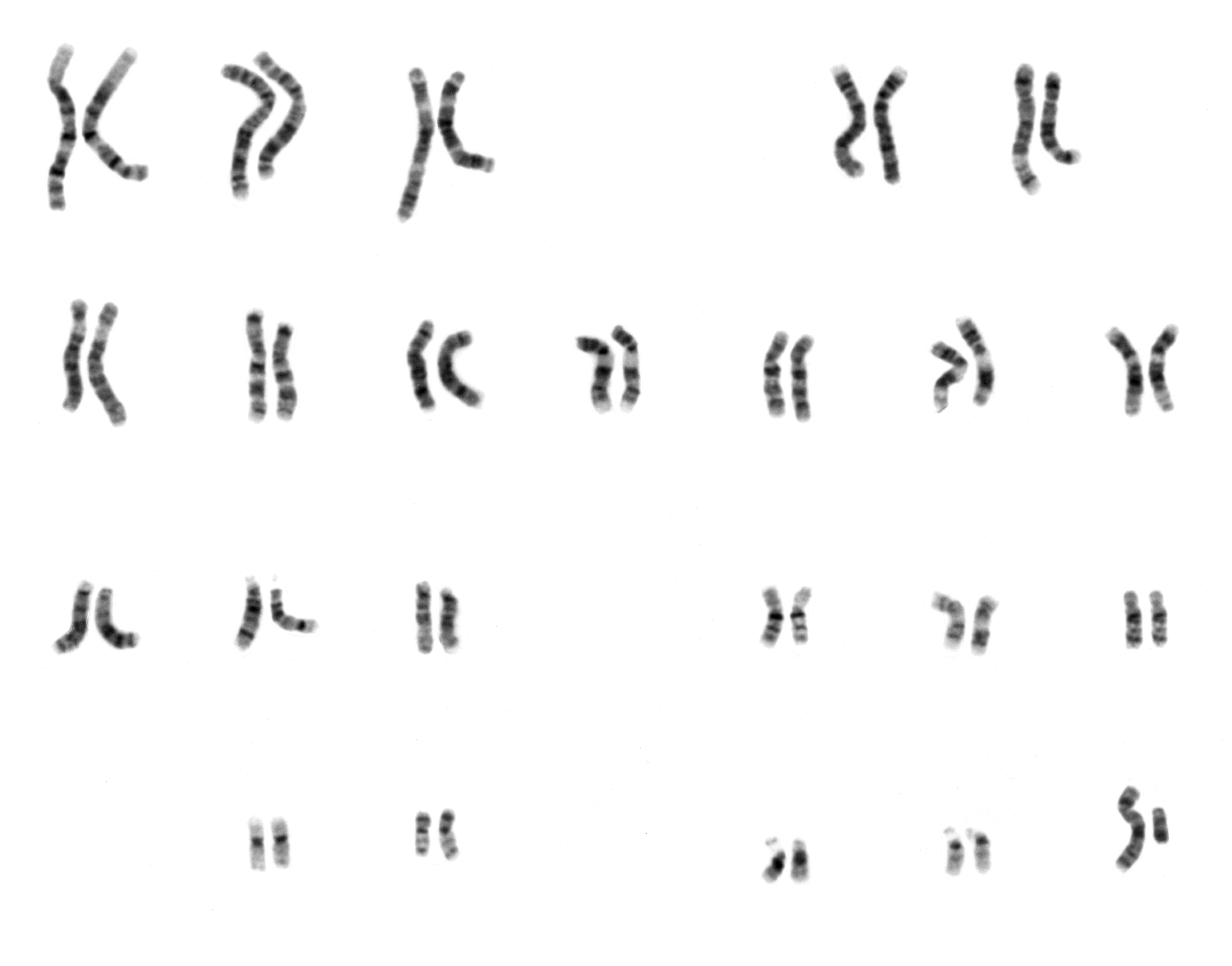
Karyotyping
A karyotype is the general appearance of the complete set of chromosomes in the cells of a species or in an individual organism, mainly including their sizes, numbers, and shapes. Karyotyping is the process by which a karyotype is discerned by determining the chromosome complement of an individual, including the number of chromosomes and any abnormalities. A karyogram or idiogram is a graphical depiction of a karyotype, wherein chromosomes are generally organized in pairs, ordered by size and position of centromere for chromosomes of the same size. Karyotyping generally combines light microscopy and photography in the metaphase of the cell cycle, and results in a photomicrographic (or simply micrographic) karyogram. In contrast, a schematic karyogram is a designed graphic representation of a karyotype. In schematic karyograms, just one of the sister chromatids of each chromosome is generally shown for brevity, and in reality they are generally so close together that they look as ... [...More Info...] [...Related Items...] OR: [Wikipedia] [Google] [Baidu] |