|
Choparts Joints
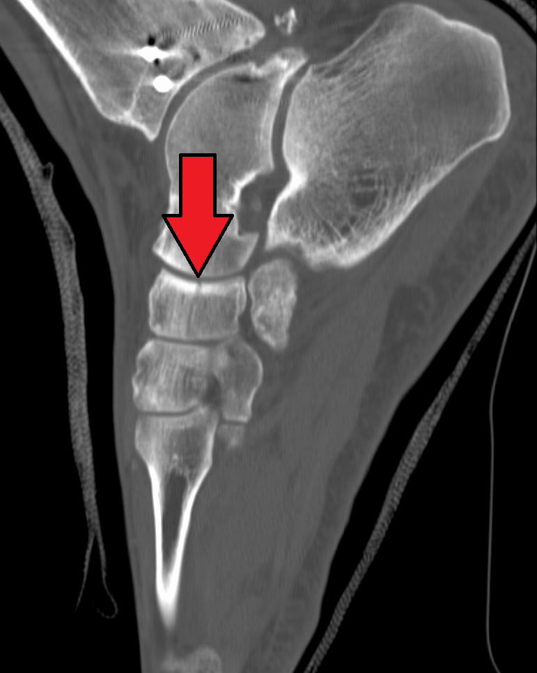
The transverse tarsal joint or midtarsal joint or Chopart's joint is formed by the articulation of the calcaneus with the cuboid (the calcaneocuboid joint), and the articulation of the talus with the navicular (the talocalcaneonavicular joint). The movement which takes place in this joint is more extensive than that in the other tarsal joints, and consists of a sort of rotation by means of which the foot may be slightly flexed or extended, the sole being at the same time carried medially (inverted) or laterally ( everted). The term ''Chopart's joint'' is named after the French surgeon François Chopart François Chopart (20 October 1743 – 9 June 1795) was a French surgeon born in Paris. He was trained in medicine at the Hôtel-Dieu, Pitié and the Bicêtre hospitals. In 1771 he became a professor of practical surgery at the '' École prat .... References Further reading * External links Diagram at ouhsc.edu Joints {{musculoskeletal-stub ... [...More Info...] [...Related Items...] OR: [Wikipedia] [Google] [Baidu] |
Calcaneus
In humans and many other primates, the calcaneus (; from the Latin ''calcaneus'' or ''calcaneum'', meaning heel; : calcanei or calcanea) or heel bone is a bone of the Tarsus (skeleton), tarsus of the foot which constitutes the heel. In some other animals, it is the point of the hock (anatomy), hock. Structure In humans, the calcaneus is the largest of the tarsal bones and the largest bone of the foot. Its long axis is pointed forwards and laterally. The talus bone, calcaneus, and navicular bone are considered the proximal row of tarsal bones. In the calcaneus, several important structures can be distinguished:Platzer (2004), p 216 There is a large calcaneal tuberosity located posteriorly on plantar surface with medial and lateral tubercles on its surface. Besides, there is another peroneal tubercle on its lateral surface. On its lower edge on either side are its lateral and medial processes (serving as the origins of the Abductor hallucis muscle, abductor hallucis and Abductor di ... [...More Info...] [...Related Items...] OR: [Wikipedia] [Google] [Baidu] |
Cuboid Bone
In the human body, the cuboid bone is one of the seven tarsal bones of the foot. Structure The cuboid bone is the most lateral of the bones in the distal row of the tarsus. It is roughly cubical in shape, and presents a prominence in its inferior (or plantar) surface, the tuberosity of the cuboid. The bone provides a groove where the tendon of the peroneus longus muscle passes to reach its insertion in the first metatarsal and medial cuneiform bones. Surfaces The dorsal surface, directed upward and lateralward, is rough, for the attachment of ligaments. The plantar surface presents in front a deep groove, the peroneal sulcus, which runs obliquely forward and medialward; it lodges the tendon of the peroneus longus, and is bounded behind by a prominent ridge, to which the long plantar ligament is attached. The ridge ends laterally in an eminence, the tuberosity, the surface of which presents an oval facet; on this facet glides the sesamoid bone or cartilage frequently found ... [...More Info...] [...Related Items...] OR: [Wikipedia] [Google] [Baidu] |
Calcaneocuboid Joint
The calcaneocuboid joint is the joint between the calcaneus and the cuboid bone. Structure The calcaneocuboid joint is a type of saddle joint between the calcaneus and the cuboid bone. Ligaments There are five ligaments connecting the calcaneus and the cuboid bone, forming parts of the articular capsule: * the dorsal calcaneocuboid ligament. * part of the bifurcated ligament. * the long plantar ligament. * and the plantar calcaneocuboid ligament. Function The calcaneocuboid joint is conventionally described as among the least mobile joints in the human foot. The articular surfaces of the two bones are relatively flat with some irregular undulations, which seem to suggest movement limited to a single rotation and some translation. However, the cuboid rotates as much as 25° about an oblique axis during inversion- eversion in a movement that could be called involution. Clinical significance The calcaneocuboid joint may be affected by a calcaneal fracture. This may be a s ... [...More Info...] [...Related Items...] OR: [Wikipedia] [Google] [Baidu] |
Talus Bone
The talus (; Latin for ankle or ankle bone; : tali), talus bone, astragalus (), or ankle bone is one of the group of Foot#Structure, foot bones known as the tarsus (skeleton), tarsus. The tarsus forms the lower part of the ankle joint. It transmits the entire weight of the body from the lower legs to the foot.Platzer (2004), p 216 The talus has joints with the two bones of the lower leg, the tibia and thinner fibula. These leg bones have two prominences (the Lateral malleolus, lateral and Medial malleolus, medial malleoli) that articulation (anatomy), articulate with the talus. At the foot end, within the tarsus, the talus articulates with the calcaneus (heel bone) below, and with the curved navicular bone in front; together, these foot articulations form the Ball-and-socket joint, ball-and-socket-shaped talocalcaneonavicular joint. The talus is the second largest of the Tarsus (skeleton), tarsal bones; it is also one of the bones in the human body with the highest percentage of i ... [...More Info...] [...Related Items...] OR: [Wikipedia] [Google] [Baidu] |
Navicular
The navicular bone is a small bone found in the feet of most mammals. Human anatomy The navicular bone in humans is one of the tarsus (skeleton), tarsal bones, found in the foot. Its name derives from the human bone's resemblance to a small boat, caused by the strongly concave Anatomical terms of location#Proximal and distal, proximal joint, articular surface. The term ''navicular bone'' or ''hand navicular bone'' was formerly used for the scaphoid bone, one of the Carpal bones, carpal bones of the wrist. The navicular bone in humans is located on the Anatomical terms of location#Relative directions, medial side of the foot, and articulates proximally with the Talus bone, talus, Anatomical terms of location#Relative directions, distally with the three cuneiform bones, and Anatomical terms of location#Relative directions, laterally with the Cuboid bone, cuboid. It is the last of the foot bones to start ossification and does not tend to do so until the end of the third year in ... [...More Info...] [...Related Items...] OR: [Wikipedia] [Google] [Baidu] |
Talocalcaneonavicular Joint
The talocalcaneonavicular joint is a ball and socket joint in the foot; the rounded head of the talus is received into the concavity formed by the posterior surface of the navicular, the anterior articular surface of the calcaneus, and the upper surface of the plantar calcaneonavicular ligament. Structure As its shape suggests, this joint is a synovial ball-and-socket joint. It is composed of three articular surfaces: * The articulation between the medial talar articular surface on the sustentaculum tali of the superior calcaneus and the corresponding medial facet found inferiorly on the talus neck * The articulation between the anterior talar articular surface of the superior calcaneus and the anterior facet of the corresponding talus found inferiorly on the talar head * The articulation between the articular surface of navicular and the head of talus (talonavicular joint) Ligaments The plantar calcaneonavicular ligament The plantar calcaneonavicular ligament (also known as ... [...More Info...] [...Related Items...] OR: [Wikipedia] [Google] [Baidu] |
Evert
Evert is a Dutch and Swedish short form of the Germanic masculine name "Everhard" (alternative Eberhard). at the Meertens Institute database of given names in the Netherlands. It is also used as surname. Notable people with the name include: Given name * Evert van Aelst (1602–1657), Dutch still life painter * Evert Andersen (1772–1809), Norwegian naval officer * Evert Augustus Duyckinck (1816–1878), American publisher and bi ...[...More Info...] [...Related Items...] OR: [Wikipedia] [Google] [Baidu] |
Surgeon
In medicine, a surgeon is a medical doctor who performs surgery. Even though there are different traditions in different times and places, a modern surgeon is a licensed physician and received the same medical training as physicians before specializing in surgery. In some countries and jurisdictions, the title of 'surgeon' is restricted to maintain the integrity of the craft group in the medical profession. A specialist regarded as a legally recognized surgeon includes podiatry, dentistry, and veterinary medicine. It is estimated that surgeons perform over 300 million surgical procedures globally each year. History The first person to document a surgery was the 6th century BC Indian physician-surgeon, Sushruta. He specialized in cosmetic plastic surgery and even documented an open rhinoplasty procedure.Papel, Ira D. and Frodel, John (2008) ''Facial Plastic and Reconstructive Surgery''. Thieme Medical Pub. His Masterpiece, magnum opus ''Suśruta-saṃhitā'' is one of the m ... [...More Info...] [...Related Items...] OR: [Wikipedia] [Google] [Baidu] |
François Chopart
François Chopart (20 October 1743 – 9 June 1795) was a French surgeon born in Paris. He was trained in medicine at the Hôtel-Dieu, Pitié and the Bicêtre hospitals. In 1771 he became a professor of practical surgery at the '' École pratique'' in Paris, and in 1782 succeeded Toussaint Bordenave (1728–1782) as chair of physiology. Chopart was a pioneer of urological surgery, putting emphasis on dealing with the urinary tract as a whole. In 1791/92 he published the two-volume ''Traité des maladies des voies urinaires''. at Who Named It With Pierre-Joseph Desault (1744–1795), he was a ... [...More Info...] [...Related Items...] OR: [Wikipedia] [Google] [Baidu] |


