|
Ball (foot)
The ball of the foot is the padded portion of the sole between the toes and the arch, underneath the heads of the metatarsal bones. In comparative foot morphology, the ball is most analogous to the metacarpal (forepaw) or metatarsal (hindpaw) pad in many mammals with paws, and serves mostly the same functions. The ball of the foot is of utmost importance when playing sports. Many sports, such as tennis, requires the player to stand on the balls of their feet for increased agility. The ball is a common area in which people develop pain, known as metatarsalgia. People who frequently wear high heels often develop pain in the balls of their feet from the immense amount of pressure that is placed on them for long periods of time, due to the inclination of the shoes. To remedy this, there is a market for ball-of-foot or general foot cushions that are placed into shoes to relieve some of the pressure. Alternately, people can have a procedure done in which a dermal filler is inje ... [...More Info...] [...Related Items...] OR: [Wikipedia] [Google] [Baidu] |
Sole (foot)
The sole is the bottom of the foot. In humans the sole of the foot is anatomically referred to as the plantar aspect. Structure The glabrous skin on the sole of the foot lacks the hair and pigmentation found elsewhere on the body, and it has a high concentration of sweat pores. The sole contains the thickest layers of skin on the body due to the weight that is continually placed on it. It is crossed by a set of creases that form during the early stages of embryonic development. Like those of the palm, the sweat pores of the sole lack sebaceous glands. The sole is a sensory organ by which we can perceive the ground while standing and walking. The subcutaneous tissue in the sole has adapted to deal with the high local compressive forces on the heel and the ball (between the toes and the arch) by developing a system of "pressure chambers." Each chamber is composed of internal fibrofatty tissue covered by external collagen connective tissue. The septa (internal walls) ... [...More Info...] [...Related Items...] OR: [Wikipedia] [Google] [Baidu] |
Toes
Toes are the digits (fingers) of the foot of a tetrapod. Animal species such as cats that walk on their toes are described as being '' digitigrade''. Humans, and other animals that walk on the soles of their feet, are described as being '' plantigrade''; ''unguligrade'' animals are those that walk on hooves at the tips of their toes. Structure There are normally five toes present on each human foot. Each toe consists of three phalanx bones, the proximal, middle, and distal, with the exception of the big toe ( la, hallux). For a minority of people, the little toe also is missing a middle bone. The hallux only contains two phalanx bones, the proximal and distal. The joints between each phalanx are the interphalangeal joints. The proximal phalanx bone of each toe articulates with the metatarsal bone of the foot at the metatarsophalangeal joint. Each toe is surrounded by skin, and present on all five toes is a toenail. The toes are, from medial to lateral: * the first to ... [...More Info...] [...Related Items...] OR: [Wikipedia] [Google] [Baidu] |
Arches Of The Foot
The arches of the foot, formed by the tarsal and metatarsal bones, strengthened by ligaments and tendons, allow the foot to support the weight of the body in the erect posture with the least weight. They are categorized as longitudinal and transverse arches. Structure Longitudinal arches The longitudinal arches of the foot can be divided into medial and lateral arches. Medial arch The medial arch is higher than the lateral longitudinal arch. It is made up by the calcaneus, the talus, the navicular, the three cuneiforms (medial, intermediate, and lateral), and the first, second, and third metatarsals. Its summit is at the superior articular surface of the talus, and its two extremities or piers, on which it rests in standing, are the tuberosity on the plantar surface of the calcaneus posteriorly and the heads of the first, second, and third metatarsal bones anteriorly. The chief characteristic of this arch is its elasticity, due to its height and to the number of small ... [...More Info...] [...Related Items...] OR: [Wikipedia] [Google] [Baidu] |
Comparative Foot Morphology
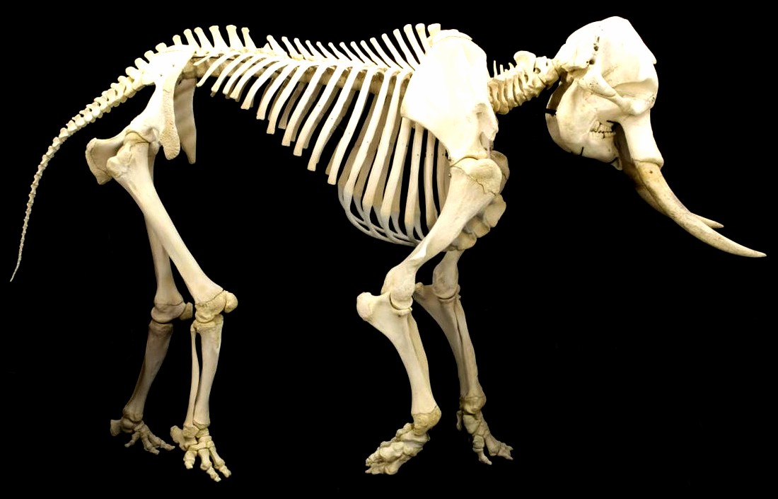
Comparative foot morphology involves comparing the form of distal limb structures of a variety of terrestrial vertebrates. Understanding the role that the foot plays for each type of organism must take account of the differences in body type, foot shape, arrangement of structures, loading conditions and other variables. However, similarities also exist among the feet of many different terrestrial vertebrates. The paw of the dog, the hoof of the horse, the ''manus'' (forefoot) and ''pes'' (hindfoot) of the elephant, and the foot of the human all share some common features of structure, organization and function. Their foot structures function as the load-transmission platform which is essential to balance, standing and types of locomotion (such as walking, trotting, galloping and running). The discipline of biomimetics applies the information gained by comparing the foot morphology of a variety of terrestrial vertebrates to human-engineering problems. For instance, it may prov ... [...More Info...] [...Related Items...] OR: [Wikipedia] [Google] [Baidu] |
Metatarsalgia
Metatarsalgia, literally metatarsal pain and colloquially known as a stone bruise, is any painful foot condition affecting the metatarsal region of the foot. This is a common problem that can affect the joints and bones of the metatarsals. Metatarsalgia is most often localized to the first metatarsal head – the ball of the foot just behind the big toe. There are two small sesamoid bones under the first metatarsal head. The next most frequent site of metatarsal head pain is under the second metatarsal. This can be due to either too short a first metatarsal bone or to "hypermobility of the first ray" – metatarsal bone and medial cuneiform bone behind it – both of which result in excess pressure being transmitted into the second metatarsal head. Signs and Symptoms Metatarsalgia is characterized by a sharp pain in the ball of the foot. Causes One cause of metatarsalgia is Morton's neuroma. When toes are squeezed together too often and for too long, the nerve that runs betw ... [...More Info...] [...Related Items...] OR: [Wikipedia] [Google] [Baidu] |
High-heeled Footwear
High-heeled shoes, also known as high heels, are a type of shoe with an angled sole. The heel in such shoes is raised above the ball of the foot. High heels cause the legs to appear longer, make the wearer appear taller, and accentuate the calf muscle. There are many types of heels in varying colors, materials, styles, and heights. High heels have been used in various ways to communicate nationality, professional affiliation, gender, and social status. High heels have been important in the West. In early 17th century Europe, for example, high heels were a sign of masculinity and high social status. It wasn't until the end of the century that this trend spread to women's fashion. By the 18th century, high-heeled shoes had split along gender lines. By this time, heels for men's shoes were chunky squares attached to riding boots or tall formal dress boots while women's high heels were narrow and pointy and often attached to slipper-like dress shoes (similar to modern heels). B ... [...More Info...] [...Related Items...] OR: [Wikipedia] [Google] [Baidu] |
Shoe Insert
A removable shoe insert, otherwise known as a foot orthosis, insole or inner sole accomplishes many purposes, including daily wear comfort, height enhancement, plantar fasciitis treatment, arch support, foot and joint pain relief from arthritis, overuse, injuries, leg length discrepancy, and other causes such as orthopedic correction and athletic performance. Medical use of foot orthoses has been criticized as lacking evidence of benefit, and practice is very inconsistent: reputed podiatrists prescribe completely different orthoses for a single patient. Further, effect of a given design of orthosis varies significantly by patient, and standard practice to personalize prescription is not available. However, evidence is mixed: patients often report at least short-term improvements in comfort, and other studies have found effectiveness. Fitting patients There are three standard methods for fitting patients: plaster casts, foam box impressions, or three-dimensional computer imaging ... [...More Info...] [...Related Items...] OR: [Wikipedia] [Google] [Baidu] |
Metatarsus
The metatarsal bones, or metatarsus, are a group of five long bones in the foot, located between the tarsal bones of the hind- and mid-foot and the phalanges of the toes. Lacking individual names, the metatarsal bones are numbered from the medial side (the side of the great toe): the first, second, third, fourth, and fifth metatarsal (often depicted with Roman numerals). The metatarsals are analogous to the metacarpal bones of the hand. The lengths of the metatarsal bones in humans are, in descending order, second, third, fourth, fifth, and first. Structure The five metatarsals are dorsal convex long bones consisting of a shaft or body, a base (proximally), and a head (distally).Platzer 2004, p. 220 The body is prismoid in form, tapers gradually from the tarsal to the phalangeal extremity, and is curved longitudinally, so as to be concave below, slightly convex above. The base or posterior extremity is wedge-shaped, articulating proximally with the tarsal bones, an ... [...More Info...] [...Related Items...] OR: [Wikipedia] [Google] [Baidu] |
Metatarsalgia
Metatarsalgia, literally metatarsal pain and colloquially known as a stone bruise, is any painful foot condition affecting the metatarsal region of the foot. This is a common problem that can affect the joints and bones of the metatarsals. Metatarsalgia is most often localized to the first metatarsal head – the ball of the foot just behind the big toe. There are two small sesamoid bones under the first metatarsal head. The next most frequent site of metatarsal head pain is under the second metatarsal. This can be due to either too short a first metatarsal bone or to "hypermobility of the first ray" – metatarsal bone and medial cuneiform bone behind it – both of which result in excess pressure being transmitted into the second metatarsal head. Signs and Symptoms Metatarsalgia is characterized by a sharp pain in the ball of the foot. Causes One cause of metatarsalgia is Morton's neuroma. When toes are squeezed together too often and for too long, the nerve that runs betw ... [...More Info...] [...Related Items...] OR: [Wikipedia] [Google] [Baidu] |
Tactile Pad
A tactile pad is an area of skin that is particularly sensitive to pressure, temperature, or pain. Tactile pads are characterized by high concentrations of free nerve endings. In primate Primates are a diverse order of mammals. They are divided into the strepsirrhines, which include the lemurs, galagos, and lorisids, and the haplorhines, which include the tarsiers and the simians ( monkeys and apes, the latter including ...s, the last phalanges in the fingers and toes have tactile pads, allowing very accurate manipulation of objects. This precision grip was an important evolutionary advance in primates. References somatosensory system {{Anatomy-stub ... [...More Info...] [...Related Items...] OR: [Wikipedia] [Google] [Baidu] |
Fat Pad
A fat pad (aka haversian gland) is a mass of closely packed fat cells surrounded by fibrous tissue septa.TheFreeDictionary > Fat padCiting: Mosby's Medical Dictionary, 8th edition. 2009 They may be extensively supplied with capillaries and nerve endings. Examples are: * ''Intraarticular fat pads''. These are also covered by a layer of synovial cells. A fat pad sign is an elevation of the anterior and posterior fat pads of the elbow joint, and suggests the presence of an occult fracture. * Buccal fat pad can be seen in nursing babies. * The fat pad of the labia majora, which can be used as a graft, often as a so-called "Martius labial fat pad graft", which can be used, for example, in urethrolysis. *Fat pads within the heels which when they get inflamed can cause heel pad syndrome Heel pad syndrome is a pain that occurs in the center of the heel. It is typically due to atrophy of the fat pad which makes up the heel. Risk factors include obesity. Other conditions with similar symp ... [...More Info...] [...Related Items...] OR: [Wikipedia] [Google] [Baidu] |


