|
Anal Triangle
The anal triangle is the posterior part of the perineum. It contains the anal canal. Structure The anal triangle can be defined either by its vertices or its sides. * ''Vertices'' ** one vertex at the coccyx bone ** the two ischial tuberosities of the pelvic bone * ''Sides'' ** perineal membrane (posterior border of perineal membrane forms anterior border of anal triangle) ** the two sacrotuberous ligaments Contents Some components of the anal triangle include:Daftary, Shirish; Chakravarti, Sudip (2011). Manual of Obstetrics, 3rd Edition. Elsevier. pp. 1-16. . * Ischioanal fossa * Anococcygeal body * Sacrotuberous ligament * Sacrospinous ligament * Pudendal nerve * Internal pudendal artery and Internal pudendal vein * Anal canal * Muscles ** Sphincter ani externus muscle ** Gluteus maximus muscle ** Obturator internus muscle ** Levator ani muscle ** Coccygeus muscle Additional images Image:Gray320.png, Articulations of pelvis. Posterior view. Image:Gray542.png, The sup ... [...More Info...] [...Related Items...] OR: [Wikipedia] [Google] [Baidu] |
Perineum
The perineum in humans is the space between the anus and scrotum in the male, or between the anus and the vulva in the female. The perineum is the region of the body between the pubic symphysis (pubic arch) and the coccyx (tail bone), including the perineal body and surrounding structures. There is some variability in how the boundaries are defined. The perineal raphe is visible and pronounced to varying degrees. The perineum is an erogenous zone. The word perineum entered English from late Latin via Greek περίναιος ~ περίνεος ''perinaios, perineos'', itself from περίνεος, περίνεοι 'male genitals' and earlier περίς ''perís'' 'penis' through influence from πηρίς ''pērís'' 'scrotum'. The term was originally understood as a purely male body-part with the perineal raphe seen as a continuation of the scrotal septum since masculinization causes the development of a large anogenital distance in men, in comparison to the corresponding lac ... [...More Info...] [...Related Items...] OR: [Wikipedia] [Google] [Baidu] |
Internal Pudendal Artery
The internal pudendal artery is one of the three pudendal arteries. It branches off the internal iliac artery, and provides blood to the external genitalia. Structure The internal pudendal artery is the terminal branch of the anterior trunk of the internal iliac artery. It is smaller in the female than in the male. Path It arises from the anterior division of internal iliac artery. It runs on the lateral pelvic wall. It exits the pelvic cavity through the greater sciatic foramen, inferior to the piriformis muscle, to enter the gluteal region. It then curves around the sacrospinous ligament to enter the perineum through the lesser sciatic foramen. It travels through the pudendal canal with the internal pudendal veins and the pudendal nerve. Branches The internal pudendal artery gives off the following branches: The deep artery of clitoris is a branch of the internal pudendal artery and supplies the clitoral crura. Another branch of the internal pudendal artery is the ... [...More Info...] [...Related Items...] OR: [Wikipedia] [Google] [Baidu] |
Perineum
The perineum in humans is the space between the anus and scrotum in the male, or between the anus and the vulva in the female. The perineum is the region of the body between the pubic symphysis (pubic arch) and the coccyx (tail bone), including the perineal body and surrounding structures. There is some variability in how the boundaries are defined. The perineal raphe is visible and pronounced to varying degrees. The perineum is an erogenous zone. The word perineum entered English from late Latin via Greek περίναιος ~ περίνεος ''perinaios, perineos'', itself from περίνεος, περίνεοι 'male genitals' and earlier περίς ''perís'' 'penis' through influence from πηρίς ''pērís'' 'scrotum'. The term was originally understood as a purely male body-part with the perineal raphe seen as a continuation of the scrotal septum since masculinization causes the development of a large anogenital distance in men, in comparison to the corresponding lac ... [...More Info...] [...Related Items...] OR: [Wikipedia] [Google] [Baidu] |
Sacrum
The sacrum (plural: ''sacra'' or ''sacrums''), in human anatomy, is a large, triangular bone at the base of the spine that forms by the fusing of the sacral vertebrae (S1S5) between ages 18 and 30. The sacrum situates at the upper, back part of the pelvic cavity, between the two wings of the pelvis. It forms joints with four other bones. The two projections at the sides of the sacrum are called the alae (wings), and articulate with the ilium at the L-shaped sacroiliac joints. The upper part of the sacrum connects with the last lumbar vertebra (L5), and its lower part with the coccyx (tailbone) via the sacral and coccygeal cornua. The sacrum has three different surfaces which are shaped to accommodate surrounding pelvic structures. Overall it is concave (curved upon itself). The base of the sacrum, the broadest and uppermost part, is tilted forward as the sacral promontory internally. The central part is curved outward toward the posterior, allowing greater room for the p ... [...More Info...] [...Related Items...] OR: [Wikipedia] [Google] [Baidu] |
Coccygeus Muscle
The coccygeus muscle or ischiococcygeus is a muscle of the pelvic floor, located posterior to levator ani and anterior to the sacrospinous ligament. Structure The coccygeus muscle is posterior to levator ani and anterior to the sacrospinous ligament in the pelvic floor. It is a triangular plane of muscular and tendinous fibers. It arises by its apex from the spine of the ischium and sacrospinous ligament. It is inserted by its base into the margin of the coccyx and into the side of the lowest piece of the sacrum. In combination with the levator ani, it forms the pelvic diaphragm. The pudendal nerve runs between the coccygeus muscle and the piriformis muscle, superficial to the coccygeus muscle. Nerve supply The coccygeus muscle is innervated by the pudendal nerve, which runs between it and the piriformis muscle. Function The coccygeus muscle assists the levator ani and piriformis muscle in closing in the back part of the outlet of the pelvis. This helps to ... [...More Info...] [...Related Items...] OR: [Wikipedia] [Google] [Baidu] |
Levator Ani Muscle
The levator ani is a broad, thin muscle group, situated on either side of the pelvis. It is formed from three muscle components: the pubococcygeus, the iliococcygeus, and the puborectalis. It is attached to the inner surface of each side of the lesser pelvis, and these unite to form the greater part of the pelvic floor. The coccygeus muscle completes the pelvic floor, which is also called the ''pelvic diaphragm''. It supports the viscera in the pelvic cavity, and surrounds the various structures that pass through it. The levator ani is the main pelvic floor muscle and painfully contracts during vaginismus. It also contracts rhythmically during orgasm. Structure The levator ani is made up of 3 parts: * Iliococcygeus muscle * Pubococcygeus muscle * Puborectalis muscle The iliococcygeus arises from the inner side of the ischium (the lower and back part of the hip bone) and from the posterior part of the tendinous arch of the obturator fascia, and is attached to the coccyx an ... [...More Info...] [...Related Items...] OR: [Wikipedia] [Google] [Baidu] |
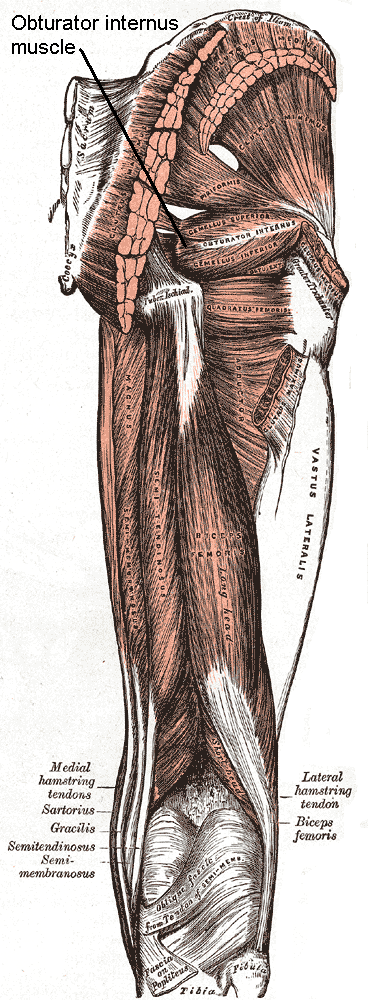
Obturator Internus Muscle
The internal obturator muscle or obturator internus muscle originates on the medial surface of the obturator membrane, the ischium near the membrane, and the rim of the pubis. It exits the pelvic cavity through the lesser sciatic foramen. The internal obturator is situated partly within the lesser pelvis, and partly at the back of the hip-joint. It functions to help laterally rotate femur with hip extension and abduct femur with hip flexion, as well as to steady the femoral head in the acetabulum. Structure Origin The internal obturator muscle arises from the inner surface of the antero-lateral wall of the pelvis. It surrounds the obturator foramen. It is attached to the inferior pubic ramus and ischium, and at the side to the inner surface of the hip bone below and behind the pelvic brim. It reaches from the upper part of the greater sciatic foramen above and behind to the obturator foramen below and in front. It also arises from the pelvic surface of the obturator membr ... [...More Info...] [...Related Items...] OR: [Wikipedia] [Google] [Baidu] |
Gluteus Maximus Muscle
The gluteal muscles, often called glutes are a group of three muscles which make up the gluteal region commonly known as the buttocks: the gluteus maximus, gluteus medius and gluteus minimus. The three muscles originate from the ilium and sacrum and insert on the femur. The functions of the muscles include extension, abduction, external rotation, and internal rotation of the hip joint. Structure The gluteus maximus is the largest and most superficial of the three gluteal muscles. It makes up a large part of the shape and appearance of the hips. It is a narrow and thick fleshy mass of a quadrilateral shape, and forms the prominence of the buttocks. The gluteus medius is a broad, thick, radiating muscle, situated on the outer surface of the pelvis. It lies profound to the gluteus maximus and its posterior third is covered by the gluteus maximus, its anterior two-thirds by the gluteal aponeurosis, which separates it from the superficial fascia and skin. The gluteus minimus is th ... [...More Info...] [...Related Items...] OR: [Wikipedia] [Google] [Baidu] |
Sphincter Ani Externus Muscle
The external anal sphincter (or sphincter ani externus ) is a flat plane of skeletal muscle fibers, elliptical in shape and intimately adherent to the skin surrounding the margin of the anus. Anatomy The external anal sphincter measures about 8 to 10 cm in length, from its anterior to its posterior extremity, and is about 2.5 cm opposite the anus, the sphincter muscle retracts on defecating. It consists of two layers: ''superficial'' and ''deep''. * The superficial layer, constitutes the main portion of the muscle, and arises from a narrow tendinous band, the anococcygeal raphe, which stretches from the tip of the coccyx to the posterior margin of the anus; it forms two flattened planes of muscular tissue, which encircle the anus and meet in front to be inserted into the central tendinous point of the perineum, joining with the superficial transverse perineal muscle, the levator ani, and the bulbospongiosus muscle also known as the bulbocavernosus. * The deeper laye ... [...More Info...] [...Related Items...] OR: [Wikipedia] [Google] [Baidu] |
Anal Canal
The anal canal is the part that connects the rectum to the anus, located below the level of the pelvic diaphragm. It is located within the anal triangle of the perineum, between the right and left ischioanal fossa. As the final functional segment of the bowel, it functions to regulate release of excrement by two muscular sphincter complexes. The anus is the aperture at the terminal portion of the anal canal. Structure In humans, the anal canal is approximately long, from the anorectal junction to the anus. It is directed downwards and backwards. It is surrounded by inner involuntary and outer voluntary sphincters which keep the lumen closed in the form of an anteroposterior slit. The canal is differentiated from the rectum by a transition along the internal surface from endodermal to skin-like ectodermal tissue. The anal canal is traditionally divided into two segments, upper and lower, separated by the pectinate line (also known as the dentate line): * upper zone (zo ... [...More Info...] [...Related Items...] OR: [Wikipedia] [Google] [Baidu] |
Internal Pudendal Vein
The internal pudendal veins (internal pudic veins) are a set of veins in the pelvis. They are the venae comitantes of the internal pudendal artery. Internal pudendal veins are enclosed by pudendal canal, with internal pudendal artery and pudendal nerve. They begin in the deep veins of the vulva and of the penis, issuing from the bulb of the vestibule and the bulb of the penis, respectively. They accompany the internal pudendal artery, and unite to form a single vessel, which ends in the internal iliac vein. They receive the veins from the urethral bulb, the perineal and inferior hemorrhoidal veins. The deep dorsal vein of the penis Deep or The Deep may refer to: Places United States * Deep Creek (Appomattox River tributary), Virginia * Deep Creek (Great Salt Lake), Idaho and Utah * Deep Creek (Mahantango Creek tributary), Pennsylvania * Deep Creek (Mojave River tributary) ... communicates with the internal pudendal veins, but ends mainly in the pudendal plexus. Refere ... [...More Info...] [...Related Items...] OR: [Wikipedia] [Google] [Baidu] |
Pudendal Nerve
The pudendal nerve is the main nerve of the perineum. It carries sensation from the external genitalia of both sexes and the skin around the anus and perineum, as well as the motor supply to various pelvic muscles, including the male or female external urethral sphincter and the external anal sphincter. If damaged, most commonly by childbirth, lesions may cause sensory loss or fecal incontinence. The nerve may be temporarily blocked as part of an anaesthetic procedure. The pudendal canal that carries the pudendal nerve is also known by the eponymous term "Alcock's canal", after Benjamin Alcock, an Irish anatomist who documented the canal in 1836. Structure The pudendal nerve is paired, meaning there are two nerves, one on the left and one on the right side of the body. Each is formed as three roots immediately converge above the upper border of the sacrotuberous ligament and the coccygeus muscle. The three roots become two cords when the middle and lower root join to ... [...More Info...] [...Related Items...] OR: [Wikipedia] [Google] [Baidu] |


