|
Amorphosynthesis
Amorphosynthesis, also called a hemi-sensory deficit, is a neuropsychological condition in which a patient experiences unilateral inattention to sensory input. This phenomenon is frequently associated with damage to the right cerebral hemisphere resulting in severe sensory deficits that are observed on the contralesional (left) side of the body. A right-sided deficit is less commonly observed and the effects are reported to be temporary and minor. Evidence suggests that the right cerebral hemisphere has a dominant role in attention and awareness to somatic sensations through ipsilateral and contralateral stimulation. In contrast, the left cerebral hemisphere is activated only by contralateral stimuli. Thus, the left and right cerebral hemispheres exhibit redundant processing to the right-side of the body and a lesion to the left cerebral hemisphere can be compensated by the ipsiversive processes of the right cerebral hemisphere. For this reason, right-sided amorphosynthesis is less o ... [...More Info...] [...Related Items...] OR: [Wikipedia] [Google] [Baidu] |
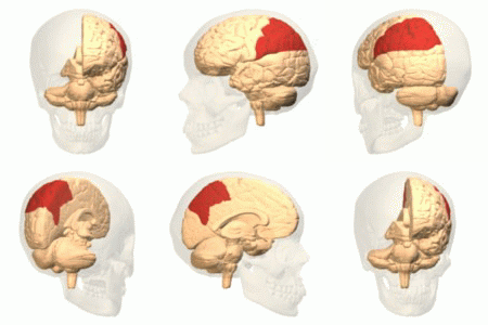
Parietal Lobe
The parietal lobe is one of the four major lobes of the cerebral cortex in the brain of mammals. The parietal lobe is positioned above the temporal lobe and behind the frontal lobe and central sulcus. The parietal lobe integrates sensory information among various modalities, including spatial sense and navigation (proprioception), the main sensory receptive area for the sense of touch in the somatosensory cortex which is just posterior to the central sulcus in the postcentral gyrus, and the dorsal stream of the visual system. The major sensory inputs from the skin ( touch, temperature, and pain receptors), relay through the thalamus to the parietal lobe. Several areas of the parietal lobe are important in language processing. The somatosensory cortex can be illustrated as a distorted figure – the cortical homunculus (Latin: "little man") in which the body parts are rendered according to how much of the somatosensory cortex is devoted to them. The superior parietal lobule a ... [...More Info...] [...Related Items...] OR: [Wikipedia] [Google] [Baidu] |
Cerebral Hemispheres
The vertebrate cerebrum (brain) is formed by two cerebral hemispheres that are separated by a groove, the longitudinal fissure. The brain can thus be described as being divided into left and right cerebral hemispheres. Each of these hemispheres has an outer layer of grey matter, the cerebral cortex, that is supported by an inner layer of white matter. In eutherian (placental) mammals, the hemispheres are linked by the corpus callosum, a very large bundle of nerve fibers. Smaller commissures, including the anterior commissure, the posterior commissure and the fornix, also join the hemispheres and these are also present in other vertebrates. These commissures transfer information between the two hemispheres to coordinate localized functions. There are three known poles of the cerebral hemispheres: the ''occipital pole'', the '' frontal pole'', and the ''temporal pole''. The central sulcus is a prominent fissure which separates the parietal lobe from the frontal lobe and the pri ... [...More Info...] [...Related Items...] OR: [Wikipedia] [Google] [Baidu] |
Hyperextension
Motion, the process of movement, is described using specific anatomical terms. Motion includes movement of organs, joints, limbs, and specific sections of the body. The terminology used describes this motion according to its direction relative to the anatomical position of the body parts involved. Anatomists and others use a unified set of terms to describe most of the movements, although other, more specialized terms are necessary for describing unique movements such as those of the hands, feet, and eyes. In general, motion is classified according to the anatomical plane it occurs in. ''Flexion'' and ''extension'' are examples of ''angular'' motions, in which two axes of a joint are brought closer together or moved further apart. ''Rotational'' motion may occur at other joints, for example the shoulder, and are described as ''internal'' or ''external''. Other terms, such as ''elevation'' and ''depression'', describe movement above or below the horizontal plane. Many anatomic ... [...More Info...] [...Related Items...] OR: [Wikipedia] [Google] [Baidu] |
Hermann Oppenheim
Hermann Oppenheim (1 January 1858 – 5 May 1919) was one of the leading neurologists in Germany. Life and work Oppenheim is the son of Juda Oppenheim (1824–1891), the long-time rabbi of the Warburg synagogue community , and his wife, Cäcilie, née Steeg (1822–1898). He studied medicine at the Universities of Berlin, Göttingen and Bonn. He started his career at the Charité-Hospital in Berlin as an assistant to Karl Westphal (1833–1890). In 1891 Oppenheim opened a successful private hospital in Berlin. In 1894, Oppenheim was the author of a textbook on nervous diseases titled ''Lehrbuch der Nervenkrankheiten für Ärzte und Studierende'', a book that soon became a standard in his profession. It was published in several editions and languages, and is considered one of the best textbooks on neurology ever written. He also published significant works on tabes dorsalis, alcoholism, anterior poliomyelitis, syphilis, multiple sclerosis and traumatic neurosis. In the fiel ... [...More Info...] [...Related Items...] OR: [Wikipedia] [Google] [Baidu] |
Stereognosis
Stereognosis (also known as haptic perception or tactile gnosis) is the ability to perceive and recognize the form of an object in the absence of visual and auditory information, by using tactile information to provide cues from texture, size, spatial properties, and temperature, etc. In humans, this sense, along with tactile spatial acuity, vibration perception, texture discrimination and proprioception, is mediated by the dorsal column-medial lemniscus pathway of the central nervous system. Stereognosis tests determine whether or not the parietal lobe of the brain is intact. Typically, these tests involved having the patient identify common objects (e.g. keys, comb, safety pins) placed in their hand without any visual cues. Stereognosis is a higher cerebral associative cortical function. Astereognosis is the failure to identify or recognize objects by palpation in the absence of visual or auditory information, even though tactile, proprioceptive, and thermal sensations may be un ... [...More Info...] [...Related Items...] OR: [Wikipedia] [Google] [Baidu] |
Hemiparesis
Hemiparesis, or unilateral paresis, is weakness of one entire side of the body ('' hemi-'' means "half"). Hemiplegia is, in its most severe form, complete paralysis of half of the body. Hemiparesis and hemiplegia can be caused by different medical conditions, including congenital causes, trauma, tumors, or stroke.Detailed article about hemiparesis at Disabled-World.com Signs and symptoms Depending on the type of hemiparesis diagnosed, different bodily functions can be affected. Some effects are expected (e.g., partial paralysis of a limb on the affected side). Other impairments, though, can at first seem completely non-related to the limb weakness but are, in fact, a direct result of the damage to the affected side of the brain. Loss of ...
|
Scintigraphy
Scintigraphy (from Latin ''scintilla'', "spark"), also known as a gamma scan, is a diagnostic test in nuclear medicine, where radioisotopes attached to drugs that travel to a specific organ or tissue (radiopharmaceuticals) are taken internally and the emitted gamma radiation is captured by external detectors ( gamma cameras) to form two-dimensional images in a similar process to the capture of x-ray images. In contrast, SPECT and '' positron emission tomography'' (PET) form 3-dimensional images and are therefore classified as separate techniques from scintigraphy, although they also use gamma cameras to detect internal radiation. Scintigraphy is unlike a diagnostic X-ray where external radiation is passed through the body to form an image. Process Scintillography is an imaging method of nuclear events provoked by collisions or charged current interactions among nuclear particles or ionizing radiation and atoms which result in a brief, localised pulse of electromagnetic radi ... [...More Info...] [...Related Items...] OR: [Wikipedia] [Google] [Baidu] |
Angiography
Angiography or arteriography is a medical imaging technique used to visualize the inside, or lumen, of blood vessels and organs of the body, with particular interest in the arteries, veins, and the heart chambers. Modern angiography is performed by injecting a radio-opaque contrast agent into the blood vessel and imaging using X-ray based techniques such as fluoroscopy. The word itself comes from the Greek words ἀγγεῖον ''angeion'' 'vessel' and γράφειν ''graphein'' 'to write, record'. The film or image of the blood vessels is called an ''angiograph'', or more commonly an ''angiogram''. Though the word can describe both an arteriogram and a venogram, in everyday usage the terms angiogram and arteriogram are often used synonymously, whereas the term venogram is used more precisely. The term angiography has been applied to radionuclide angiography and newer vascular imaging techniques such as CO2 angiography, CT angiography and MR angiography. The term ''iso ... [...More Info...] [...Related Items...] OR: [Wikipedia] [Google] [Baidu] |
Constructional Apraxia
Constructional apraxia is characterized by an inability or difficulty to build, assemble, or draw objects. Apraxia is a neurological disorder in which people are unable to perform tasks or movements even though they understand the task, are willing to complete it, and have the physical ability to perform the movements. Constructional apraxia may be caused by lesions in the parietal lobe following stroke or it may serve as an indicator for Alzheimer's disease. Signs and symptoms A key deficit in constructional apraxia patients is the inability to correctly copy or draw an image. There are qualitative differences between patients with left hemisphere damage, right hemisphere damage, and Alzheimer's disease. Left hemisphere damage Patients with damage to their left hemisphere tend to preserve items, oversimplify drawing features and omit details when drawing from memory. In addition, left hemisphere patients are less likely to systematically arrange the parts of their drawing. R ... [...More Info...] [...Related Items...] OR: [Wikipedia] [Google] [Baidu] |
Anosognosia
Anosognosia is a condition in which a person with a disability is cognitively unaware of having it due to an underlying physical or psychological (e.g., PTSD, Stockholm syndrome, schizophrenia, bipolar disorder, dementia) condition. Anosognosia can result from ''physiological damage'' to brain structures, typically to the parietal lobe or a diffuse lesion on the fronto-temporal-parietal area in the right hemisphere, and is thus a neuropsychiatric disorder. A deficit of self-awareness, it was first named by the neurologist Joseph Babinski in 1914. Phenomenologically, anosognosia has similarities to denial, which is a psychological defense mechanism; attempts have been made at a unified explanation. Anosognosia is sometimes accompanied by asomatognosia, a form of neglect in which patients deny ownership of body parts such as their limbs. The term is from Ancient Greek ἀ- ''a-'', 'without', νόσος ''nosos'', 'disease' and γνῶσις ''gnōsis'', 'knowledge'. I ... [...More Info...] [...Related Items...] OR: [Wikipedia] [Google] [Baidu] |
Electroencephalogram
Electroencephalography (EEG) is a method to record an electrogram of the spontaneous electrical activity of the brain. The biosignals detected by EEG have been shown to represent the postsynaptic potentials of pyramidal neurons in the neocortex and allocortex. It is typically non-invasive, with the EEG electrodes placed along the scalp (commonly called "scalp EEG") using the International 10-20 system, or variations of it. Electrocorticography, involving surgical placement of electrodes, is sometimes called "intracranial EEG". Clinical interpretation of EEG recordings is most often performed by visual inspection of the tracing or quantitative EEG analysis. Voltage fluctuations measured by the EEG bioamplifier and electrodes allow the evaluation of normal brain activity. As the electrical activity monitored by EEG originates in neurons in the underlying brain tissue, the recordings made by the electrodes on the surface of the scalp vary in accordance with their orientation ... [...More Info...] [...Related Items...] OR: [Wikipedia] [Google] [Baidu] |
Extinction (neurology)
Extinction is a neurological disorder that impairs the ability to perceive multiple stimuli of the same type simultaneously. Extinction is usually caused by damage resulting in lesions on one side of the brain. Those who are affected by extinction have a lack of awareness in the contralesional side of space (towards the left side space following a right lesion) and a loss of exploratory search and other actions normally directed toward that side. Effect of the laterality of the sensory inputs Unilateral lesions of various brain structures can cause a failure to sense contralesional stimuli in the absence of obvious sensory losses. This failure is defined as unilateral extinction if it occurs solely in the case of simultaneous bilateral sensory stimulations. Unilateral extinction can occur with bilateral visual, auditory and tactile stimuli, as well as with bilateral cross-modal stimulations of these sensory systems, and is more frequent following right hemisphere brain damage (RHD ... [...More Info...] [...Related Items...] OR: [Wikipedia] [Google] [Baidu] |

_-_inferiror_view.png)


