|
Apple Domain
PAN domains have significant functional versatility fulfilling diverse biological roles by mediating protein-protein and protein-carbohydrate interactions. These domains contain a hair-pin loop like structure, similar to that found in knottins but with a different pattern of disulfide bonds. It has been shown that the N-terminal domains of members of the plasminogen/hepatocyte growth factor family, the apple domains of the plasma prekallikrein/ coagulation factor XI family, and domains of various nematode proteins belong to the same module superfamily, the PAN module. The PAN domain contains a conserved core of three disulfide bridges. In some members of the family there is an additional fourth disulfide bridge that links the N- and C-termini of the domain. The apple domain, as well as other examples of the PAN domain, consists of seven β-strands that fold into a curved antiparallel sheet cradling an α-helix. Two disulfide bonds lock the helix onto the central β4 and � ... [...More Info...] [...Related Items...] OR: [Wikipedia] [Google] [Baidu] |
Eimeria Tenella
''Eimeria tenella'' is a species of ''Eimeria'' that causes hemorrhagic cecal coccidiosis in young poultry. It is found worldwide. Description This species has a monoxenous life cycle with the only definitive host as chickens; it is extremely host-specific. Acquired via fecal contamination of food and water (oral-fecal route), it undergoes endogenous merogony in the crypts of Lieberkuhn (intestinal ceca of chicken) and gametogony in epithelial cells of the small intestines. Fusion of microgamete and macrogamete forms results in unsporulated zygotes, which are released with feces of chicken. The zygote sporulates after one to five days, and becomes infective. Diagnosis is based on finding oocysts in feces. While no effective treatment exists, the rate of infection can be reduced via prophylactics, anticoccidial drugs and vaccination of baby chicks. Life cycle ''Eimeria tenella'' has a monogenetic life cycle, that is, the life cycle involves a single host. Various stages ... [...More Info...] [...Related Items...] OR: [Wikipedia] [Google] [Baidu] |
Prekallikrein
Prekallikrein (PK), also known as Fletcher factor, is an 85,000 Mr serine protease that complexes with high-molecular-weight kininogen. PK is the precursor of plasma kallikrein, which is a serine protease that activates kinins. PK is cleaved to produce kallikrein by activated Factor XII (Hageman factor). Structure Prekallikrein is homologous to factor XI, and similarly consists of four apple domains and a fifth, catalytic serine protease domain. The four apple domains create a disk-like platform around the base of the catalytic domain. However, unlike factor XI, prekallikrein does not form dimers. Prekallikrein is activated to form kallikrein Kallikreins are a subgroup of serine proteases, enzymes capable of cleaving peptide bonds in proteins. In humans, plasma kallikrein (encoded by ''KLKB1 gene'') has no known paralogue, while tissue kallikrein-related peptidases (''KLKs'') encode a f ... by factor XII cleavage of a bond homologous to the corresponding bond cleaved dur ... [...More Info...] [...Related Items...] OR: [Wikipedia] [Google] [Baidu] |
α-helix
An alpha helix (or α-helix) is a sequence of amino acids in a protein that are twisted into a coil (a helix). The alpha helix is the most common structural arrangement in the Protein secondary structure, secondary structure of proteins. It is also the most extreme type of local structure, and it is the local structure that is most easily predicted from a sequence of amino acids. The alpha helix has a right-handed helix conformation in which every backbone amino, N−H group hydrogen bonds to the backbone carbonyl, C=O group of the amino acid that is four residue (biochemistry), residues earlier in the protein sequence. Other names The alpha helix is also commonly called a: * Pauling–Corey–Branson α-helix (from the names of three scientists who described its structure) * 3.613-helix because there are 3.6 amino acids in one ring, with 13 atoms being involved in the ring formed by the hydrogen bond (starting with amidic hydrogen and ending with carbonyl oxygen) Discovery ... [...More Info...] [...Related Items...] OR: [Wikipedia] [Google] [Baidu] |
Beta Sheet
The beta sheet (β-sheet, also β-pleated sheet) is a common motif of the regular protein secondary structure. Beta sheets consist of beta strands (β-strands) connected laterally by at least two or three backbone hydrogen bonds, forming a generally twisted, pleated sheet. A β-strand is a stretch of polypeptide chain typically 3 to 10 amino acids long with backbone in an extended conformation. The supramolecular association of β-sheets has been implicated in the formation of the fibrils and protein aggregates observed in amyloidosis, Alzheimer's disease and other proteinopathies. History The first β-sheet structure was proposed by William Astbury in the 1930s. He proposed the idea of hydrogen bonding between the peptide bonds of parallel or antiparallel extended β-strands. However, Astbury did not have the necessary data on the bond geometry of the amino acids in order to build accurate models, especially since he did not then know that the peptide bond was planar. ... [...More Info...] [...Related Items...] OR: [Wikipedia] [Google] [Baidu] |
C-terminus
The C-terminus (also known as the carboxyl-terminus, carboxy-terminus, C-terminal tail, carboxy tail, C-terminal end, or COOH-terminus) is the end of an amino acid chain (protein Proteins are large biomolecules and macromolecules that comprise one or more long chains of amino acid residue (biochemistry), residues. Proteins perform a vast array of functions within organisms, including Enzyme catalysis, catalysing metab ... or polypeptide), terminated by a free carboxyl group (-COOH). When the protein is translated from messenger RNA, it is created from N-terminus to C-terminus. The convention for writing peptide sequences is to put the C-terminal end on the right and write the sequence from N- to C-terminus. Chemistry Each amino acid has a carboxyl group and an amine group. Amino acids link to one another to form a chain by a dehydration reaction which joins the amine group of one amino acid to the carboxyl group of the next. Thus polypeptide chains have an end with an ... [...More Info...] [...Related Items...] OR: [Wikipedia] [Google] [Baidu] |
N-terminus
The N-terminus (also known as the amino-terminus, NH2-terminus, N-terminal end or amine-terminus) is the start of a protein or polypeptide, referring to the free amine group (-NH2) located at the end of a polypeptide. Within a peptide, the amine group is bonded to the carboxylic group of another amino acid, making it a chain. That leaves a free carboxylic group at one end of the peptide, called the C-terminus, and a free amine group on the other end called the N-terminus. By convention, peptide sequences are written N-terminus to C-terminus, left to right (in LTR writing systems). This correlates the translation direction to the text direction, because when a protein is translated from messenger RNA, it is created from the N-terminus to the C-terminus, as amino acids are added to the carboxyl end of the protein. Chemistry Each amino acid has an amine group and a carboxylic group. Amino acids link to one another by peptide bonds which form through a dehydration reaction that ... [...More Info...] [...Related Items...] OR: [Wikipedia] [Google] [Baidu] |
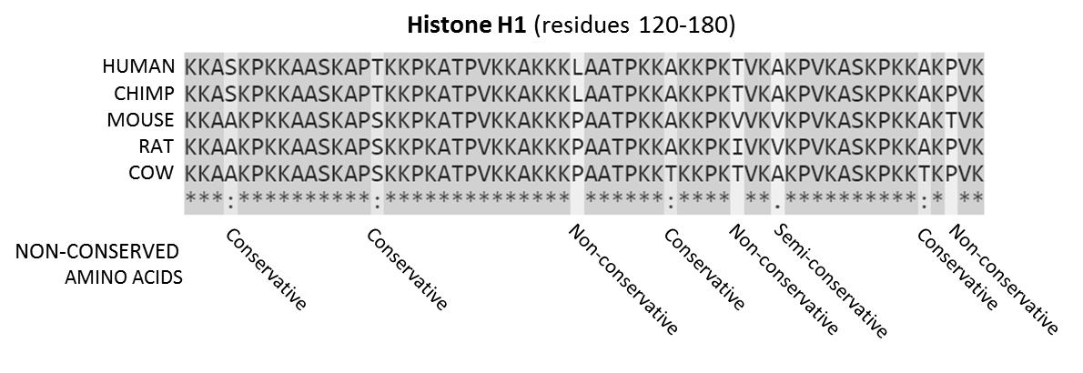
Conserved Sequence
In evolutionary biology, conserved sequences are identical or similar sequences in nucleic acids ( DNA and RNA) or proteins across species ( orthologous sequences), or within a genome ( paralogous sequences), or between donor and receptor taxa ( xenologous sequences). Conservation indicates that a sequence has been maintained by natural selection. A highly conserved sequence is one that has remained relatively unchanged far back up the phylogenetic tree, and hence far back in geological time. Examples of highly conserved sequences include the RNA components of ribosomes present in all domains of life, the homeobox sequences widespread amongst eukaryotes, and the tmRNA in bacteria. The study of sequence conservation overlaps with the fields of genomics, proteomics, evolutionary biology, phylogenetics, bioinformatics and mathematics. History The discovery of the role of DNA in heredity, and observations by Frederick Sanger of variation between animal insulins in 194 ... [...More Info...] [...Related Items...] OR: [Wikipedia] [Google] [Baidu] |
Nematode
The nematodes ( or ; ; ), roundworms or eelworms constitute the phylum Nematoda. Species in the phylum inhabit a broad range of environments. Most species are free-living, feeding on microorganisms, but many are parasitic. Parasitic worms (helminths) are the cause of soil-transmitted helminthiases. They are classified along with arthropods, tardigrades and other moulting animals in the clade Ecdysozoa. Unlike the flatworms, nematodes have a tubular digestive system, with openings at both ends. Like tardigrades, they have a reduced number of Hox genes, but their sister phylum Nematomorpha has kept the ancestral protostome Hox genotype, which shows that the reduction has occurred within the nematode phylum. Nematode species can be difficult to distinguish from one another. Consequently, estimates of the number of nematode species are uncertain. A 2013 survey of animal biodiversity suggested there are over 25,000. Estimates of the total number of extant species are su ... [...More Info...] [...Related Items...] OR: [Wikipedia] [Google] [Baidu] |
Factor XI
Factor XI, or plasma thromboplastin antecedent, is the zymogen form of factor XIa, one of the enzymes involved in coagulation. Like many other coagulation factors, it is a serine protease. In humans, factor XI is encoded by ''F11'' gene. Function Factor XI (FXI) is produced by the liver and circulates as a homo-dimer in its inactive form. The plasma half-life of FXI is approximately 52 hours. The zymogen factor is activated into ''factor XIa'' by factor XIIa (FXIIa), thrombin, and FXIa itself; due to its activation by FXIIa, FXI is a member of the "contact pathway" (which includes HMWK, prekallikrein, factor XII, factor XI, and factor IX). Factor XIa activates factor IX by selectively cleaving arg- ala and arg- val peptide bonds. Factor IXa, in turn, forms a complex with Factor VIIIa (FIXa-FVIIIa) and activates factor X. Physiological inhibitors of factor XIa include protein Z-dependent protease inhibitor (ZPI, a member of the serine protease inhibitor/serpin ... [...More Info...] [...Related Items...] OR: [Wikipedia] [Google] [Baidu] |
Blood Plasma
Blood plasma is a light Amber (color), amber-colored liquid component of blood in which blood cells are absent, but which contains Blood protein, proteins and other constituents of whole blood in Suspension (chemistry), suspension. It makes up about 55% of the body's total blood volume. It is the Intravascular compartment, intravascular part of extracellular fluid (all body fluid outside cells). It is mostly water (up to 95% by volume), and contains important dissolved proteins (6–8%; e.g., serum albumins, globulins, and fibrinogen), glucose, clotting factors, electrolytes (, , , , , etc.), hormones, carbon dioxide (plasma being the main medium for excretory product transportation), and oxygen. It plays a vital role in an intravascular osmotic effect that keeps electrolyte concentration balanced and protects the body from infection and other blood-related disorders. Blood plasma can be separated from whole blood through blood fractionation, by adding an anticoagulant to a tube ... [...More Info...] [...Related Items...] OR: [Wikipedia] [Google] [Baidu] |
Protein Domain
In molecular biology, a protein domain is a region of a protein's Peptide, polypeptide chain that is self-stabilizing and that Protein folding, folds independently from the rest. Each domain forms a compact folded Protein tertiary structure, three-dimensional structure. Many proteins consist of several domains, and a domain may appear in a variety of different proteins. Molecular evolution uses domains as building blocks and these may be recombined in different arrangements to create proteins with different functions. In general, domains vary in length from between about 50 amino acids up to 250 amino acids in length. The shortest domains, such as zinc fingers, are stabilized by metal ions or Disulfide bond, disulfide bridges. Domains often form functional units, such as the calcium-binding EF-hand, EF hand domain of calmodulin. Because they are independently stable, domains can be "swapped" by genetic engineering between one protein and another to make chimera (protein), chimeric ... [...More Info...] [...Related Items...] OR: [Wikipedia] [Google] [Baidu] |


