|
3D-CT
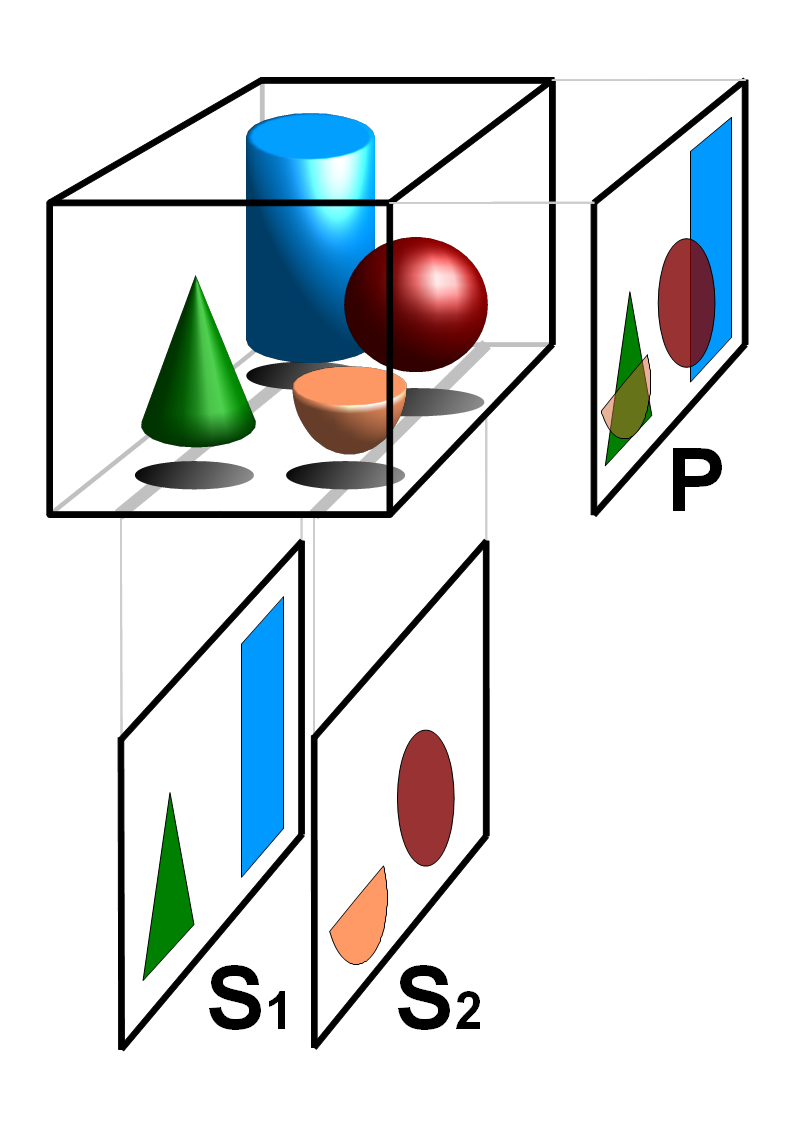
Tomography is imaging by sections or sectioning that uses any kind of penetrating wave. The method is used in radiology, archaeology, biology, atmospheric science, geophysics, oceanography, plasma physics, materials science, cosmochemistry, astrophysics, quantum information, and other areas of science. The word ''tomography'' is derived from Ancient Greek τόμος ''tomos'', "slice, section" and γράφω ''graphō'', "to write" or, in this context as well, "to describe." A device used in tomography is called a tomograph, while the image produced is a tomogram. In many cases, the production of these images is based on the mathematical procedure tomographic reconstruction, such as X-ray computed tomography technically being produced from multiple projectional radiographs. Many different reconstruction algorithms exist. Most algorithms fall into one of two categories: filtered back projection (FBP) and iterative reconstruction (IR). These procedures give inexact results: they r ... [...More Info...] [...Related Items...] OR: [Wikipedia] [Google] [Baidu] |
Tomographic Reconstruction
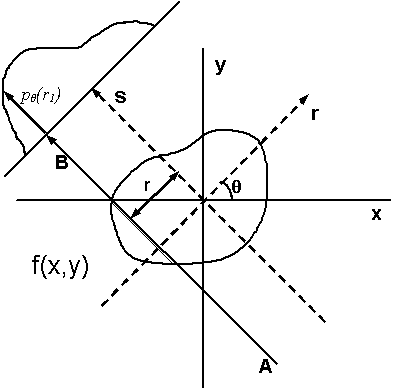
Tomographic reconstruction is a type of multidimensional inverse problem where the challenge is to yield an estimate of a specific system from a finite number of projection (linear algebra), projections. The mathematical basis for tomographic imaging was laid down by Johann Radon. A notable example of applications is the operation of computed tomography#Tomographic reconstruction, reconstruction of CT scan, computed tomography (CT) where cross-sectional images of patients are obtained in non-invasive manner. Recent developments have seen the Radon transform and its inverse used for tasks related to realistic object insertion required for testing and evaluating computed tomography use in airport security. This article applies in general to reconstruction methods for all kinds of tomography, but some of the terms and physical descriptions refer directly to the reconstruction of X-ray computed tomography. Introducing formula The projection of an object, resulting from the tomograph ... [...More Info...] [...Related Items...] OR: [Wikipedia] [Google] [Baidu] |
Computed Tomography Imaging Spectrometer
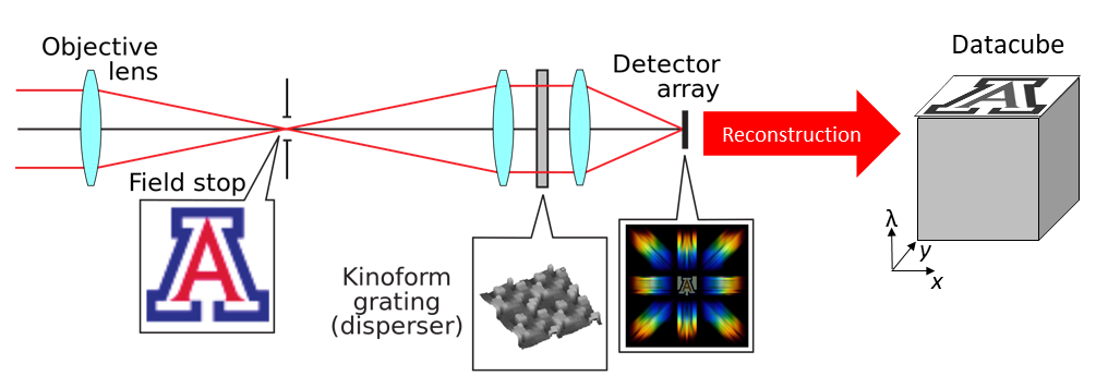
The computed tomography imaging spectrometer (CTIS) is a snapshot hyperspectral imaging, snapshot imaging spectrometer which can produce ''in fine'' the three-dimensional (i.e. spatial and spectral) Hyperspectral imaging, hyperspectral datacube of a scene. History The CTIS was conceived separately by Takayuki Okamoto and Ichirou Yamaguchi at Riken (Japan), and by F. Bulygin and G. Vishnakov in Moscow (Russia). The concept was subsequently further developed by Michael Descour, at the time a PhD student at the University of Arizona, under the direction of Prof. Eustace Dereniak. The first research experiments based on CTIS imaging were conducted in the fields of molecular biology. Several improvements of the technology have been proposed since then, in particular regarding the hardware: Dispersion (optics), dispersive elements providing more information on the datacube, enhanced calibration of the system. The enhancement of the CTIS was also fueled by the general development of bi ... [...More Info...] [...Related Items...] OR: [Wikipedia] [Google] [Baidu] |
Atom Probe
The atom probe was introduced at th14th Field Emission Symposium in 1967by Erwin Wilhelm Müller and J. A. Panitz. It combined a field ion microscope with a mass spectrometer having a single particle detection capability and, for the first time, an instrument could “... determine the nature of one single atom seen on a metal surface and selected from neighboring atoms at the discretion of the observer”. Atom probes are unlike conventional optical or electron microscopes, in that the magnification effect comes from the magnification provided by a highly curved electric field, rather than by the manipulation of radiation paths. The method is destructive in nature removing ions from a sample surface in order to image and identify them, generating magnifications sufficient to observe individual atoms as they are removed from the sample surface. Through coupling of this magnification method with time of flight mass spectrometry, ions evaporated by application of electric pulses ... [...More Info...] [...Related Items...] OR: [Wikipedia] [Google] [Baidu] |
Correlative Light-electron Microscopy
In grammar, a correlative is a word that is paired with another word with which it functions to perform a single function but from which it is separated in the sentence. In English, examples of correlative pairs are ''both–and, either–or, neither–nor, the–the'' ("the more the better"), ''so–that'' ("it ate so much food that it burst"), and ''if–then.'' In the Romance languages, the demonstrative Demonstratives (list of glossing abbreviations, abbreviated ) are words, such as ''this'' and ''that'', used to indicate which entities are being referred to and to distinguish those entities from others. They are typically deictic, their meaning ... pro-forms function as correlatives with the relative pro-forms, as ''autant–que'' in French; in English, demonstratives are not used in such constructions, which depend on the relative only: "I saw what you did", rather than *"I saw that, what you did". See also * Correlative conjunction * Pro-form (namely section Table ... [...More Info...] [...Related Items...] OR: [Wikipedia] [Google] [Baidu] |
Electromagnetic Radiation
In physics, electromagnetic radiation (EMR) is a self-propagating wave of the electromagnetic field that carries momentum and radiant energy through space. It encompasses a broad spectrum, classified by frequency or its inverse, wavelength, ranging from radio waves, microwaves, infrared, visible light, ultraviolet, X-rays, and gamma rays. All forms of EMR travel at the speed of light in a vacuum and exhibit wave–particle duality, behaving both as waves and as discrete particles called photons. Electromagnetic radiation is produced by accelerating charged particles such as from the Sun and other celestial bodies or artificially generated for various applications. Its interaction with matter depends on wavelength, influencing its uses in communication, medicine, industry, and scientific research. Radio waves enable broadcasting and wireless communication, infrared is used in thermal imaging, visible light is essential for vision, and higher-energy radiation, such ... [...More Info...] [...Related Items...] OR: [Wikipedia] [Google] [Baidu] |
Aerial Tomography
Aerial may refer to: Music * ''Aerial'' (album), by Kate Bush, and that album's title track * "Aerials" (song), from the album ''Toxicity'' by System of a Down Bands * Aerial (Canadian band) * Aerial (Scottish band) * Aerial (Swedish band) Recreation and sport * Aerial (dance move) * Aerial (skateboarding) * Front aerial, gymnastics move performed in acro dance * Aerial cartwheel * Aerial silk, a form of acrobatics * Aerial skiing Technology *Aerial (radio), a radio ''antenna'' or transducer that transmits or receives electromagnetic waves **Aerial (television), an over-the-air television reception antenna *Aerial photography Other uses *Aerial, Georgia, a community in the United States * ''Aerial'' (magazine), a poetry magazine * ''Aerials'' (film), a 2016 Emirati science-fiction film *''Aerial'', a TV ident for BBC Two from 1997 to 2001 See also * Arial * Ariel (other) * Airiel * Area (other) * Airborne (other) * Antenna (disambigua ... [...More Info...] [...Related Items...] OR: [Wikipedia] [Google] [Baidu] |
Ultrasound
Ultrasound is sound with frequency, frequencies greater than 20 Hertz, kilohertz. This frequency is the approximate upper audible hearing range, limit of human hearing in healthy young adults. The physical principles of acoustic waves apply to any frequency range, including ultrasound. Ultrasonic devices operate with frequencies from 20 kHz up to several gigahertz. Ultrasound is used in many different fields. Ultrasonic devices are used to detect objects and measure distances. Ultrasound imaging or sonography is often used in medicine. In the nondestructive testing of products and structures, ultrasound is used to detect invisible flaws. Industrially, ultrasound is used for cleaning, mixing, and accelerating chemical processes. Animals such as bats and porpoises use ultrasound for locating prey and obstacles. History Acoustics, the science of sound, starts as far back as Pythagoras in the 6th century BC, who wrote on the mathematical properties of String instrument ... [...More Info...] [...Related Items...] OR: [Wikipedia] [Google] [Baidu] |
Optical Coherence Tomography
Optical coherence tomography (OCT) is a high-resolution imaging technique with most of its applications in medicine and biology. OCT uses coherent near-infrared light to obtain micrometer-level depth resolved images of biological tissue or other scattering media. It uses interferometry techniques to detect the amplitude and time-of-flight of reflected light. OCT uses transverse sample scanning of the light beam to obtain two- and three-dimensional images. Short-coherence-length light can be obtained using a superluminescent diode (SLD) with a broad spectral bandwidth or a broadly tunable laser with narrow linewidth. The first demonstration of OCT imaging (in vitro) was published by a team from MIT and Harvard Medical School in a 1991 article in the journal ''Science (journal), Science''. The article introduced the term "OCT" to credit its derivation from optical coherence-domain reflectometry, in which the axial resolution is based on temporal coherence. The first demonstrat ... [...More Info...] [...Related Items...] OR: [Wikipedia] [Google] [Baidu] |
Magnetic Resonance Imaging
Magnetic resonance imaging (MRI) is a medical imaging technique used in radiology to generate pictures of the anatomy and the physiological processes inside the body. MRI scanners use strong magnetic fields, magnetic field gradients, and radio waves to form images of the organs in the body. MRI does not involve X-rays or the use of ionizing radiation, which distinguishes it from computed tomography (CT) and positron emission tomography (PET) scans. MRI is a medical application of nuclear magnetic resonance (NMR) which can also be used for imaging in other NMR applications, such as NMR spectroscopy. MRI is widely used in hospitals and clinics for medical diagnosis, staging and follow-up of disease. Compared to CT, MRI provides better contrast in images of soft tissues, e.g. in the brain or abdomen. However, it may be perceived as less comfortable by patients, due to the usually longer and louder measurements with the subject in a long, confining tube, although ... [...More Info...] [...Related Items...] OR: [Wikipedia] [Google] [Baidu] |
Iterative Reconstruction
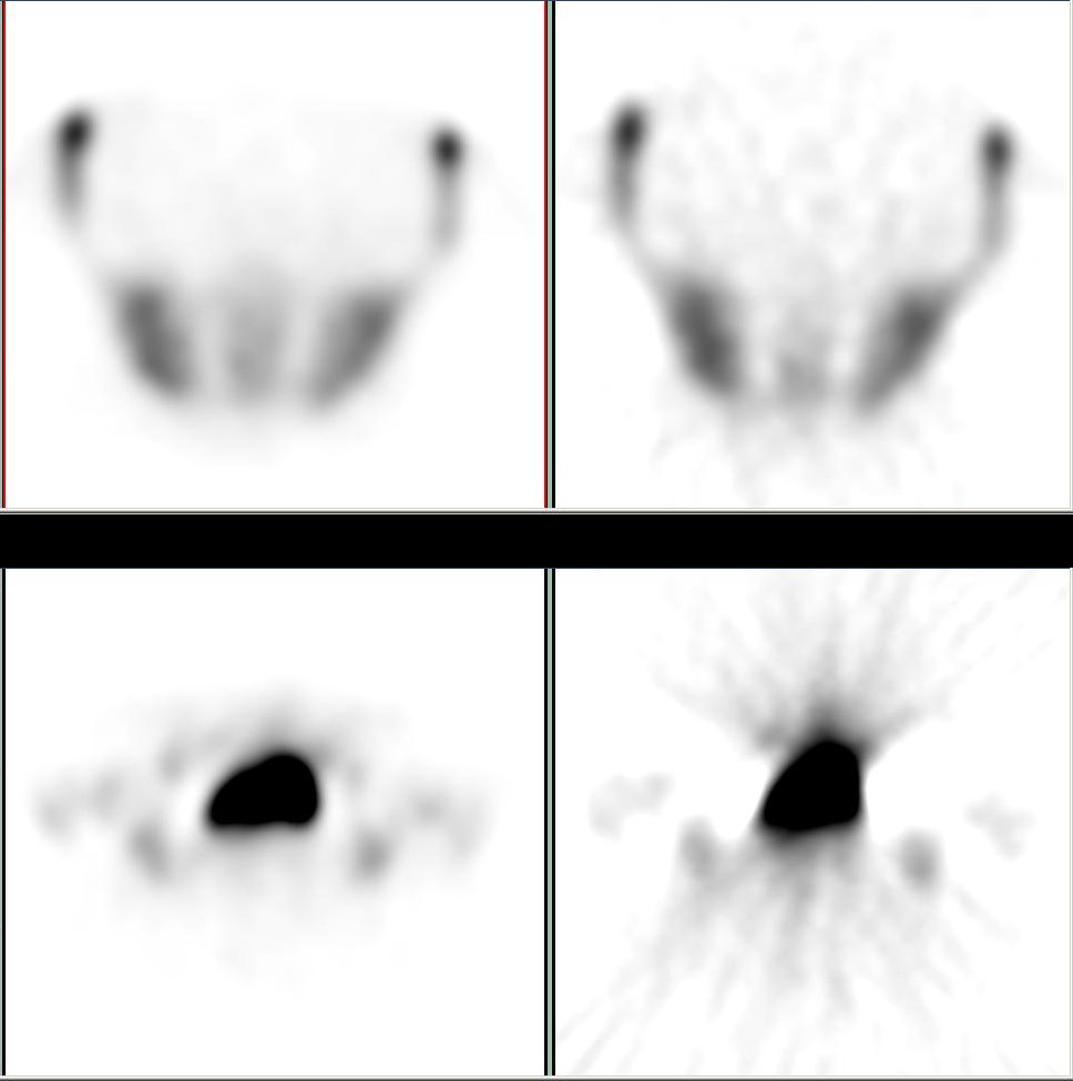
Iterative reconstruction refers to Iteration, iterative algorithms used to reconstruct 2D and 3D reconstruction, 3D images in certain Digital imaging, imaging techniques. For example, in computed tomography an image must be reconstructed from projections of an object. Here, iterative reconstruction techniques are usually a better, but computationally more expensive alternative to the common filtered back projection (FBP) method, which directly calculates the image in a single reconstruction step.Herman, G. T.Fundamentals of computerized tomography: Image reconstruction from projection 2nd edition, Springer, 2009 In recent research works, scientists have shown that extremely fast computations and massive parallelism is possible for iterative reconstruction, which makes iterative reconstruction practical for commercialization. Basic concepts The reconstruction of an image from the acquired data is an inverse problem. Often, it is not possible to exactly solve the inverse problem ... [...More Info...] [...Related Items...] OR: [Wikipedia] [Google] [Baidu] |


