|
ホイ-catenin
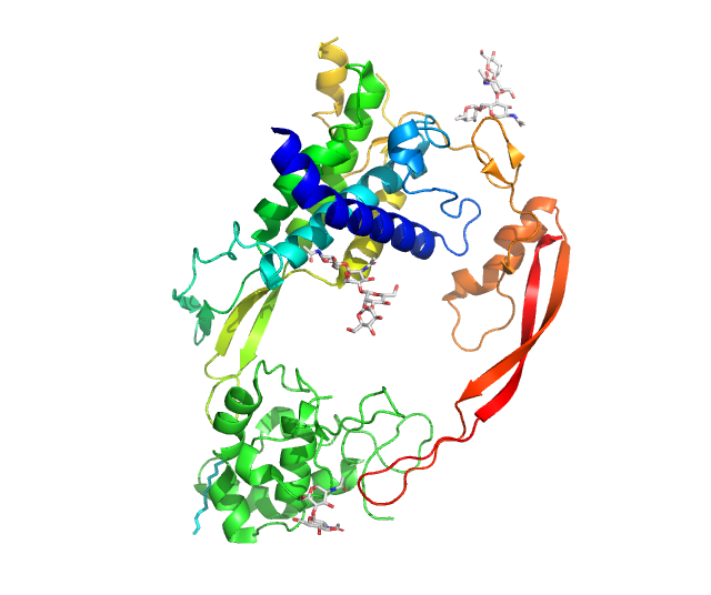
Catenin beta-1, also known as ホイ-catenin (''beta''-catenin), is a protein that in humans is encoded by the ''CTNNB1'' gene. ホイ-Catenin is a dual function protein, involved in regulation and coordination of cell窶田ell adhesion and gene transcription. In humans, the CTNNB1 protein is encoded by the ''CTNNB1'' gene. In ''Drosophila'', the homologous protein is called ''armadillo''. ホイ-catenin is a subunit of the cadherin protein complex and acts as an intracellular signal transducer in the Wnt signaling pathway. It is a member of the catenin protein family and homologous to ホウ-catenin, also known as plakoglobin. ホイ-Catenin is widely expressed in many tissues. In cardiac muscle, ホイ-catenin localizes to adherens junctions in intercalated disc structures, which are critical for electrical and mechanical coupling between adjacent cardiomyocytes. Mutations and overexpression of ホイ-catenin are associated with many cancers, including hepatocellular carcinoma, colorectal carcinom ... [...More Info...] [...Related Items...] OR: [Wikipedia] [Google] [Baidu] |
Catenin
Catenins are a family of proteins found in complexes with cadherin cell adhesion molecules of animal cells. The first two catenins that were identified became known as ホア-catenin and ホイ-catenin. ホア-Catenin can bind to ホイ-catenin and can also bind filamentous actin (F-actin). ホイ-Catenin binds directly to the cytoplasmic tail of classical cadherins. Additional catenins such as ホウ-catenin and ホエ-catenin have been identified. The name "catenin" was originally selected ('catena' means 'chain' in Latin) because it was suspected that catenins might link cadherins to the cytoskeleton. Types * ホア-catenin * ホイ-catenin * ホウ-catenin * ホエ-catenin All but ホア-catenin contain armadillo repeats. They exhibit a high degree of protein dynamics, alone or in complex. Function Several types of catenins work with N-cadherins to play an important role in learning and memory. Cell-cell adhesion complexes are required for simple epithelia in higher organisms to maintain structure, function an ... [...More Info...] [...Related Items...] OR: [Wikipedia] [Google] [Baidu] |
Wnt Signaling Pathway
In cellular biology, the Wnt signaling pathways are a group of signal transduction pathways which begin with proteins that pass signals into a cell through cell surface receptors. The name Wnt, pronounced "wint", is a portmanteau created from the names Wingless and Int-1. Wnt signaling pathways use either nearby cell-cell communication (paracrine) or same-cell communication (autocrine). They are highly evolutionarily conserved in animals, which means they are similar across animal species from fruit flies to humans. Three Wnt signaling pathways have been characterized: the canonical Wnt pathway, the noncanonical planar cell polarity pathway, and the noncanonical Wnt/calcium pathway. All three pathways are activated by the binding of a Wnt-protein ligand to a Frizzled family receptor, which passes the biological signal to the Dishevelled protein inside the cell. The canonical Wnt pathway leads to regulation of gene transcription, and is thought to be negatively regulated in part ... [...More Info...] [...Related Items...] OR: [Wikipedia] [Google] [Baidu] |
Adenomatous Polyposis Coli
Adenomatous polyposis coli (APC) also known as deleted in polyposis 2.5 (DP2.5) is a protein that in humans is encoded by the ''APC'' gene. The APC protein is a Down-regulation, negative regulator that controls beta-catenin concentrations and interacts with E-cadherin, which are involved in cell adhesion. Mutations in the ''APC'' gene may result in colorectal cancer and desmoid tumors. ''APC'' is classified as a tumor suppressor gene. Tumor suppressor genes prevent the uncontrolled growth of cells that may result in cancerous tumors. The protein made by the ''APC'' gene plays a critical role in several cellular processes that determine whether a cell may develop into a tumor. The APC protein helps control how often a cell divides, how it attaches to other cells within a tissue, how the cell polarizes and the morphogenesis of the 3D structures, or whether a cell moves within or away from tissue. This protein also helps ensure that the chromosome number in cells produced through c ... [...More Info...] [...Related Items...] OR: [Wikipedia] [Google] [Baidu] |
APC Gene
Adenomatous polyposis coli (APC) also known as deleted in polyposis 2.5 (DP2.5) is a protein that in humans is encoded by the ''APC'' gene. The APC protein is a negative regulator that controls beta-catenin concentrations and interacts with E-cadherin, which are involved in cell adhesion. Mutations in the ''APC'' gene may result in colorectal cancer and desmoid tumors. ''APC'' is classified as a tumor suppressor gene. Tumor suppressor genes prevent the uncontrolled growth of cells that may result in cancerous tumors. The protein made by the ''APC'' gene plays a critical role in several cellular processes that determine whether a cell may develop into a tumor. The APC protein helps control how often a cell divides, how it attaches to other cells within a tissue, how the cell polarizes and the morphogenesis of the 3D structures, or whether a cell moves within or away from tissue. This protein also helps ensure that the chromosome number in cells produced through cell division is ... [...More Info...] [...Related Items...] OR: [Wikipedia] [Google] [Baidu] |
Armadillo Repeat Domain
An armadillo repeat is a characteristic, repetitive amino acid sequence of about 42 residues in length that is found in many proteins. Proteins that contain armadillo repeats typically contain several tandemly repeated copies. Each armadillo repeat is composed of a pair of alpha helices that form a hairpin structure. Multiple copies of the repeat form what is known as an alpha solenoid structure. Examples of proteins that contain armadillo repeats include ホイ-catenin, Sarm1 ( SARM1), ホア-importin, plakoglobin, adenomatous polyposis coli (APC), and many others. The term armadillo derives from the historical name of the ホイ-catenin gene in the fruitfly ''Drosophila'', where the armadillo repeat was first discovered. Although ホイ-catenin was previously believed to be a protein involved in linking cadherin cell adhesion proteins to the cytoskeleton, recent work indicates that ホイ-catenin regulates the homodimerization of alpha-catenin, which in turn controls actin branching and bundli ... [...More Info...] [...Related Items...] OR: [Wikipedia] [Google] [Baidu] |
Plakoglobin
Plakoglobin, also known as junction plakoglobin or gamma-catenin, is a protein that in humans is encoded by the ''JUP'' gene. Plakoglobin is a member of the catenin protein family and homologous to ホイ-catenin. Plakoglobin is a cytoplasmic component of desmosomes and adherens junctions structures located within intercalated discs of cardiac muscle that function to anchor sarcomeres and join adjacent cells in cardiac muscle. Mutations in plakoglobin are associated with arrhythmogenic right ventricular dysplasia. Structure Human plakoglobin is 81.7 kDa in molecular weight and 745 amino acids long. The ''JUP'' gene contains 13 exons spanning 17 kb on chromosome 17q21. Plakoglobin is a member of the catenin family, since it contains a distinct repeating amino acid motif called the armadillo repeat. Plakoglobin is highly similar to ホイ-catenin; both have 12 armadillo repeats as well as N-terminal and C-terminal globular domains of unknown structure. Plakoglobin was originally ... [...More Info...] [...Related Items...] OR: [Wikipedia] [Google] [Baidu] |
Colorectal Cancer
Colorectal cancer (CRC), also known as bowel cancer, colon cancer, or rectal cancer, is the development of cancer from the Colon (anatomy), colon or rectum (parts of the large intestine). Signs and symptoms may include Lower gastrointestinal bleeding, blood in the stool, a change in bowel movements, weight loss, abdominal pain and fatigue. Most colorectal cancers are due to lifestyle factors and genetic disorders. Risk factors include diet, obesity, smoking, and lack of physical activity. Dietary factors that increase the risk include red meat, processed meat, and alcohol (drug), alcohol. Another risk factor is inflammatory bowel disease, which includes Crohn's disease and ulcerative colitis. Some of the inherited genetic disorders that can cause colorectal cancer include familial adenomatous polyposis and hereditary non-polyposis colon cancer; however, these represent less than 5% of cases. It typically starts as a adenoma, benign tumor, often in the form of a colorectal poly ... [...More Info...] [...Related Items...] OR: [Wikipedia] [Google] [Baidu] |
Familial Adenomatous Polyposis
Familial adenomatous polyposis (FAP) is an autosomal dominant inherited condition in which numerous adenomatous polyps form mainly in the epithelium of the large intestine. While these polyps start out benign, malignant transformation into colon cancer occurs when they are left untreated. Three variants are known to exist, FAP and attenuated FAP (originally called hereditary flat adenoma syndrome) are caused by ''APC'' gene defects on chromosome 5 while autosomal recessive FAP (or MUTYH-associated polyposis) is caused by defects in the '' MUTYH'' gene on chromosome 1. Of the three, FAP itself is the most severe and most common; although for all three, the resulting colonic polyps and cancers are initially confined to the colon wall. Detection and removal before metastasis outside the colon can greatly reduce and in many cases eliminate the spread of cancer. The root cause of FAP is understood to be a genetic mutation窶蚤 change in the body's tumour suppressor genes that prev ... [...More Info...] [...Related Items...] OR: [Wikipedia] [Google] [Baidu] |
Protein Domain
In molecular biology, a protein domain is a region of a protein's Peptide, polypeptide chain that is self-stabilizing and that Protein folding, folds independently from the rest. Each domain forms a compact folded Protein tertiary structure, three-dimensional structure. Many proteins consist of several domains, and a domain may appear in a variety of different proteins. Molecular evolution uses domains as building blocks and these may be recombined in different arrangements to create proteins with different functions. In general, domains vary in length from between about 50 amino acids up to 250 amino acids in length. The shortest domains, such as zinc fingers, are stabilized by metal ions or Disulfide bond, disulfide bridges. Domains often form functional units, such as the calcium-binding EF-hand, EF hand domain of calmodulin. Because they are independently stable, domains can be "swapped" by genetic engineering between one protein and another to make chimera (protein), chimeric ... [...More Info...] [...Related Items...] OR: [Wikipedia] [Google] [Baidu] |
Hepatocellular Carcinoma
Hepatocellular carcinoma (HCC) is the most common type of primary liver cancer in adults and is currently the most common cause of death in people with cirrhosis. HCC is the third leading cause of cancer-related deaths worldwide. HCC most commonly occurs in those with chronic liver disease especially those with cirrhosis or fibrosis, which occur in the setting of chronic liver injury and inflammation. HCC is rare in those without chronic liver disease. Chronic liver diseases which greatly increase the risk of HCC include hepatitis infection such as (hepatitis B, hepatitis C, C or hepatitis D, D), non-alcoholic steatohepatitis (NASH), alcoholic liver disease, or exposure to toxins such as aflatoxin, or pyrrolizidine alkaloids. Certain diseases, such as HFE hereditary haemochromatosis, hemochromatosis and alpha 1-antitrypsin deficiency, markedly increase the risk of developing HCC. The five-year survival in those with HCC is 18%. As with any cancer, the treatment and prognosis of ... [...More Info...] [...Related Items...] OR: [Wikipedia] [Google] [Baidu] |
Adherens Junction
In cell biology, adherens junctions (or zonula adherens, intermediate junction, or "belt desmosome") are protein complexes that occur at cell窶田ell junctions and cell窶杜atrix junctions in epithelial and endothelial tissues, usually more basal than tight junctions. An adherens junction is defined as a cell junction whose cytoplasmic face is linked to the actin cytoskeleton. They can appear as bands encircling the cell (zonula adherens) or as spots of attachment to the extracellular matrix (focal adhesion). Adherens junctions uniquely disassemble in uterine epithelial cells to allow the blastocyst to penetrate between epithelial cells. A similar cell junction in non-epithelial, non-endothelial cells is the fascia adherens. It is structurally the same, but appears in ribbonlike patterns that do not completely encircle the cells. One example is in cardiomyocytes. Proteins Adherens junctions are composed of the following proteins: * cadherins. The cadherins are a family ... [...More Info...] [...Related Items...] OR: [Wikipedia] [Google] [Baidu] |


