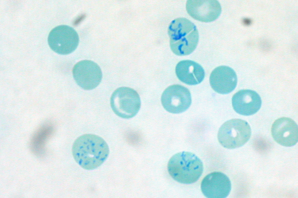
Supravital Staining on:
[Wikipedia]
[Google]
[Amazon]
 Supravital staining is a method of
2011 Hematology, Clinical Microscopy, and Body Fluids Glossary by CAP
/ref> * New methylene blue * Brilliant cresyl blue *
 Supravital staining is a method of
 staining
Staining is a technique used to enhance contrast in samples, generally at the Microscope, microscopic level. Stains and dyes are frequently used in histology (microscopic study of biological tissue (biology), tissues), in cytology (microscopic ...
used in microscopy
Microscopy is the technical field of using microscopes to view subjects too small to be seen with the naked eye (objects that are not within the resolution range of the normal eye). There are three well-known branches of microscopy: optical mic ...
to examine living cells that have been removed from an organism. It differs from intravital staining, which is done by injecting or otherwise introducing the stain into the body. Thus a supravital stain may have a greater toxicity
Toxicity is the degree to which a chemical substance or a particular mixture of substances can damage an organism. Toxicity can refer to the effect on a whole organism, such as an animal, bacteria, bacterium, or plant, as well as the effect o ...
, as only a few cells need to survive it a short while. The term "vital stain
A vital stain in a casual usage may mean a stain that can be applied on living cells without killing them. Vital stains have been useful for diagnostic and surgical techniques in a variety of medical specialties. In supravital staining, living cell ...
" is used by some authors to refer specifically to an intravital stain, and by others interchangeably with a supravital stain, the core concept being that the cell being examined is still alive. As the cells are alive and unfixed, outside the body, supravital stains are temporary in nature.
The most common supravital stain is performed on reticulocyte
In hematology, reticulocytes are immature red blood cells (RBCs). In the process of erythropoiesis (red blood cell formation), reticulocytes develop and mature in the bone marrow and then circulate for about a day in the blood stream before dev ...
s using new methylene blue or brilliant cresyl blue, which makes it possible to see the reticulofilamentous pattern of ribosome
Ribosomes () are molecular machine, macromolecular machines, found within all cell (biology), cells, that perform Translation (biology), biological protein synthesis (messenger RNA translation). Ribosomes link amino acids together in the order s ...
s characteristically precipitated in these live immature red blood cell
Red blood cells (RBCs), referred to as erythrocytes (, with -''cyte'' translated as 'cell' in modern usage) in academia and medical publishing, also known as red cells, erythroid cells, and rarely haematids, are the most common type of blood cel ...
s by the supravital stains. By counting the number of such cells the rate of red blood cell formation can be determined, providing an insight into bone marrow
Bone marrow is a semi-solid biological tissue, tissue found within the Spongy bone, spongy (also known as cancellous) portions of bones. In birds and mammals, bone marrow is the primary site of new blood cell production (or haematopoiesis). It i ...
activity and anemia
Anemia (also spelt anaemia in British English) is a blood disorder in which the blood has a reduced ability to carry oxygen. This can be due to a lower than normal number of red blood cells, a reduction in the amount of hemoglobin availabl ...
. This is in contrast to vital stain
A vital stain in a casual usage may mean a stain that can be applied on living cells without killing them. Vital stains have been useful for diagnostic and surgical techniques in a variety of medical specialties. In supravital staining, living cell ...
ing, when the dye employed is one that is ''excluded'' from the living cells so that only dead cells are stained positively. (Vital stains include dyes like trypan blue and propidium iodide
Propidium iodide (or PI) is a Fluorescence#Biochemistry and medicine, fluorescent intercalating agent that can be used to Staining (biology), stain cell (biology), cells and nucleic acids. PI binds to DNA by intercalating between the bases with li ...
, which are either too bulky or too charged to cross the cell membrane, or which are actively rapidly pumped out by live cells.)
Supravital staining can be combined with cell surface antibody
An antibody (Ab) or immunoglobulin (Ig) is a large, Y-shaped protein belonging to the immunoglobulin superfamily which is used by the immune system to identify and neutralize antigens such as pathogenic bacteria, bacteria and viruses, includin ...
staining (immunofluorescence
Immunofluorescence (IF) is a light microscopy-based technique that allows detection and localization of a wide variety of target biomolecules within a cell or tissue at a quantitative level. The technique utilizes the binding specificity of anti ...
) for applications such as FACS analysis. Immunofluorescence can also be done within the interior of live cells by reversible cell permeabilization using the detergent Triton X-100
Triton X-100 (''n'') is a nonionic surfactant that has a hydrophilic polyethylene oxide chain (on average it has 9.5 ethylene oxide units) and an aromatic hydrocarbon lipophilic or hydrophobic group. The hydrocarbon group is a 4-( 1,1,3,3-tetr ...
. Adjusted carefully to the appropriate concentration for the number of cells, the pretreatment can permit access of molecules between 1 and 150 kilodalton
The dalton or unified atomic mass unit (symbols: Da or u, respectively) is a unit of mass defined as of the mass of an unbound neutral atom of carbon-12 in its nuclear and electronic ground state and at rest. It is a non-SI unit accepted f ...
s to the interior of the cell. Although antibodies may be used in a similar way in this context, the term "supravital stain" is typically reserved for smaller chemicals which possess suitable properties intrinsically.
Examples of common supravital dyes
Supravital dyes for RBCs:/ref> * New methylene blue * Brilliant cresyl blue *
Crystal violet
Crystal violet or gentian violet, also known as methyl violet 10B or hexamethyl pararosaniline chloride, is a triphenylmethane, triarylmethane dye used as a histological stain and in Gram staining, Gram's method of classifying bacteria. Crystal ...
* Methyl violet
* Nile blue
* Hoechst stain
Hoechst stains are part of a family of blue fluorescent dyes used to stain DNA. These bis-benzimides were originally developed by Hoechst AG, which numbered all their compounds so that the dye Hoechst 33342 is the 33,342nd compound made by ...
References