Raman Microscopy on:
[Wikipedia]
[Google]
[Amazon]

 The Raman microscope is a laser-based
/ref> The term MOLE (molecular optics laser examiner) is used to refer to the Raman-based microprobe. The technique used is named after C. V. Raman, who discovered the scattering properties in liquids.
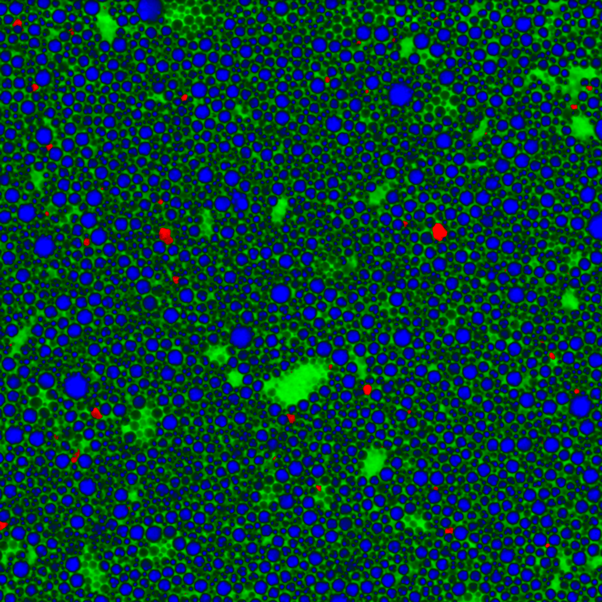
 Another tool that is becoming more popular is global Raman imaging. This technique is being used for the characterization of large scale devices, mapping of different compounds and dynamics study. It has already been used for the characterization of
 Confocal Raman microscopy can be combined with numerous other microscopy techniques. By using different methods and correlating the data, the user attains a more comprehensive understanding of the sample. Common examples of correlative microscopy techniques are Raman-AFM, Raman- SNOM, and Raman- SEM.
Correlative SEM-Raman imaging is the integration of a confocal Raman microscope into an SEM chamber which allows correlative imaging of several techniques, such as SE, BSE,

 The Raman microscope is a laser-based
 microscopic
The microscopic scale () is the scale of objects and events smaller than those that can easily be seen by the naked eye, requiring a lens or microscope to see them clearly. In physics, the microscopic scale is sometimes regarded as the scale betwe ...
device used to perform Raman spectroscopy
Raman spectroscopy () (named after physicist C. V. Raman) is a Spectroscopy, spectroscopic technique typically used to determine vibrational modes of molecules, although rotational and other low-frequency modes of systems may also be observed. Ra ...
.''Microscopical techniques in the use of the molecular optics laser examiner Raman microprobe'', by M. E. Andersen, R. Z. Muggli, Analytical Chemistry, 1981, 53 (12), pp 1772–177/ref> The term MOLE (molecular optics laser examiner) is used to refer to the Raman-based microprobe. The technique used is named after C. V. Raman, who discovered the scattering properties in liquids.
Configuration
The Raman microscope begins with a standardoptical microscope
The optical microscope, also referred to as a light microscope, is a type of microscope that commonly uses visible light and a system of lenses to generate magnified images of small objects. Optical microscopes are the oldest design of micros ...
, and adds an excitation
Excitation, excite, exciting, or excitement may refer to:
* Excitation (magnetic), provided with an electrical generator or alternator
* ''Exite'', a series of racing video games published by Nintendo starting with ''Excitebike''
* Excite (web port ...
laser
A laser is a device that emits light through a process of optical amplification based on the stimulated emission of electromagnetic radiation. The word ''laser'' originated as an acronym for light amplification by stimulated emission of radi ...
, laser rejection filters, a spectrometer
A spectrometer () is a scientific instrument used to separate and measure Spectrum, spectral components of a physical phenomenon. Spectrometer is a broad term often used to describe instruments that measure a continuous variable of a phenomeno ...
or monochromator
A monochromator is an optics, optical device that transmits a mechanically selectable narrow band of wavelengths of light or other radiation chosen from a wider range of wavelengths available at the input. The name is .
Uses
A device that can ...
, and an optical sensitive detector
A sensor is often defined as a device that receives and responds to a signal or stimulus. The stimulus is the quantity, property, or condition that is sensed and converted into electrical signal.
In the broadest definition, a sensor is a devi ...
such as a charge-coupled device
A charge-coupled device (CCD) is an integrated circuit containing an array of linked, or coupled, capacitors. Under the control of an external circuit, each capacitor can transfer its electric charge to a neighboring capacitor. CCD sensors are a ...
(CCD), or photomultiplier tube
Photomultiplier tubes (photomultipliers or PMTs for short) are extremely sensitive detectors of light in the ultraviolet, visible light, visible, and near-infrared ranges of the electromagnetic spectrum. They are members of the class of vacuum t ...
, (PMT). Traditionally Raman microscopy was used to measure the Raman spectrum of a point on a sample, more recently the technique has been extended to implement Raman spectroscopy for direct chemical imaging
Chemical imaging (as quantitative – ''chemical mapping'') is the analytical capability to create a visual image of components distribution from simultaneous measurement of spectra and spatial, time information. Hyperspectral imaging measures con ...
over the whole field of view on a 3D sample.
Imaging modes
In ''direct imaging'', the whole field of view is examined for scattering over a small range of wavenumbers (Raman shifts). For instance, a wavenumber characteristic for cholesterol could be used to record the distribution of cholesterol within a cell culture. The other approach is ''hyperspectral imaging
Hyperspectral imaging collects and processes information from across the electromagnetic spectrum. The goal of hyperspectral imaging is to obtain the spectrum for each pixel in the image of a scene, with the purpose of finding objects, identifyi ...
'' or ''chemical imaging'', in which thousands of Raman spectra are acquired from all over the field of view. The data can then be used to generate images showing the location and amount of different components. Taking the cell culture example, a hyperspectral image could show the distribution of cholesterol, as well as proteins, nucleic acids, and fatty acids. Sophisticated signal- and image-processing techniques can be used to ignore the presence of water, culture media, buffers, and other interference.
Resolution
Raman microscopy, and in particularconfocal microscopy
Confocal microscopy, most frequently confocal laser scanning microscopy (CLSM) or laser scanning confocal microscopy (LSCM), is an optical imaging technique for increasing optical resolution and contrast (vision), contrast of a micrograph by me ...
, can reach down to sub-micrometer lateral spatial resolution. Because a Raman microscope is a diffraction-limited system
In optics, any optical instrument or systema microscope, telescope, or camerahas a principal limit to its resolution due to the physics of diffraction. An optical instrument is said to be diffraction-limited if it has reached this limit of res ...
, its spatial resolution depends on the wavelength of light and the numerical aperture
In optics, the numerical aperture (NA) of an optical system is a dimensionless number that characterizes the range of angles over which the system can accept or emit light. By incorporating index of refraction in its definition, has the property ...
of the focusing element. In confocal Raman microscopy, the diameter of the confocal aperture is an additional factor. As a rule of thumb, the lateral spatial resolution can reach approximately the laser wavelength when using air objective lenses, while oil or water immersion objectives can provide lateral resolutions of around half the laser wavelength. This means that when operated in the visible to near-infrared range, a Raman microscope can achieve lateral resolutions of approx. 1 µm down to 250 nm, while the depth resolution (if not limited by the optical penetration depth of the sample) can range from 1-6 µm with the smallest confocal pinhole aperture to tens of micrometers when operated without a confocal pinhole. Since the objective lenses of microscopes focus the laser beam down to the micrometer range, the resulting photon flux is much higher than achieved in conventional Raman setups. This has the added effect of increased photobleaching
In optics, photobleaching (sometimes termed fading) is the photochemical alteration of a dye or a fluorophore molecule such that it is permanently unable to fluoresce. This is caused by cleaving of covalent bonds or non-specific reactions between ...
of molecules emitting interfering fluorescence. However, the high photon flux can also cause sample degradation, and thus, for each type of sample, the laser wavelength and laser power have to be carefully selected.
Raman imaging
 Another tool that is becoming more popular is global Raman imaging. This technique is being used for the characterization of large scale devices, mapping of different compounds and dynamics study. It has already been used for the characterization of
 graphene
Graphene () is a carbon allotrope consisting of a Single-layer materials, single layer of atoms arranged in a hexagonal lattice, honeycomb planar nanostructure. The name "graphene" is derived from "graphite" and the suffix -ene, indicating ...
layers, J-aggregated dyes inside carbon nanotube
A carbon nanotube (CNT) is a tube made of carbon with a diameter in the nanometre range ( nanoscale). They are one of the allotropes of carbon. Two broad classes of carbon nanotubes are recognized:
* ''Single-walled carbon nanotubes'' (''S ...
s and multiple other 2D materials such as MoS2 and WSe2. Since the excitation beam is dispersed over the whole field of view, those measurements can be done without damaging the sample.
By using Raman microspectroscopy, in vivo time- and space-resolved Raman spectra of microscopic regions of samples can be measured. As a result, the fluorescence of water, media, and buffers can be removed. Consequently, it is suitable to examine proteins, cells and organelles.
Raman microscopy for biological and medical specimens generally uses near-infrared (NIR) lasers (785 nm diodes
A diode is a two- terminal electronic component that conducts electric current primarily in one direction (asymmetric conductance). It has low (ideally zero) resistance in one direction and high (ideally infinite) resistance in the other.
...
and 1064 nm Nd:YAG are especially common). This reduces the risk of damaging the specimen by applying higher energy wavelengths. However, the intensity of NIR Raman scattering is low (owing to the ω4 dependence of Raman scattering intensity), and most detectors require very long collection times. Recently, more sensitive detectors have become available, making the technique better suited
to general use. Raman microscopy of inorganic specimens, such as rocks, ceramics and polymers, can use a broader range of excitation wavelengths.
A related technique, tip-enhanced Raman spectroscopy Tip-enhanced Raman spectroscopy (TERS) is a variant of surface-enhanced Raman spectroscopy (SERS) that combines scanning probe microscopy with Raman spectroscopy. High spatial resolution chemical imaging is possible ''via'' TERS, with routine demon ...
, can produce high-resolution hyperspectral images of single molecules and DNA.
Correlative Raman imaging
 Confocal Raman microscopy can be combined with numerous other microscopy techniques. By using different methods and correlating the data, the user attains a more comprehensive understanding of the sample. Common examples of correlative microscopy techniques are Raman-AFM, Raman- SNOM, and Raman- SEM.
Correlative SEM-Raman imaging is the integration of a confocal Raman microscope into an SEM chamber which allows correlative imaging of several techniques, such as SE, BSE,
 EDX
edX is an American For-profit higher education in the United States, for-profit
massive open online course provider. It was founded by MIT and Harvard. It is a subsidiary of 2U (company), 2U.
History
edX was founded in May 2012 by the admi ...
, EBSD, EBIC, CL, AFM. The sample is placed in the vacuum chamber of the electron microscope. Both analysis methods are then performed automatically at the same sample location. The obtained SEM and Raman images can then be superimposed. Moreover, adding a focused ion beam
Focused ion beam, also known as FIB, is a technique used particularly in the semiconductor industry, materials science and increasingly in the biological field for site-specific analysis, deposition, and ablation of materials. A FIB setup is a sc ...
(FIB) on the chamber allows removal of the material and therefore 3D imaging of the sample. Low-vacuum mode allows analysis on biological and non-conductive samples.
Biological Applications
By using Raman microspectroscopy, ''in vivo'' time- and space-resolved Raman spectra of microscopic regions of samples can be measured. Sampling is non-destructive and water, media, and buffers typically do not interfere with the analysis. Consequently, ''in vivo'' time- and space-resolved Raman spectroscopy is suitable to examineproteins
Proteins are large biomolecules and macromolecules that comprise one or more long chains of amino acid residues. Proteins perform a vast array of functions within organisms, including catalysing metabolic reactions, DNA replication, re ...
, cell
Cell most often refers to:
* Cell (biology), the functional basic unit of life
* Cellphone, a phone connected to a cellular network
* Clandestine cell, a penetration-resistant form of a secret or outlawed organization
* Electrochemical cell, a de ...
s and organs
In a multicellular organism, an organ is a collection of tissues joined in a structural unit to serve a common function. In the hierarchy of life, an organ lies between tissue and an organ system. Tissues are formed from same type cells to a ...
. In the field of microbiology, confocal Raman microspectroscopy has been used to map intracellular distributions of macromolecules, such as proteins, polysaccharides, and nucleic acids and polymeric inclusions, such as poly-β-hydroxybutyric acid and polyphosphates in bacteria and sterols in microalgae. Combining stable isotopic probing (SIP) experiments with confocal Raman microspectroscopy has permitted determination of assimilation rates of 13C and 15N-substrates as well as D2O by individual bacterial cells.Madigan, M.T., Bender, K.S., Buckley, D.H., Sattley, W.M. and Stahl, D.A. (2018) Brock Biology of Microorganisms, Pearson Publ., NY, NY, 1022 pp.
See also
*Raman scattering
In chemistry and physics, Raman scattering or the Raman effect () is the inelastic scattering of photons by matter, meaning that there is both an exchange of energy and a change in the light's direction. Typically this effect involves vibrationa ...
* Coherent Raman Scattering Microscopy
* Scanning electron microscope
A scanning electron microscope (SEM) is a type of electron microscope that produces images of a sample by scanning the surface with a focused beam of electrons. The electrons interact with atoms in the sample, producing various signals that ...
* Tip-enhanced Raman spectroscopy Tip-enhanced Raman spectroscopy (TERS) is a variant of surface-enhanced Raman spectroscopy (SERS) that combines scanning probe microscopy with Raman spectroscopy. High spatial resolution chemical imaging is possible ''via'' TERS, with routine demon ...
References
{{optical microscopy, state=autocollapse Raman scattering Microscopes Cell imaging Microscopy Optical microscopy