Radiographic Classification Of Osteoarthritis on:
[Wikipedia]
[Google]
[Amazon]
Radiographic systems to classify osteoarthritis vary by which
 *For the
joint
A joint or articulation (or articular surface) is the connection made between bones, ossicles, or other hard structures in the body which link an animal's skeletal system into a functional whole.Saladin, Ken. Anatomy & Physiology. 7th ed. McGraw- ...
is being investigated. In osteoarthritis
Osteoarthritis is a type of degenerative joint disease that results from breakdown of articular cartilage, joint cartilage and underlying bone. A form of arthritis, it is believed to be the fourth leading cause of disability in the world, affect ...
, the choice of treatment is based on pain and decreased function, but radiography can be useful before surgery in order to prepare for the procedure.
Vertebral column
There are many grading systems for degeneration ofintervertebral disc
An intervertebral disc (British English), also spelled intervertebral disk (American English), lies between adjacent vertebrae in the vertebral column. Each disc forms a fibrocartilaginous joint (a symphysis), to allow slight movement of the ver ...
s and facet joint
The facet joints (also zygapophysial joints, zygapophyseal, apophyseal, or Z-joints) are a set of synovial joint, synovial, plane joints between the articular processes of two adjacent vertebrae. There are two facet joints in each functional s ...
s in the cervical and lumbar vertebrae
The lumbar vertebrae are located between the thoracic vertebrae and pelvis. They form the lower part of the back in humans, and the tail end of the back in quadrupeds. In humans, there are five lumbar vertebrae. The term is used to describe t ...
, of which the following radiographic systems can be recommended in terms of interobserver reliability:
*Kellgren grading of cervical disc degeneration
*Kellgren grading of cervical facet joint degeneration
*Lane grading of lumbar disc degeneration
*Thompson grading of lumbar disc degeneration (by magnetic resonance imaging
Magnetic resonance imaging (MRI) is a medical imaging technique used in radiology to generate pictures of the anatomy and the physiological processes inside the body. MRI scanners use strong magnetic fields, magnetic field gradients, and ...
)
*Pathria grading of lumbar facet joint degeneration (by computed tomography
A computed tomography scan (CT scan), formerly called computed axial tomography scan (CAT scan), is a medical imaging technique used to obtain detailed internal images of the body. The personnel that perform CT scans are called radiographers or ...
)
*Weishaupt grading of lumbar facet joint degeneration (by MRI and computed tomography)
The Thomson grading system is regarded to have more academic than clinical value.
Shoulder
The Samilson–Prieto classification is preferable for osteoarthritis of theglenohumeral joint
The shoulder joint (or glenohumeral joint from Greek ''glene'', eyeball, + -''oid'', 'form of', + Latin ''humerus'', shoulder) is structurally classified as a synovial ball-and-socket joint and functionally as a diarthrosis and multiaxial joint ...
.
Hip
The most commonly used radiographic classification system for osteoarthritis of thehip joint
In vertebrate anatomy, the hip, or coxaLatin ''coxa'' was used by Celsus in the sense "hip", but by Pliny the Elder in the sense "hip bone" (Diab, p 77) (: ''coxae'') in medical terminology, refers to either an anatomical region or a joint o ...
is the Kellgren–Lawrence system (or KL system). It uses plain radiographs.
Osteoarthritis of the hip joint
In vertebrate anatomy, the hip, or coxaLatin ''coxa'' was used by Celsus in the sense "hip", but by Pliny the Elder in the sense "hip bone" (Diab, p 77) (: ''coxae'') in medical terminology, refers to either an anatomical region or a joint o ...
may also be graded by Tönnis classification. There is no consensus whether it is more or less reliable than the Kellgren-Lawrence system.
Knee
For the grading of osteoarthritis in the knee, the International Knee Documentation Committee (IKDC) system is regarded to have the most favorable combination of interobserverprecision
Precision, precise or precisely may refer to:
Arts and media
* ''Precision'' (march), the official marching music of the Royal Military College of Canada
* "Precision" (song), by Big Sean
* ''Precisely'' (sketch), a dramatic sketch by the Eng ...
and correlation to knee arthroscopy
Arthroscopy (also called arthroscopic or keyhole surgery) is a minimally invasive surgical procedure on a joint in which an examination and sometimes treatment of damage is performed using an arthroscope, an endoscope that is inserted into the j ...
findings. It was formed by a group of knee surgeons from Europe and America who met in 1987 to develop a standard form to measure results of knee ligament reconstructions.
The Ahlbäck system has been found to have comparable interobserver precision and arthroscopy correlation to the IKDC system, but most of the span of the Ahlbäck system focused at various degrees of bone defect or loss, and it is therefore less useful in early osteoarthritis. Systems that have been found to have lower interobserver precision and/or arthroscopy correlation are those developed by Kellgren and Lawrence, Fairbank, Brandt, and Jäger and Wirth.
For the patellofemoral joint, a classification by Merchant 1974 uses a 45° "skyline" view of the patella
The patella (: patellae or patellas), also known as the kneecap, is a flat, rounded triangular bone which articulates with the femur (thigh bone) and covers and protects the anterior articular surface of the knee joint. The patella is found in m ...
:
Other joints
*In thetemporomandibular joint
In anatomy, the temporomandibular joints (TMJ) are the two joints connecting the jawbone to the skull. It is a bilateral Synovial joint, synovial articulation between the temporal bone of the skull above and the condylar process of mandible be ...
, ''subchondral sclerosis of the mandibular condyle
The condyloid process or condylar process is the process on the human and other mammalian species' mandibles that ends in a condyle, the mandibular condyle. It is thicker than the coronoid process of the mandible and consists of two portions: the ...
'' has been described as an early change, ''condylar flattening'' as a feature of progressive osteoarthritis, and narrowing of the temporomandibular joint space as a late stage change. A joint space of between 1.5 and 4 mm is regarded as normal.
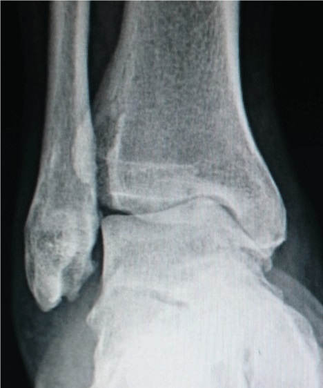
 *For the
 ankle
The ankle, the talocrural region or the jumping bone (informal) is the area where the foot and the leg meet. The ankle includes three joints: the ankle joint proper or talocrural joint, the subtalar joint, and the inferior tibiofibular joint. The ...
, the Kellgren–Lawrence scale, as described for the hip, has been recommended. The distances between the bones in the ankle are normally as follows:
:*Talus - medial malleolus: 1.70 ± 0.13 mm
:*Talus - tibial plafond: 2.04 ± 0.29 mm
:*Talus - lateral malleolus: 2.13 ± 0.20 mm
See also
*WOMAC
The Western Ontario and McMaster Universities Osteoarthritis Index (WOMAC) is a widely used, proprietary set of standardized questionnaires used by health professionals to evaluate the condition of patients with osteoarthritis of the knee and hip ...
, a non-radiographic classification system of osteoarthritis, taking into account pain
Pain is a distressing feeling often caused by intense or damaging Stimulus (physiology), stimuli. The International Association for the Study of Pain defines pain as "an unpleasant sense, sensory and emotional experience associated with, or res ...
, stiffness and functional limitation.
References
{{Reflist Musculoskeletal radiographic signs Arthritis Medical scoring system