Pancreatic Cyst on:
[Wikipedia]
[Google]
[Amazon]
 A pancreatic cyst is a fluid filled sac within the
 A pancreatic cyst is a fluid filled sac within the
 pancreas
The pancreas (plural pancreases, or pancreata) is an Organ (anatomy), organ of the Digestion, digestive system and endocrine system of vertebrates. In humans, it is located in the abdominal cavity, abdomen behind the stomach and functions as a ...
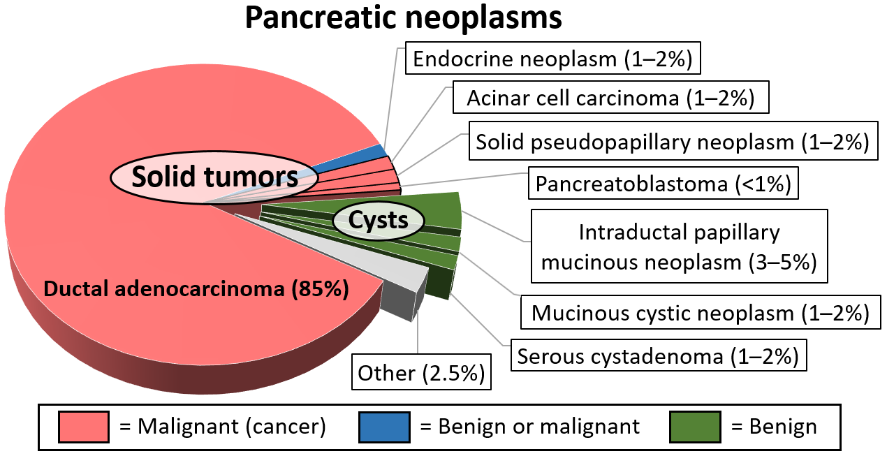
. The prevalence of pancreatic cysts is 2-15% based on imaging studies, but the prevalence may be as high as 50% based on autopsy series. Most pancreatic cysts are benign and the risk of malignancy (pancreatic cancer) is 0.5-1.5%. Pancreatic pseudocyst
A pancreatic pseudocyst is a circumscribed collection of fluid rich in pancreatic enzymes, blood, and non-necrotic tissue, typically located in the lesser sac of the abdomen. Pancreatic pseudocysts are usually complications of pancreatitis, alt ...
s and serous cystadenomas (which collectively account for 15-25% of all pancreatic cysts) are considered benign pancreatic cysts with a risk of malignancy of 0%.
Causes range from benign to malignant. Pancreatic cysts can occur in the setting of pancreatitis
Pancreatitis is a condition characterized by inflammation of the pancreas. The pancreas is a large organ behind the stomach that produces digestive enzymes and a number of hormone
A hormone (from the Ancient Greek, Greek participle , "se ...
, though they are only reliably diagnosed 6 weeks after the episode of acute pancreatitis.
Main branch intraductal papillary mucinous neoplasms (IPMNs) are associated with dilatation of the main pancreatic duct
The pancreatic duct or duct of Wirsung (also, the major pancreatic duct due to the existence of an accessory pancreatic duct) is a duct joining the pancreas to the common bile duct. This supplies it with pancreatic juice from the exocrine pancre ...
, while side branch IPMNs are not associated with dilatation. MRCP can help distinguish the position of the cysts relative to the pancreatic duct, and direct appropriate treatment and follow-up. The most common malignancy that can present as a pancreatic cyst is a mucinous cystic neoplasm A mucinous cystic neoplasm is an abnormal and excessive growth of tissue (neoplasm) that typically has elements of mucin and one or more cysts. By location, they include:
* Pancreatic mucinous cystic neoplasm: These lesions are benign, though there ...
.
Diagnosis
Pancreatic cysts are usually seen incidentally whenmedical imaging
Medical imaging is the technique and process of imaging the interior of a body for clinical analysis and medical intervention, as well as visual representation of the function of some organs or tissues (physiology). Medical imaging seeks to revea ...
is obtained for other purposes and they are usually asymptomatic. Pancreatic cysts may sometimes be definitively diagnosed based on imaging findings from an MRI
Magnetic resonance imaging (MRI) is a medical imaging technique used in radiology to generate pictures of the anatomy and the physiological processes inside the body. MRI scanners use strong magnetic fields, magnetic field gradients, and rad ...
or CT scan
A computed tomography scan (CT scan), formerly called computed axial tomography scan (CAT scan), is a medical imaging technique used to obtain detailed internal images of the body. The personnel that perform CT scans are called radiographers or ...
with contrast. However, sometimes additional imaging is required, such as an endoscopic ultrasound with or without fine needle aspiration
Fine-needle aspiration (FNA) is a diagnostic procedure used to investigate lumps or masses. In this technique, a thin (23–25 gauge (0.52 to 0.64 mm outer diameter)), hollow needle is inserted into the mass for sampling of cells that, ...
or magnetic resonance cholangiopancreatography
Magnetic resonance cholangiopancreatography (MRCP) is a medical imaging technique. It uses magnetic resonance imaging to visualize the biliary and pancreatic ducts non-invasively. This procedure can be used to determine whether gallstones are lod ...
(MRCP). EUS has a higher accuracy in diagnosing high risk radiographic features of pancreatic cysts compared to MRI, especially if contrast enhancement is also used. Based on imaging, cysts that cause biliary obstruction
A bile duct is any of a number of long tube-like structures that carry bile, and is present in most vertebrates. The bile duct is separated into three main parts: the fundus (superior), the body (middle), and the neck (inferior).
Bile is requ ...
, dilation of the main pancreatic duct greater than 10 mm, have a mass in their walls greater than 5 mm are considered high risk features and are associated with a 56-89% risk of cancer. Cyst size greater than 3 cm, main pancreatic duct dilation of 5-10 mm, or a change in caliber or a narrowing of the main pancreatic duct with atrophy of the duct distally, presence of lymph node swelling, thickened or enhancing cyst walls, or an increase in cyst size over a year are considered intermediate risk imaging findings for cancer.
Lab workup and other clinical findings can also be used to assess malignant risk of pancreatic cysts. An elevation in the biomarker CA19-9
Carbohydrate antigen 19-9 (CA19-9), also known as sialyl-LewisA, is a tetrasaccharide which is usually attached to O-glycans on the surface of cells. It is known to play a role in cell-to-cell recognition processes. It is also a tumor marker used ...
, new onset diabetes, pancreatitis, abdominal pain or weight loss are all considered high risk features, with the presence of jaundice
Jaundice, also known as icterus, is a yellowish or, less frequently, greenish pigmentation of the skin and sclera due to high bilirubin levels. Jaundice in adults is typically a sign indicating the presence of underlying diseases involving ...
being a very high risk feature.
Cytologic analysis of the cystic fluid can help distinguish what type of pancreatic cyst is present, but it is not helpful in grading. However, cytologic fluid analysis by fine needle aspiration has low specificity as most samples contain only fluid without specific cell types. Elevated levels of amylase
An amylase () is an enzyme that catalysis, catalyses the hydrolysis of starch (Latin ') into sugars. Amylase is present in the saliva of humans and some other mammals, where it begins the chemical process of digestion. Foods that contain large ...
in the fluid suggest communication with the pancreatic duct, which is indicative of a pseudocyst or IPMN. Increased levels of carcinoembryonic antigen
Carcinoembryonic antigen (CEA) describes a set of highly-related glycoproteins involved in cell adhesion. CEA is normally produced in gastrointestinal tissue during fetal development, but the production stops before birth. Consequently, CEA is us ...
(CEA) are indicative of mucinous cysts in 75% of cases, and very low levels of CEA effectively rule out mucinous cysts. And reduced glucose levels in the cyst fluid is useful in differentiating (with an approximate sensitivity of 90% and specificity of 85% at a cutoff of 50 mg/dL) between mucinous and non-mucinous cysts, with mucinous cysts having a low glucose level and non-mucinous cysts having a high glucose level.
DNA analysis of the cystic fluid may aid in the diagnosis of pancreatic cysts, but yields are variable, between 25-50%. VHL tumor suppressor gene mutations (associated with Von Hippel-Lindau disease) are associated with simple cysts, serous cystadenomas and less commonly pancreatic neuroendocrine tumors. KRAS
''KRAS'' ( Kirsten rat sarcoma virus) is a gene that provides instructions for making a protein called K-Ras, a part of the RAS/MAPK pathway. The protein relays signals from outside the cell to the cell's nucleus. These signals instruct the ce ...
mutations are associated with mucinous cysts, GNAS mutations are associated with IPMNs, CTNNB1
Catenin beta-1, also known as β-catenin (''beta''-catenin), is a protein that in humans is encoded by the ''CTNNB1'' gene.
β-Catenin is a dual function protein, involved in regulation and coordination of cell–cell adhesion and gene transcr ...
mutations are associated with solid pseudopapillary tumour
A solid pseudopapillary tumour is a low-grade malignant neoplasm of the pancreas of :wikt:papilla, papillary architecture that typically afflicts young women.
Signs and symptoms
Solid pseudopapillary tumours are often asymptomatic and are identif ...
s.
Types
Pancreatic pseudocysts are benign, with a risk of malignant progression of 0%. Pseudocysts are associated with acute orchronic pancreatitis
Chronic pancreatitis is a long-standing inflammation of the pancreas that alters the organ's normal structure and functions. It can present as episodes of acute inflammation in a previously injured pancreas, or as chronic damage with persistent p ...
and the cysts usually commnicate with the main pancreatic duct. They usually resolve spontaneously and are unilocular (not septated; ie. do not have walls separating parts of the cyst) and may be solitary or multiple.
Serous cystadenomas are benign as well, with a risk of malignant potential of 0% and they usually present in the 5-7th decade of life with 60% of instances being in women. They do not communicate with the main pancreatic duct. Their imaging characteristics vary and have been described as muti-cystic with a honeycomb appearance, with less common variants being solid, macrocystic or unilocular. Serous systadenomas may have a central scar in the cyst seen on CT or MRI, and this is characteristic of the type, but is only seen in 30% of cases.
Intraductal papillary mucinous neoplasms (IPMN) involve the main pancreatic duct (main ductal IPMN) or its branches (branch duct IPMN) or both (mixed type IPMN). IPMNs are pre-malignant with main duct IPMNs having a 33-85% malignant potential and branch duct IPMNs having a 15% risk of malignancy at 15 years. IPMNs usually occur during the 5-th decade of life and have an equal male and female incidence. They may present as solitary or multiple lesions, and they may cause pancreatitis as the pancreatic duct is blocked by mucin.
Pancreatic mucinous cystic neoplasm
Pancreatic mucinous cystic neoplasm (MCN) is a type of cystic lesion that occurs in the pancreas. Amongst individuals undergoing surgical resection of a pancreatic cyst, about 23 percent were mucinous cystic neoplasms. These lesions are benign, t ...
usually involve the tail of the pancreas and 90% of cases involve women and they usually present in the 4-6th decade of life. They have a malignant potential of 10-34%. They do not communicate with the pancreatic ducts and they usually present as single, thick walled, non-loculated (having a single chamber) lesions. They characteristically contain ovarian type stromal cells.
Pancreatic neuroendocrine tumors
Neuroendocrine tumors (NETs) are neoplasms that arise from cells of the endocrine (hormonal) and nervous systems. They most commonly occur in the intestine, where they are often called carcinoid tumors, but they are also found in the pancreas, lu ...
may sometimes undergo cystic degeneration forming cysts. These types of tumors arise from pancreatic endocrine cells, and 10% are functional, being able to secrete hormones.They are characterized on imaging by their thick walls. 80% of these tumors express somatostatin
Somatostatin, also known as growth hormone-inhibiting hormone (GHIH) or by #Nomenclature, several other names, is a peptide hormone that regulates the endocrine system and affects neurotransmission and cell proliferation via interaction with G ...
receptors thus allowing them to be visualized on Gallium DOTA scans.
Follow up guidelines
Cysts from 1–5 mm on CT orultrasound
Ultrasound is sound with frequency, frequencies greater than 20 Hertz, kilohertz. This frequency is the approximate upper audible hearing range, limit of human hearing in healthy young adults. The physical principles of acoustic waves apply ...
are typically too small to characterize and considered benign. No further imaging follow-up is recommended for these lesions. Cysts from 6–9 mm require a single follow-up in 2–3 years, preferably with magnetic resonance cholangiopancreatography (MRCP) to better evaluate the pancreatic duct. If stable at follow-up, no further imaging follow-up is recommended. For cysts from 1–1.9 cm follow-up is suggested with MRCP or multiphasic CT in 1–2 years. If stable at follow-up, the interval of imaging follow-up is increased to 2–3 years. Cysts from 2–2.9 cm have more malignant potential, and a baseline endoscopic ultrasound is suggested, followed by MRCP or multiphasic CT in 6–12 months. If patients are young, surgery may be considered to avoid the need for prolonged surveillance. If these cysts are stable at follow-up, interval imaging follow-up can be done in 1–2 years.
References
External links
* Scholten L, van Huijgevoort N, C, M, van Hooft J, E, Besselink M, G, Del Chiaro M: Pancreatic Cystic Neoplasms: Different Types, Different Management, New Guidelines. Visc Med 2018;34:173-177. doi: 10.1159/000489641 Review article {{Authority control Pancreas disorders Cysts