Ferroptosis Human Prostate Cancer Model on:
[Wikipedia]
[Google]
[Amazon]
Ferroptosis is a type of
 Small molecules such as erastin,
 Neural connections are constantly changing within the nervous system. Synaptic connections that are used more often are kept intact and promoted, while synaptic connections that are rarely used are subject to degradation. Elevated levels of synaptic connection loss and degradation of neurons are linked to neurodegenerative diseases. More recently, ferroptosis has been linked to diverse brain diseases. Two new studies show that ferroptosis contributes to neuronal death after intracerebral hemorrhage. Neurons that are degraded through ferroptosis release lipid metabolites from inside the cell body. The lipid metabolites are harmful to surrounding neurons, causing inflammation in the brain. Inflammation is a pathological feature of
 Preliminary reports suggest that ferroptosis may be a means through which tumor cells can be killed. Ferroptosis has been implicated in several types of cancer, including:
*
KEGG pathway entry
Programmed cell death Medical aspects of death Cellular processes
programmed cell death
Programmed cell death (PCD; sometimes referred to as cellular suicide) is the death of a cell (biology), cell as a result of events inside of a cell, such as apoptosis or autophagy. PCD is carried out in a biological process, which usually confers ...
dependent on iron and characterized by the accumulation of lipid peroxides, and is genetically and biochemically distinct from other forms of regulated cell death such as apoptosis. Ferroptosis is initiated by the failure of the glutathione
Glutathione (GSH, ) is an antioxidant in plants, animals, fungi, and some bacteria and archaea. Glutathione is capable of preventing damage to important cellular components caused by sources such as reactive oxygen species, free radicals, pe ...
-dependent antioxidant defenses, resulting in unchecked lipid peroxidation and eventual cell death. Lipophilic antioxidants and iron chelators can prevent ferroptotic cell death. Although the connection between iron and lipid peroxidation has been appreciated for years, it was not until 2012 that Brent Stockwell and Scott J. Dixon coined the term ferroptosis and described several of its key features.
Researchers have identified roles in which ferroptosis can contribute to the medical field, such as the development of cancer therapies. Ferroptosis activation plays a regulatory role on growth of tumor cells in the human body. However, the positive effects of ferroptosis could be potentially neutralized by its disruption of metabolic pathways and disruption of homeostasis
In biology, homeostasis (British English, British also homoeostasis) Help:IPA/English, (/hɒmɪə(ʊ)ˈsteɪsɪs/) is the state of steady internal, physics, physical, and chemistry, chemical conditions maintained by organism, living systems. Thi ...
in the human body. Since ferroptosis is a form of regulated cell death, some of the molecules that regulate ferroptosis are involved in metabolic pathways that regulate cysteine exploitation, glutathione state, nicotinamide adenine dinucleotide phosphate (NADP) function, lipid peroxidation, and iron homeostasis.
Mechanism
The hallmark feature of ferroptosis is the iron-dependent accumulation of oxidatively damaged phospholipids (i.e., lipid peroxides). This change occurs when free radicals abstractelectron
The electron (, or in nuclear reactions) is a subatomic particle with a negative one elementary electric charge. Electrons belong to the first generation of the lepton particle family,
and are generally thought to be elementary partic ...
s from a lipid molecule (typically affecting polyunsaturated fatty acids), thereby promoting their oxidation. The primary cellular mechanism of protection against ferroptosis is mediated by glutathione peroxidase 4 (GPX4), a glutathione-dependent hydroperoxidase that converts lipid peroxides into non-toxic lipid alcohols. Recently, a second parallel protective pathway was independently discovered by two labs that involves the oxidoreductase FSP1 (also known as AIFM2). Their findings indicate that FSP1 enzymatically reduces non-mitochondrial coenzyme Q10, thereby generating a potent lipophilic antioxidant that suppresses the propagation of lipid peroxides. A similar mechanism for a cofactor moonlighting as a diffusable antioxidant was discovered in the same year for tetrahydrobiopterin (BH4), a product of the rate-limiting enzyme GCH1.
 Small molecules such as erastin,
 sulfasalazine
Sulfasalazine, sold under the brand name Azulfidine among others, is a medication used to treat rheumatoid arthritis, ulcerative colitis, and Crohn's disease. It is considered by some to be a first-line treatment in rheumatoid arthritis. It is ...
, sorafenib
Sorafenib, sold under the brand name Nexavar, is a kinase inhibitor drug approved for the treatment of primary kidney cancer (advanced renal cell carcinoma), advanced primary liver cancer (hepatocellular carcinoma), FLT3-ITD positive AML and ra ...
, (1''S'', 3''R'')-RSL3, ML162, and ML210 are known inhibitors of tumor cell growth via induction of ferroptosis. These compounds do not trigger apoptosis and therefore do not cause chromatin
Chromatin is a complex of DNA and protein found in eukaryote, eukaryotic cells. The primary function is to package long DNA molecules into more compact, denser structures. This prevents the strands from becoming tangled and also plays important ...
margination or poly (ADP-ribose) polymerase
Poly (ADP-ribose) polymerase (PARP) is a family of proteins involved in a number of cellular processes such as DNA repair, genomic stability, and programmed cell death.
Members of PARP family
The PARP family comprises 17 members (10 putat ...
(PARP) cleavage. Instead, ferroptosis causes changes in mitochondrial phenotype. Iron
Iron () is a chemical element with symbol Fe (from la, ferrum) and atomic number 26. It is a metal that belongs to the first transition series and group 8 of the periodic table. It is, by mass, the most common element on Earth, right in ...
is also necessary for small-molecule ferroptosis induction; therefore, these compounds can be inhibited by iron chelators. Erastin acts through inhibition of the cystine/glutamate transporter, thus causing decreased intracellular glutathione (GSH) levels. Given that GSH is necessary for GPX4 function, depletion of this cofactor can lead to ferroptotic cell death. Ferroptosis can also be induced through inhibition of GPX4, as is the molecular mechanism of action of RSL3, ML162, and ML210. In some cells, FSP1 compensates for loss of GPX4 activity, and both GPX4 and FSP1 must be inhibited simultaneously to induce ferroptosis.
Live-cell imaging
Live-cell imaging is the study of living cells using time-lapse microscopy. It is used by scientists to obtain a better understanding of biological function through the study of cellular dynamics. Live-cell imaging was pioneered in the first de ...
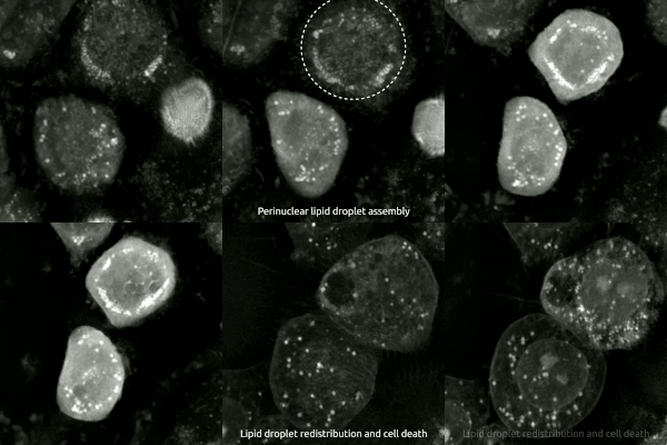
has been used to observe the morphological changes that cells undergo during ferroptosis. Initially the cell contracts and then begins to swell. Perinuclear lipid assembly is observed immediately before ferroptosis occurs. After the process is complete, lipid droplets are redistributed throughout the cell (see GIF on right side).
Comparison to apoptosis in the nervous system
Another form of cell death that occurs in the nervous system is apoptosis, which results in cell breakage into small, apoptotic bodies taken up throughphagocytosis
Phagocytosis () is the process by which a cell uses its plasma membrane to engulf a large particle (≥ 0.5 μm), giving rise to an internal compartment called the phagosome. It is one type of endocytosis. A cell that performs phagocytosis i ...
. This process occurs continuously within mammalian nervous system processes that begin at fetal development and continue through adult life. Apoptotic death is crucial for the correct population size of neuronal and glial
Glia, also called glial cells (gliocytes) or neuroglia, are non-neuronal cells in the central nervous system (brain and spinal cord) and the peripheral nervous system that do not produce electrical impulses. They maintain homeostasis, form m ...
cells. Similarly to ferroptosis, deficiencies in apoptotic processes can result in many health complications, including neurodegeneration
A neurodegenerative disease is caused by the progressive loss of structure or function of neurons, in the process known as neurodegeneration. Such neuronal damage may ultimately involve cell death. Neurodegenerative diseases include amyotrophic ...
.
Within the study of neuronal apoptosis, most research has been conducted on the neurons of the superior cervical ganglion
The superior cervical ganglion (SCG) is part of the autonomic nervous system (ANS); more specifically, it is part of the sympathetic nervous system, a division of the ANS most commonly associated with the fight or flight response. The ANS is co ...
. In order for these neurons to survive and innervate their target tissues, they must have nerve growth factor
Nerve growth factor (NGF) is a neurotrophic factor and neuropeptide primarily involved in the regulation of growth, maintenance, proliferation, and survival of certain target neurons. It is perhaps the prototypical growth factor, in that it was on ...
(NGF). Normally, NGF binds to a tyrosine kinase receptor, TrkA
Tropomyosin receptor kinase A (TrkA), also known as high affinity nerve growth factor receptor, neurotrophic tyrosine kinase receptor type 1, or TRK1-transforming tyrosine kinase protein is a protein that in humans is encoded by the ''NTRK1'' gen ...
, which activates phosphatidylinositol 3-kinase-Akt ( PI3K-Akt) and extracellular signal-regulated kinase (Raf-MEK-ERK) signaling pathways. This occurs during normal development which promotes neuronal growth in the sympathetic nervous system
The sympathetic nervous system (SNS) is one of the three divisions of the autonomic nervous system, the others being the parasympathetic nervous system and the enteric nervous system. The enteric nervous system is sometimes considered part of ...
.
During embryonic development, the absence of NGF activates apoptosis by decreasing the activity of the signaling pathways normally activated by NGF. Without NGF, the neurons of the sympathetic nervous system begin to atrophy, glucose uptake rates fall, and the rates of protein synthesis and gene expression slow. Apoptotic death from NGF withdrawal also requires caspase
Caspases (cysteine-aspartic proteases, cysteine aspartases or cysteine-dependent aspartate-directed proteases) are a family of protease enzymes playing essential roles in programmed cell death. They are named caspases due to their specific cyst ...
activity. Upon NGF withdrawal, caspase-3
Caspase-3 is a caspase protein that interacts with caspase-8 and caspase-9. It is encoded by the ''CASP3'' gene. ''CASP3'' orthologs have been identified in numerous mammals for which complete genome data are available. Unique orthologs are also ...
activation occurs through an in-vitro pathway beginning with the release of cytochrome c
The cytochrome complex, or cyt ''c'', is a small hemeprotein found loosely associated with the inner membrane of the mitochondrion. It belongs to the cytochrome c family of proteins and plays a major role in cell apoptosis. Cytochrome c is hig ...
from the mitochondria. In a surviving sympathetic neuron, the overexpression of anti-apoptotic B-cell CLL/lymphoma 2 (Bcl-2) proteins prevents NGF withdrawal-induced death. However, overexpression of a separate, pro-apoptotic Bcl-2 gene, Bax, stimulates the release of cytochrome c2. Cytochrome c promotes the activation of caspase-9
Caspase-9 is an enzyme that in humans is encoded by the CASP9 gene. It is an initiator caspase, critical to the apoptotic pathway found in many tissues. Caspase-9 homologs have been identified in all mammals for which they are known to exist, such ...
through the formation of the apoptosome. Once caspase-9 is activated, it can cleave and activate caspase-3 resulting in cell death. Notably, apoptosis does not release intracellular fluid as neurons that are degraded though ferroptosis do. During ferroptosis, neurons release lipid metabolites from inside the cell body. This is a key difference between ferroptosis and apoptosis.
In neurons
 Neural connections are constantly changing within the nervous system. Synaptic connections that are used more often are kept intact and promoted, while synaptic connections that are rarely used are subject to degradation. Elevated levels of synaptic connection loss and degradation of neurons are linked to neurodegenerative diseases. More recently, ferroptosis has been linked to diverse brain diseases. Two new studies show that ferroptosis contributes to neuronal death after intracerebral hemorrhage. Neurons that are degraded through ferroptosis release lipid metabolites from inside the cell body. The lipid metabolites are harmful to surrounding neurons, causing inflammation in the brain. Inflammation is a pathological feature of
 Alzheimer’s disease
Alzheimer's disease (AD) is a neurodegenerative disease that usually starts slowly and progressively worsens. It is the cause of 60–70% of cases of dementia. The most common early symptom is difficulty in remembering recent events. As ...
and intracerebral hemorrhage.
In a study performed using mice, it was found that the absence of Gpx4 promoted ferroptosis. Foods high in vitamin E
Vitamin E is a group of eight fat soluble compounds that include four tocopherols and four tocotrienols. Vitamin E deficiency, which is rare and usually due to an underlying problem with digesting dietary fat rather than from a diet low in vitami ...
promote Gpx4 activity, consequently inhibiting ferroptosis and preventing inflammation in brain regions. In the experimental group of mice that were manipulated to have decreased Gpx4 levels, mice were observed to have cognitive impairment and neurodegeneration of hippocampal neurons, again linking ferroptosis to neurodegenerative diseases.
Similarly, presence of transcription factors, specifically ATF4
Activating transcription factor 4 (tax-responsive enhancer element B67), also known as ATF4, is a protein that in humans is encoded by the ''ATF4'' gene.
Function
This gene encodes a transcription factor that was originally identified as a wi ...
, can influence how readily a neuron undergoes cell death. The presence of ATF4 promotes resistance in cells against ferroptosis. However, this resistance can cause other diseases, such as cancer, to progress and become malignant. While ATF4 provides resistance ferroptosis, an abundance of ATF4 causes neurodegeneration.
Recent studies have suggested that ferroptosis contributes to neuronal cell death after traumatic brain injury.
Potential role in cancer treatment
 Preliminary reports suggest that ferroptosis may be a means through which tumor cells can be killed. Ferroptosis has been implicated in several types of cancer, including:
*
 Breast
The breast is one of two prominences located on the upper ventral region of a primate's torso. Both females and males develop breasts from the same embryological tissues.
In females, it serves as the mammary gland, which produces and s ...
* Acute myeloid leukemia
Acute myeloid leukemia (AML) is a cancer of the myeloid line of blood cells, characterized by the rapid growth of abnormal cells that build up in the bone marrow and blood and interfere with haematopoiesis, normal blood cell production. Sympto ...
* Pancreatic ductal adenocarcinoma
The pancreas is an organ of the digestive system and endocrine system of vertebrates. In humans, it is located in the abdomen behind the stomach and functions as a gland. The pancreas is a mixed or heterocrine gland, i.e. it has both an endoc ...
* Ovarian
* B-cell lymphoma
The B-cell lymphomas are types of lymphoma affecting B cells. Lymphomas are "blood cancers" in the lymph nodes. They develop more frequently in older adults and in immunocompromised individuals.
B-cell lymphomas include both Hodgkin's lymphoma ...
* Renal cell carcinoma
Renal cell carcinoma (RCC) is a kidney cancer that originates in the lining of the proximal convoluted tubule, a part of the very small tubes in the kidney that transport primary urine. RCC is the most common type of kidney cancer in adults, res ...
s
* Lung
* Glioblastoma
Glioblastoma, previously known as glioblastoma multiforme (GBM), is one of the most aggressive types of cancer that begin within the brain. Initially, signs and symptoms of glioblastoma are nonspecific. They may include headaches, personality cha ...
These forms of cancer have been hypothesized to be highly sensitive to ferroptosis induction. An upregulation of iron levels has also been seen to induce ferroptosis in certain types of cancer, such as breast cancer. Breast cancer cells have exhibited vulnerability to ferroptosis via a combination of siramesine and lapatinib. These cells also exhibited an autophagic cycle independent of ferroptotic activity, indicating that the two different forms of cell death could be controlled to activate at specific times following treatment.
Notably, not all cancers are necessarily sensitive to ferroptosis induction. For instance, one study has demonstrated that ferroptosis in polymorphonuclear myeloid-derived suppressor cells in the tumor microenvironment
The tumor microenvironment (TME) is the environment around a Neoplasm, tumor, including the surrounding blood vessels, White blood cell, immune cells, fibroblasts, Cell signaling, signaling molecules and the extracellular matrix (ECM). The tumor ...
releases oxidized lipids that contribute to immune suppression.
See also
References
{{ReflistExternal links
KEGG pathway entry
Programmed cell death Medical aspects of death Cellular processes