|
Womb
The uterus (from Latin ''uterus'', : uteri or uteruses) or womb () is the organ in the reproductive system of most female mammals, including humans, that accommodates the embryonic and fetal development of one or more fertilized eggs until birth. The uterus is a hormone-responsive sex organ that contains glands in its lining that secrete uterine milk for embryonic nourishment. (The term ''uterus'' is also applied to analogous structures in some non-mammalian animals.) In humans, the lower end of the uterus is a narrow part known as the isthmus that connects to the cervix, the anterior gateway leading to the vagina. The upper end, the body of the uterus, is connected to the fallopian tubes at the uterine horns; the rounded part, the fundus, is above the openings to the fallopian tubes. The connection of the uterine cavity with a fallopian tube is called the uterotubal junction. The fertilized egg is carried to the uterus along the fallopian tube. It will have divided on its ... [...More Info...] [...Related Items...] OR: [Wikipedia] [Google] [Baidu] |
Vagina
In mammals and other animals, the vagina (: vaginas or vaginae) is the elastic, muscular sex organ, reproductive organ of the female genital tract. In humans, it extends from the vulval vestibule to the cervix (neck of the uterus). The #Vaginal opening and hymen, vaginal introitus is normally partly covered by a thin layer of mucous membrane, mucosal tissue called the hymen. The vagina allows for Copulation (zoology), copulation and birth. It also channels Menstruation (mammal), menstrual flow, which occurs in humans and closely related primates as part of the menstrual cycle. To accommodate smoother penetration of the vagina during sexual intercourse or other sexual activity, vaginal moisture increases during sexual arousal in human females and other female mammals. This increase in moisture provides vaginal lubrication, which reduces friction. The texture of the vaginal walls creates friction for the penis during sexual intercourse and stimulates it toward ejaculation, en ... [...More Info...] [...Related Items...] OR: [Wikipedia] [Google] [Baidu] |
Blastocyst
The blastocyst is a structure formed in the early embryonic development of mammals. It possesses an inner cell mass (ICM) also known as the ''embryoblast'' which subsequently forms the embryo, and an outer layer of trophoblast cells called the trophectoderm. This layer surrounds the inner cell mass and a fluid-filled cavity or lumen known as the blastocoel. In the late blastocyst, the trophectoderm is known as the trophoblast. The trophoblast gives rise to the chorion and amnion, the two fetal membranes that surround the embryo. The placenta derives from the embryonic chorion (the portion of the chorion that develops villi) and the underlying uterine tissue of the mother. The corresponding structure in non-mammalian animals is an undifferentiated ball of cells called the blastula. In humans, blastocyst formation begins about five days after fertilization when a fluid-filled cavity opens up in the morula, the early embryonic stage of a ball of 16 cells. The blastocyst ... [...More Info...] [...Related Items...] OR: [Wikipedia] [Google] [Baidu] |
Prenatal Development
Prenatal development () involves the development of the embryo and of the fetus during a viviparous animal's gestation. Prenatal development starts with fertilization, in the germinal stage of embryonic development, and continues in fetal development until birth. The term "prenate" is used to describe an unborn offspring at any stage of gestation. In human pregnancy, prenatal development is also called antenatal development. The development of the human embryo follows fertilization, and continues as fetal development. By the end of the tenth week of gestational age, the embryo has acquired its basic form and is referred to as a fetus. The next period is that of fetal development where many organs become fully developed. This fetal period is described both topically (by organ) and chronologically (by time) with major occurrences being listed by gestational age. The very early stages of embryonic development are the same in all mammals, but later stages of development, an ... [...More Info...] [...Related Items...] OR: [Wikipedia] [Google] [Baidu] |
Mammal
A mammal () is a vertebrate animal of the Class (biology), class Mammalia (). Mammals are characterised by the presence of milk-producing mammary glands for feeding their young, a broad neocortex region of the brain, fur or hair, and three Evolution of mammalian auditory ossicles, middle ear bones. These characteristics distinguish them from reptiles and birds, from which their ancestors Genetic divergence, diverged in the Carboniferous Period over 300 million years ago. Around 6,640 Neontology#Extant taxon, extant species of mammals have been described and divided into 27 Order (biology), orders. The study of mammals is called mammalogy. The largest orders of mammals, by number of species, are the rodents, bats, and eulipotyphlans (including hedgehogs, Mole (animal), moles and shrews). The next three are the primates (including humans, monkeys and lemurs), the Artiodactyl, even-toed ungulates (including pigs, camels, and whales), and the Carnivora (including Felidae, ... [...More Info...] [...Related Items...] OR: [Wikipedia] [Google] [Baidu] |
Endometrium
The endometrium is the inner epithelium, epithelial layer, along with its mucous membrane, of the mammalian uterus. It has a basal layer and a functional layer: the basal layer contains stem cells which regenerate the functional layer. The functional layer thickens and then is shed during menstruation in humans and some other mammals, including other apes, Old World monkeys, some species of bat, the elephant shrew and the Cairo spiny mouse. In most other mammals, the endometrium is reabsorbed in the estrous cycle. During pregnancy, the glands and blood vessels in the endometrium further increase in size and number. Vascular spaces fuse and become interconnected, forming the placenta, which supplies oxygen and nutrition to the embryo and fetus.Blue Histology - Female Reproductive System ... [...More Info...] [...Related Items...] OR: [Wikipedia] [Google] [Baidu] |
Birth
Birth is the act or process of bearing or bringing forth offspring, also referred to in technical contexts as parturition. In mammals, the process is initiated by hormones which cause the muscular walls of the uterus to contract, expelling the fetus at a developmental stage when it is ready to feed and breathe. In some species, the offspring is precocial and can move around almost immediately after birth but in others, it is altricial and completely dependent on parenting. In marsupials, the fetus is born at a very immature stage after a short gestation and develops further in its mother's womb Pouch (marsupial), pouch. It is not only mammals that give birth. Some reptiles, amphibians, fish and invertebrates carry their developing young inside them. Some of these are Ovoviviparity, ovoviviparous, with the eggs being hatched inside the mother's body, and others are Viviparity, viviparous, with the embryo developing inside their body, as in the case of mammals. Human childbirth ... [...More Info...] [...Related Items...] OR: [Wikipedia] [Google] [Baidu] |
Uterine Milk
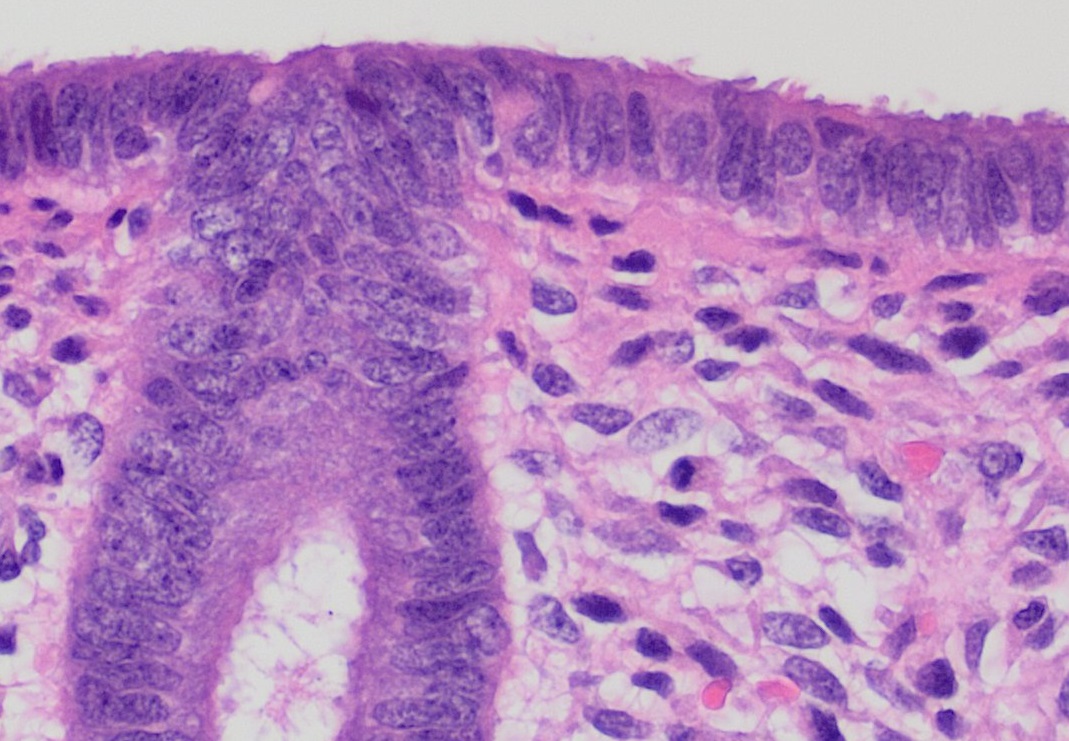
Uterine glands or endometrial glands are tubular glands, lined by a simple columnar epithelium, found in the functional layer of the endometrium that lines the uterus. Their appearance varies during the menstrual cycle. During the proliferative phase, uterine glands appear long due to estrogen secretion by the ovaries. During the secretory phase, the uterine glands become very coiled with wide lumens and produce a glycogen-rich secretion known as ''histotroph'' or ''uterine milk''. This change corresponds with an increase in blood flow to spiral arteries due to increased progesterone secretion from the corpus luteum. During the pre-menstrual phase, progesterone secretion decreases as the corpus luteum degenerates, which results in decreased blood flow to the spiral arteries. The functional layer of the uterus containing the glands becomes necrotic, and eventually sloughs off during the menstrual phase of the cycle. They are of small size in the unimpregnated uterus, but shortly af ... [...More Info...] [...Related Items...] OR: [Wikipedia] [Google] [Baidu] |
Uterine Isthmus
The uterine isthmus is the inferior-posterior part of uterus The uterus (from Latin ''uterus'', : uteri or uteruses) or womb () is the hollow organ, organ in the reproductive system of most female mammals, including humans, that accommodates the embryonic development, embryonic and prenatal development, f ..., on its cervical end — here, the uterine muscle ( myometrium) is narrower and thinner. It connects the body and cervix. The uterine isthmus can become more compressibile in pregnancy, which is a finding known as Hegar's sign. References Uterus {{genitourinary-stub, sex=female ... [...More Info...] [...Related Items...] OR: [Wikipedia] [Google] [Baidu] |
Cervix
The cervix (: cervices) or cervix uteri is a dynamic fibromuscular sexual organ of the female reproductive system that connects the vagina with the uterine cavity. The human female cervix has been documented anatomically since at least the time of Hippocrates, over 2,000 years ago. The cervix is approximately 4 cm long with a diameter of approximately 3 cm and tends to be described as a cylindrical shape, although the front and back walls of the cervix are contiguous. The size of the cervix changes throughout a woman's life cycle. For example, women in the fertile years of their reproductive cycle tend to have larger cervixes than postmenopausal women; likewise, women who have produced offspring have a larger cervix than those who have not. In relation to the vagina, the part of the cervix that opens to the uterus is called the ''internal os'' and the opening of the cervix in the vagina is called the ''external os''. Between them is a conduit commonly called the cervic ... [...More Info...] [...Related Items...] OR: [Wikipedia] [Google] [Baidu] |
Paramesonephric Duct
The paramesonephric ducts (or Müllerian ducts) are paired ducts of the embryo in the reproductive system of humans and other mammals that run down the lateral sides of the genital ridge and terminate at the sinus tubercle in the primitive urogenital sinus. They form in both sexes during 6th week of fetal development. In the female, go on to form the fallopian tubes/oviducts, uterus, cervix, and the upper one-third of the vagina. In males fetuses, they are normally made to regress by anti-Müllerian hormone which begins to be secreted by the testes during 8th week of fetal development. Each maramesonephric duct is situated just lateral to the mesonephric ducts, mesonephric ducts (Wolffian duct) of the same side. Development The female reproductive system is composed of two embryological segments: the urogenital sinus and the paramesonephric ducts. The two are conjoined at the sinus tubercle. Paramesonephric ducts are present on the embryo of both sexes. Only in females do they d ... [...More Info...] [...Related Items...] OR: [Wikipedia] [Google] [Baidu] |
Uterine Gland
Uterine glands or endometrial glands are tubular glands, lined by a simple columnar epithelium, found in the functional layer of the endometrium that lines the uterus. Their appearance varies during the menstrual cycle. During the proliferative phase, uterine glands appear long due to estrogen secretion by the ovaries. During the secretory phase, the uterine glands become very coiled with wide lumens and produce a glycogen-rich secretion known as ''histotroph'' or ''uterine milk''. This change corresponds with an increase in blood flow to spiral arteries due to increased progesterone secretion from the corpus luteum. During the pre-menstrual phase, progesterone secretion decreases as the corpus luteum degenerates, which results in decreased blood flow to the spiral arteries. The functional layer of the uterus containing the glands becomes necrotic, and eventually sloughs off during the menstrual phase of the cycle. They are of small size in the unimpregnated uterus, but shortly af ... [...More Info...] [...Related Items...] OR: [Wikipedia] [Google] [Baidu] |
Uterine Horns
The uterine horns (cornua of uterus) are the points in the upper uterus where the fallopian tubes or oviducts exit to meet the ovaries. They are one of the points of attachment for the round ligament of uterus (the other being the mons pubis). They also provide attachment to the ovarian ligament, which is located below the fallopian tube at the back, while the round ligament of uterus is located below the tube at the front. The uterine horns are far more prominent in other animals (such as cows and cats) than they are in humans. In the cat, implantation of the embryo occurs in one of the two uterine horns, not the body of the uterus itself. Occasionally, if a fallopian tube does not connect, the uterine horn will fill with blood each month, and a minor one-day surgery will be performed to remove it. Often, people who are born with this have trouble getting pregnant as both ovaries are functional and either may ovulate. The spare egg An egg is an organic vessel grown by ... [...More Info...] [...Related Items...] OR: [Wikipedia] [Google] [Baidu] |


