|
Ventricular Fibrillation
Ventricular fibrillation (V-fib or VF) is an abnormal heart rhythm in which the Ventricle (heart), ventricles of the heart Fibrillation, quiver. It is due to disorganized electrical conduction system of the heart, electrical activity. Ventricular fibrillation results in cardiac arrest with loss of consciousness and no pulse. This is followed by sudden cardiac death in the absence of treatment. Ventricular fibrillation is initially found in about 10% of people with cardiac arrest. Ventricular fibrillation can occur due to coronary heart disease, valvular heart disease, cardiomyopathy, Brugada syndrome, long QT syndrome, electric shock, or intracranial hemorrhage. Diagnosis is by an electrocardiogram (ECG) showing irregular unformed QRS complexes without any clear P wave (electrocardiography), P waves. An important differential diagnosis is torsades de pointes. Treatment is with cardiopulmonary resuscitation (CPR) and defibrillation. Defibrillation#Change to a biphasic waveform, ... [...More Info...] [...Related Items...] OR: [Wikipedia] [Google] [Baidu] |
Electrocardiography
Electrocardiography is the process of producing an electrocardiogram (ECG or EKG), a recording of the heart's electrical activity through repeated cardiac cycles. It is an electrogram of the heart which is a graph of voltage versus time of the electrical activity of the heart using electrodes placed on the skin. These electrodes detect the small electrical changes that are a consequence of cardiac muscle depolarization followed by repolarization during each cardiac cycle (heartbeat). Changes in the normal ECG pattern occur in numerous cardiac abnormalities, including: * Cardiac rhythmicity, Cardiac rhythm disturbances, such as atrial fibrillation and ventricular tachycardia; * Inadequate coronary artery blood flow, such as myocardial ischemia and myocardial infarction; * and electrolyte disturbances, such as hypokalemia. Traditionally, "ECG" usually means a 12-lead ECG taken while lying down as discussed below. However, other devices can record the electrical activity of ... [...More Info...] [...Related Items...] OR: [Wikipedia] [Google] [Baidu] |
Abnormal Heart Rhythm
Arrhythmias, also known as cardiac arrhythmias, are irregularities in the heartbeat, including when it is too fast or too slow. Essentially, this is anything but normal sinus rhythm. A resting heart rate that is too fast – above 100 beats per minute in adults – is called tachycardia, and a resting heart rate that is too slow – below 60 beats per minute – is called bradycardia. Some types of arrhythmias have no symptoms. Symptoms, when present, may include palpitations or feeling a pause between heartbeats. In more serious cases, there may be lightheadedness, passing out, shortness of breath, chest pain, or decreased level of consciousness. While most cases of arrhythmia are not serious, some predispose a person to complications such as stroke or heart failure. Others may result in sudden death. Arrhythmias are often categorized into four groups: extra beats, supraventricular tachycardias, ventricular arrhythmias and bradyarrhythmias. Extra beats include premature a ... [...More Info...] [...Related Items...] OR: [Wikipedia] [Google] [Baidu] |
Cardiopulmonary Resuscitation
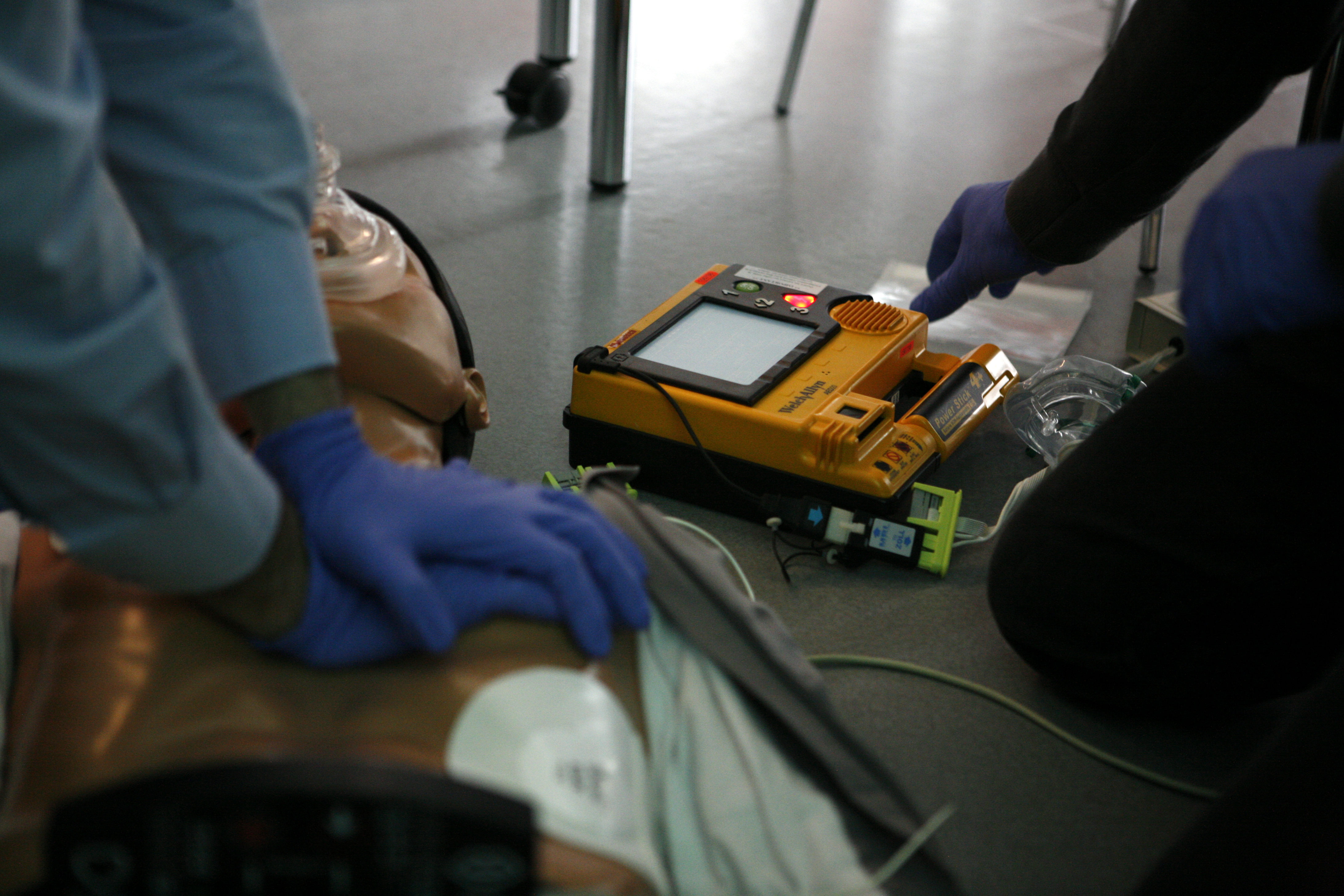
Cardiopulmonary resuscitation (CPR) is an emergency procedure used during Cardiac arrest, cardiac or Respiratory arrest, respiratory arrest that involves chest compressions, often combined with artificial ventilation, to preserve brain function and maintain circulation until spontaneous breathing and heartbeat can be restored. It is indication (medicine), recommended for those who are unresponsive with no breathing or abnormal breathing, for example, agonal respirations. CPR involves chest compressions for adults between and deep and at a rate of at least 100 to 120 per minute. The rescuer may also provide artificial ventilation by either exhaling air into the subject's mouth or nose (mouth-to-mouth resuscitation) or using a device that pushes air into the subject's lungs (mechanical ventilation). Current recommendations emphasize early and high-quality chest compressions over artificial ventilation; a simplified CPR method involving only chest compressions is recommended for ... [...More Info...] [...Related Items...] OR: [Wikipedia] [Google] [Baidu] |
Torsades De Pointes
''Torsades de pointes, torsade de pointes'' or ''torsades des pointes'' (TdP; also called ''torsades'') (, , translated as "twisting of peaks") is a specific type of abnormal heart rhythm that can lead to sudden cardiac death. It is a polymorphic ventricular tachycardia that exhibits distinct characteristics on the electrocardiogram (ECG). It was described by French physician François Dessertenne in 1966. Prolongation of the QT interval can increase a person's risk of developing this abnormal heart rhythm, occurring in between 1% and 10% of patients who receive QT-prolonging antiarrhythmic drugs. Signs and symptoms Most episodes will revert spontaneously to a normal sinus rhythm. Symptoms and consequences include palpitations, dizziness, lightheadedness (during shorter episodes), fainting (during longer episodes), and sudden cardiac death. Causes Torsades occurs as both an inherited (linked to at least 17 genes) and as an acquired form caused most often by drugs a ... [...More Info...] [...Related Items...] OR: [Wikipedia] [Google] [Baidu] |
Differential Diagnosis
In healthcare, a differential diagnosis (DDx) is a method of analysis that distinguishes a particular disease or condition from others that present with similar clinical features. Differential diagnostic procedures are used by clinicians to diagnose the specific disease in a patient, or, at least, to consider any imminently life-threatening conditions. Often, each possible disease is called a differential diagnosis (e.g., acute bronchitis could be a differential diagnosis in the evaluation of a cough, even if the final diagnosis is common cold). More generally, a differential diagnostic procedure is a systematic diagnostic method used to identify the presence of a disease entity where multiple alternatives are possible. This method may employ algorithms, akin to the process of elimination, or at least a process of obtaining information that decreases the "probabilities" of candidate conditions to negligible levels, by using evidence such as symptoms, patient history, and medi ... [...More Info...] [...Related Items...] OR: [Wikipedia] [Google] [Baidu] |
P Wave (electrocardiography)
In cardiology, the P wave on an Electrocardiography, electrocardiogram (ECG) represents atrial depolarization, which results in atrial contraction, or systole#Atrial systole, atrial systole. Physiology The P wave is a summation wave generated by the depolarization front as it transits the atria. Normally the right atrium depolarizes slightly earlier than left atrium since the depolarization wave originates in the sinoatrial node, in the high right atrium and then travels to and through the left atrium. The depolarization front is carried through the atria along semi-specialized conduction pathways including Bachmann's bundle resulting in uniform shaped waves. Depolarization originating elsewhere in the atria (atrial ectopics) result in P waves with a different morphology from normal. Pathology Peaked P waves (> 0.25 mV) suggest right atrial enlargement, cor pulmonale, (''P pulmonale'' rhythm), but have a low predictive value (~20%). A P wave with increased amplitude can in ... [...More Info...] [...Related Items...] OR: [Wikipedia] [Google] [Baidu] |
QRS Complex
The QRS complex is the combination of three of the graphical deflections seen on a typical electrocardiogram (ECG or EKG). It is usually the central and most visually obvious part of the tracing. It corresponds to the depolarization of the right and left ventricles of the heart and contraction of the large ventricular muscles. In adults, the QRS complex normally lasts ; in children it may be shorter. The Q, R, and S waves occur in rapid succession, do not all appear in all leads, and reflect a single event and thus are usually considered together. A Q wave is any downward deflection immediately following the P wave. An R wave follows as an upward deflection, and the S wave is any downward deflection after the R wave. The T wave follows the S wave, and in some cases, an additional U wave follows the T wave. To measure the QRS interval start at the end of the PR interval (or beginning of the Q wave) to the end of the S wave. Normally this interval is 0.08 to 0.10 seconds. W ... [...More Info...] [...Related Items...] OR: [Wikipedia] [Google] [Baidu] |
Coronary Heart Disease
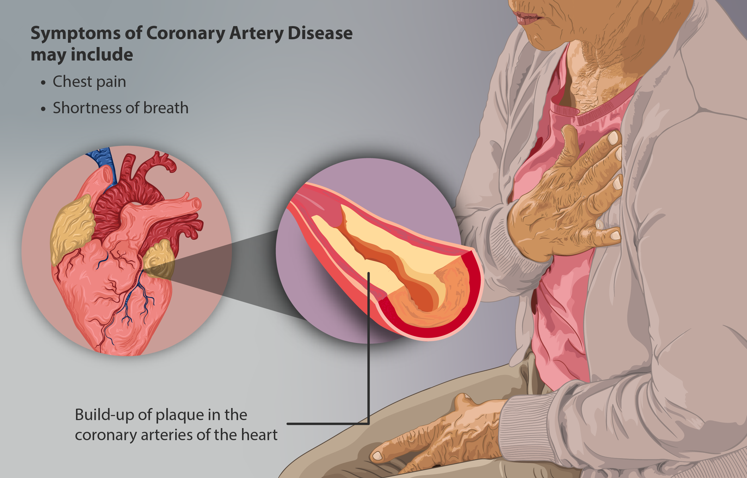
Coronary artery disease (CAD), also called coronary heart disease (CHD), or ischemic heart disease (IHD), is a type of cardiovascular disease, heart disease involving Ischemia, the reduction of blood flow to the cardiac muscle due to a build-up of atheromatous plaque in the Coronary arteries, arteries of the heart. It is the most common of the cardiovascular diseases. CAD can cause stable angina, unstable angina, myocardial ischemia, and myocardial infarction. A common symptom is angina, which is chest pain or discomfort that may travel into the shoulder, arm, back, neck, or jaw. Occasionally it may feel like heartburn. In stable angina, symptoms occur with exercise or emotional Psychological stress, stress, last less than a few minutes, and improve with rest. Shortness of breath may also occur and sometimes no symptoms are present. In many cases, the first sign is a Myocardial infarction, heart attack. Other complications include heart failure or an Heart arrhythmia, abnormal h ... [...More Info...] [...Related Items...] OR: [Wikipedia] [Google] [Baidu] |
Sudden Cardiac Death
Cardiac arrest (also known as sudden cardiac arrest ''SCA is when the heart suddenly and unexpectedly stops beating. When the heart stops beating, blood cannot properly circulate around the body and the blood flow to the brain and other organs is decreased. When the brain does not receive enough blood, this can cause a person to lose consciousness and brain cells can start to die due to lack of oxygen. Coma and persistent vegetative state may result from cardiac arrest. Cardiac arrest is also identified by a lack of central pulses and abnormal or absent breathing. Cardiac arrest and resultant hemodynamic collapse often occur due to arrhythmias (irregular heart rhythms). Ventricular fibrillation and ventricular tachycardia are most commonly recorded. However, as many incidents of cardiac arrest occur out-of-hospital or when a person is not having their cardiac activity monitored, it is difficult to identify the specific mechanism in each case. Structural heart disease, ... [...More Info...] [...Related Items...] OR: [Wikipedia] [Google] [Baidu] |
Cardiac Arrest
Cardiac arrest (also known as sudden cardiac arrest [SCA]) is when the heart suddenly and unexpectedly stops beating. When the heart stops beating, blood cannot properly Circulatory system, circulate around the body and the blood flow to the brain and other organs is decreased. When the brain does not receive enough blood, this can cause a person to lose consciousness and brain cells can start to die due to lack of oxygen. Coma and persistent vegetative state may result from cardiac arrest. Cardiac arrest is also identified by a lack of Pulse, central pulses and respiratory arrest, abnormal or absent breathing. Cardiac arrest and resultant hemodynamic collapse often occur due to arrhythmias (irregular heart rhythms). Ventricular fibrillation and ventricular tachycardia are most commonly recorded. However, as many incidents of cardiac arrest occur out-of-hospital or when a person is not having their cardiac activity monitored, it is difficult to identify the specific mechanism ... [...More Info...] [...Related Items...] OR: [Wikipedia] [Google] [Baidu] |
Electrical Conduction System Of The Heart
The cardiac conduction system (CCS, also called the electrical conduction system of the heart) transmits the Cardiac action potential, signals generated by the sinoatrial node – the heart's Cardiac pacemaker, pacemaker, to cause the heart muscle to Muscle contraction, contract, and pump blood through the body's circulatory system. The Cardiac pacemaker, pacemaking signal travels through the right atrium to the atrioventricular node, along the bundle of His, and through the bundle branches to Purkinje fibers in the Ventricle (heart), walls of the ventricles. The Purkinje fibers transmit the signals more rapidly to stimulate contraction of the ventricles. The conduction system consists of specialized Cardiomyocyte, heart muscle cells, situated within the myocardium. There is a cardiac skeleton, skeleton of fibrous tissue that surrounds the conduction system which can be seen on an ECG. Dysfunction of the conduction system can cause Heart arrhythmia, irregular heart rhythms includ ... [...More Info...] [...Related Items...] OR: [Wikipedia] [Google] [Baidu] |


