|
Ureteroscopes
Ureteroscopy is an examination of the upper urinary tract, usually performed with a endoscope, ureteroscope that is passed through the urethra and the Urinary bladder, bladder, and then directly into the ureter. The procedure is useful in the diagnosis and treatment of disorders such as kidney stones and urothelial carcinoma of the upper urinary tract. Smaller stones in the bladder or lower ureter can be removed in one piece, while bigger ones are usually broken before removal during ureteroscopy. The examination may be performed with either a flexible, semi-rigid or rigid device while the patient is under anesthesia. In specific cases, the patient is free to go home after the examination.Ureteropyeloscopy. Baylor College of Medicine. 2018 [accessed 2018 Mar 5]/ref> In pyeloscopy, the endoscope is designed to reach all the way to the renal pelvis (also called ''pyelum''), thereby allowing visualisation of the entire drainage system of the kidney. [...More Info...] [...Related Items...] OR: [Wikipedia] [Google] [Baidu] |
Kidney Stone
Kidney stone disease (known as nephrolithiasis, renal calculus disease, or urolithiasis) is a crystallopathy and occurs when there are too many minerals in the urine and not enough liquid or hydration. This imbalance causes tiny pieces of crystal to aggregate and form hard masses, or calculi (stones) in the upper urinary tract. Because renal calculi typically form in the kidney, if small enough, they are able to leave the urinary tract via the urine stream. A small calculus may pass without causing symptoms. However, if a stone grows to more than , it can cause blockage of the ureter, resulting in extremely sharp and severe pain ( renal colic) in the lower back that often radiates downward to the groin. A calculus may also result in blood in the urine, vomiting (due to severe pain), or painful urination. About half of all people who have had a kidney stone are likely to develop another within ten years. ''Renal'' is Latin for "kidney", while "nephro" is the Greek equiva ... [...More Info...] [...Related Items...] OR: [Wikipedia] [Google] [Baidu] |
Endoscope
An endoscope is an inspection instrument composed of image sensor, optical lens, light source and mechanical device, which is used to look deep into the body by way of openings such as the mouth or anus. A typical endoscope applies several modern technologies including optics, Human factors and ergonomics, ergonomics, precision mechanics, electronics, and software engineering. With an endoscope, it is possible to observe lesions that cannot be detected by X-ray, making it useful in medical diagnosis. An endoscope uses tubes only a few millimeters thick to transfer illumination in one direction and high-resolution video in the other, allowing minimally invasive surgeries. It is used to examine the internal organs like the throat or esophagus. Specialized instruments are named after their target organ. Examples include the cystoscope (bladder), Nephroscopy, nephroscope (kidney), Bronchoscopy, bronchoscope (bronchus), arthroscope (joints) and colonoscope (colon), and Laparoscopy, lapar ... [...More Info...] [...Related Items...] OR: [Wikipedia] [Google] [Baidu] |
Urethra
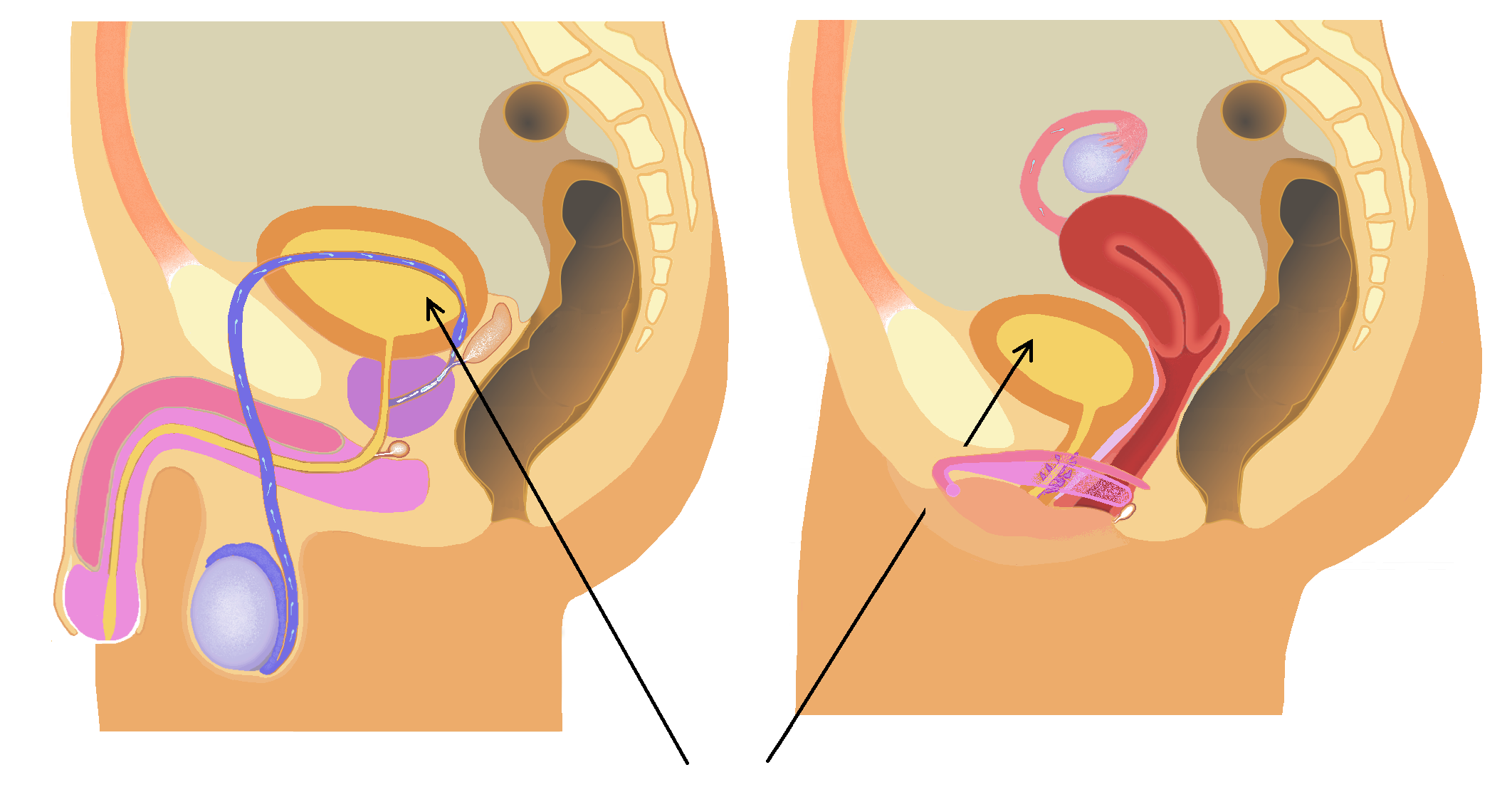
The urethra (: urethras or urethrae) is the tube that connects the urinary bladder to the urinary meatus, through which Placentalia, placental mammals Urination, urinate and Ejaculation, ejaculate. The external urethral sphincter is a striated muscle that allows voluntary control over urination. The Internal urethral sphincter, internal sphincter, formed by the involuntary smooth muscles lining the bladder neck and urethra, receives its nerve supply by the Sympathetic nervous system, sympathetic division of the autonomic nervous system. The internal sphincter is present both in males and females. Structure The urethra is a fibrous and muscular tube which connects the urinary bladder to the external urethral meatus. Its length differs between the sexes, because it passes through the penis in males. Male In the human male, the urethra is on average long and opens at the end of the external urethral meatus. The urethra is divided into four parts in men, named after the lo ... [...More Info...] [...Related Items...] OR: [Wikipedia] [Google] [Baidu] |
Urinary Bladder
The bladder () is a hollow organ in humans and other vertebrates that stores urine from the Kidney (vertebrates), kidneys. In placental mammals, urine enters the bladder via the ureters and exits via the urethra during urination. In humans, the bladder is a distensible organ that sits on the pelvic floor. The typical adult human bladder will hold between 300 and (10 and ) before the urge to empty occurs, but can hold considerably more. The Latin phrase for "urinary bladder" is ''vesica urinaria'', and the term ''vesical'' or prefix ''vesico-'' appear in connection with associated structures such as vesical veins. The modern Latin word for "bladder" – ''cystis'' – appears in associated terms such as cystitis (inflammation of the bladder). Structure In humans, the bladder is a hollow muscular organ situated at the base of the pelvis. In gross anatomy, the bladder can be divided into a broad (base), a body, an apex, and a neck. The apex (also called the vertex) is directed ... [...More Info...] [...Related Items...] OR: [Wikipedia] [Google] [Baidu] |
Ureter
The ureters are tubes composed of smooth muscle that transport urine from the kidneys to the urinary bladder. In an adult human, the ureters typically measure 20 to 30 centimeters in length and about 3 to 4 millimeters in diameter. They are lined with urothelial cells, a form of transitional epithelium, and feature an extra layer of smooth muscle in the lower third to aid in peristalsis. The ureters can be affected by a number of diseases, including urinary tract infections and kidney stone. is when a ureter is narrowed, due to for example chronic inflammation. Congenital abnormalities that affect the ureters can include the development of two ureters on the same side or abnormally placed ureters. Additionally, reflux of urine from the bladder back up the ureters is a condition commonly seen in children. The ureters have been identified for at least two thousand years, with the word "ureter" stemming from the stem relating to urinating and seen in written records since at ... [...More Info...] [...Related Items...] OR: [Wikipedia] [Google] [Baidu] |
Baylor College Of Medicine
The Baylor College of Medicine (BCM) is a private medical school in Houston, Texas, United States. Originally as the Baylor University College of Medicine from 1903 to 1969, the college became independent with the current name and has been separate from Baylor University since 1969. The college consists of four schools: the School of Medicine, the Graduate School of Biomedical Sciences, the School of Health Professions, and the National School of Tropical Medicine. The school is part owner, alongside Catholic Health Initiatives (CHI), of Baylor St. Luke's Medical Center, the flagship hospital of the CHI St. Luke's Health system. Other affiliated teaching hospitals and research institutes include Harris Health System's Ben Taub Hospital, Texas Children's Hospital, The University of Texas MD Anderson Cancer Center, TIRR Memorial Hermann, the Menninger Clinic, the Michael E. DeBakey VA Medical Center, and the Children's Hospital of San Antonio. On November 18, 2020, Bay ... [...More Info...] [...Related Items...] OR: [Wikipedia] [Google] [Baidu] |
Renal Pelvis
The renal pelvis or pelvis of the kidney is the funnel-like dilated part of the ureter in the kidney. It is formed by the convergence of the major calyces, acting as a funnel for urine flowing from the major calyces to the ureter. It has a mucous membrane and is covered with transitional epithelium and an underlying lamina propria of loose-to-dense connective tissue. The renal pelvis is situated within the renal sinus alongside the other structures of the renal sinus. Clinical significance The renal pelvis is the location of several kinds of kidney cancer and is affected by infection in pyelonephritis. A large " staghorn" kidney stone may block all or part of the renal pelvis. The size of the renal pelvis plays a major role in the grading of hydronephrosis. Normally, the anteroposterior diameter of the renal pelvis is less than 4 mm in fetuses up to 32 weeks of gestational age and 7 mm afterwards. [...More Info...] [...Related Items...] OR: [Wikipedia] [Google] [Baidu] |
Urologic Procedures
Urology (from Ancient Greek, Greek wikt:οὖρον, οὖρον ''ouron'' "urine" and ''wiktionary:-logia, -logia'' "study of"), also known as genitourinary surgery, is the branch of medicine that focuses on surgical and medical diseases of the urinary system and the reproductive organs. Organs under the domain of urology include the kidneys, adrenal glands, ureters, urinary bladder, urethra, and the male reproductive organs (testes, epididymis, epididymides, vas deferens, vasa deferentia, seminal vesicles, prostate, and Human penis, penis). The urinary and reproductive tracts are closely linked, and disorders of one often affect the other. Thus a major spectrum of the conditions managed in urology exists under the domain of genitourinary disorders. Urology combines the management of medical (i.e., non-surgical) conditions, such as urinary-tract infections and benign prostatic hyperplasia, with the management of surgical conditions such as bladder or prostate cancer, kidney st ... [...More Info...] [...Related Items...] OR: [Wikipedia] [Google] [Baidu] |


