|
Synostoses
Synostosis (; plural: synostoses) is fusion of two or more bones. It can be normal in puberty (e.g. fusion of the epiphyseal plate to become the epiphyseal line), or abnormal. When synostosis is abnormal it is a type of dysostosis. Examples of synostoses include: * craniosynostosis – an abnormal fusion of two or more cranial bones; * radioulnar synostosis – the abnormal fusion of the radius and ulna bones of the forearm; * tarsal coalition – a failure to separately form all seven bones of the tarsus (the hind part of the foot) resulting in an amalgamation of two bones; and * syndactyly – the abnormal fusion of neighboring digits. Synostosis within joints can cause ankylosis. __TOC__ Clinical significance Radioulnar synostosis is one of the more common failures of separation of parts of the upper limb. There are two general types: one is characterized by fusion of the radius and ulna at their proximal borders and the other is fused distal to the proximal radial epiphys ... [...More Info...] [...Related Items...] OR: [Wikipedia] [Google] [Baidu] |
Craniosynostosis
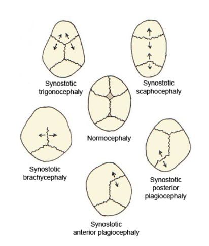
Craniosynostosis is a condition in which one or more of the fibrous sutures in a young infant's skull prematurely fuses by turning into bone (ossification), thereby changing the growth pattern of the skull. Because the skull cannot expand perpendicular to the fused suture, it compensates by growing more in the direction parallel to the closed sutures. Sometimes the resulting growth pattern provides the necessary space for the growing brain, but results in an abnormal head shape and abnormal facial features. In cases in which the compensation does not effectively provide enough space for the growing brain, craniosynostosis results in increased intracranial pressure leading possibly to visual impairment, sleeping impairment, eating difficulties, or an impairment of mental development combined with a significant reduction in IQ. Craniosynostosis occurs in one in 2000 births. Craniosynostosis is part of a syndrome in 15% to 40% of affected patients, but it usually occurs as an isol ... [...More Info...] [...Related Items...] OR: [Wikipedia] [Google] [Baidu] |
Antley–Bixler Syndrome
Antley–Bixler syndrome is a rare, severe autosomal recessive congenital disorder characterized by malformations and deformities affecting the majority of the skeleton and other areas of the body. Presentation Antley–Bixler syndrome presents itself at birth or prenatally. Features of the disorder include brachycephaly (flat forehead), craniosynostosis (complete skull-joint closure) of both coronal and lambdoid sutures, facial hypoplasia (underdevelopment); bowed ulna (forearm bone) and femur (thigh bone), synostosis of the radius (forearm bone), humerus (upper arm bone) and trapezoid (hand bone); camptodactyly (fused interphalangeal joints in the fingers), thin ilial wings (outer pelvic plate) and renal malformations. Other symptoms, such as cardiac malformations, proptotic exophthalmos (bulging eyes), arachnodactyly (spider-like fingers) as well as nasal, anal and vaginal atresia (occlusion) have been reported. Pathophysiology There are two distinct genetic mutation ... [...More Info...] [...Related Items...] OR: [Wikipedia] [Google] [Baidu] |
Bone
A bone is a rigid organ that constitutes part of the skeleton in most vertebrate animals. Bones protect the various other organs of the body, produce red and white blood cells, store minerals, provide structure and support for the body, and enable mobility. Bones come in a variety of shapes and sizes and have complex internal and external structures. They are lightweight yet strong and hard and serve multiple functions. Bone tissue (osseous tissue), which is also called bone in the uncountable sense of that word, is hard tissue, a type of specialised connective tissue. It has a honeycomb-like matrix internally, which helps to give the bone rigidity. Bone tissue is made up of different types of bone cells. Osteoblasts and osteocytes are involved in the formation and mineralisation of bone; osteoclasts are involved in the resorption of bone tissue. Modified (flattened) osteoblasts become the lining cells that form a protective layer on the bone surface. The mine ... [...More Info...] [...Related Items...] OR: [Wikipedia] [Google] [Baidu] |
Radial Head Dislocation
A pulled elbow, also known as nursemaid's elbow or a radial head subluxation, is when the ligament that wraps around the radial head slips off. Often a child will hold their arm against their body with the elbow slightly bent. They will not move the arm as this results in pain. Touching the arm, without moving the elbow, is usually not painful. A pulled elbow typically results from a sudden pull on an extended arm. This may occur when lifting or swinging a child by the arms. The underlying mechanism involves slippage of the annular ligament off of the head of the radius followed by the ligament getting stuck between the radius and humerus. Diagnosis is often based on symptoms. X-rays may be done to rule out other problems. Prevention is by avoiding potential causes. Treatment is by reduction. Moving the forearm into a palms down position with straightening at the elbow appears to be more effective than moving it into a palms up position followed by bending at the elbow. F ... [...More Info...] [...Related Items...] OR: [Wikipedia] [Google] [Baidu] |
Nievergelt Syndrome
Nievergelt is a Swiss surname. It may refer to the following: * Ernst Nievergelt (1910–1999), Swiss cyclist * Erwin Nievergelt (1929–2019), Swiss chess player {{disambig German-language surnames ... [...More Info...] [...Related Items...] OR: [Wikipedia] [Google] [Baidu] |
Limb-body Wall Complex
Limb body wall complex (LBWC) is a rare and severe syndrome of congenital malformations involving craniofacial and abdominal anomalies. LBWC emerges during early fetal development and is fatal. The cause of LBWC is unknown. Diagnosis and classification Traditionally, LBWC is diagnosed by the presence of at least two of the three Van Allen criteria: # Exencephaly or encephalocele with facial clefts # Abdominal wall defects: thoracoschisis and/or abdominoschisis # Limb defects As a component of the abdominal wall defect, the umbilical cord is shortened or absent with the fetus being directly attached to the placenta, a key feature in its prenatal diagnosis by ultrasound. Several systems have been proposed to classify LBWC cases phenotypically. Russo et al. (1993) proposed two types distinguished by the presence or absence of craniofacial defects. Sahinoglu et al. (2007) proposed three types based on the anatomical location of defects: * Type 1: Craniofacial defect and intact ... [...More Info...] [...Related Items...] OR: [Wikipedia] [Google] [Baidu] |
Hereditary Multiple Exostoses
Hereditary multiple osteochondromas (HMO), also known as hereditary multiple exostoses, is a disorder characterized by the development of multiple benign osteocartilaginous masses (exostosis, exostoses) in relation to the ends of long bones of the lower limbs such as the femurs and tibias and of the upper limbs such as the humeri and forearm bones. They are also known as osteochondromas. Additional sites of occurrence include on flat bones such as the pelvic bone and scapula. The distribution and number of these exostoses show a wide diversity among affected individuals. Exostoses usually present during childhood. The vast majority of affected individuals become clinically manifest by the time they reach adolescence. The incidence of hereditary multiple exostoses is around 1 in 50,000 individuals. Hereditary multiple osteochondromas is the preferred term used by the World Health Organization. A small percentage of affected individuals are at risk for development of sarcomas as a ... [...More Info...] [...Related Items...] OR: [Wikipedia] [Google] [Baidu] |
Hereditary Multiple Osteochondromas
Heredity, also called inheritance or biological inheritance, is the passing on of traits from parents to their offspring; either through asexual reproduction or sexual reproduction, the offspring cells or organisms acquire the genetic information of their parents. Through heredity, variations between individuals can accumulate and cause species to evolve by natural selection. The study of heredity in biology is genetics. Overview In humans, eye color is an example of an inherited characteristic: an individual might inherit the "brown-eye trait" from one of the parents. Inherited traits are controlled by genes and the complete set of genes within an organism's genome is called its genotype. The complete set of observable traits of the structure and behavior of an organism is called its phenotype. These traits arise from the interaction of the organism's genotype with the environment. As a result, many aspects of an organism's phenotype are not inherited. For example, sunt ... [...More Info...] [...Related Items...] OR: [Wikipedia] [Google] [Baidu] |
Greig Cephalopolysyndactyly Syndrome
Greig cephalopolysyndactyly syndrome is a disorder that affects development of the limbs, head, and face. The features of this syndrome are highly variable, ranging from very mild to severe. People with this condition typically have one or more extra fingers or toes (polydactyly) or an abnormally wide thumb or big toe (hallux). Presentation The skin between the fingers and toes may be fused (cutaneous syndactyly). This disorder is also characterized by widely spaced eyes (ocular hypertelorism), an abnormally large head size (macrocephaly), and a high, prominent forehead. Rarely, affected individuals may have more serious medical problems including seizures and developmental delay. Pathophysiology Greig cephalopolysyndactyly syndrome is a chromosomal condition related to chromosome 7. Mutations in the ''GLI3'' gene cause Greig cephalopolysyndactyly syndrome. The ''GLI3'' gene provides instructions for making a protein that controls gene expression, which is a process that regu ... [...More Info...] [...Related Items...] OR: [Wikipedia] [Google] [Baidu] |
Genitopatellar Syndrome
Genitopatellar syndrome is a rare disorder consisting of congenital flexion contractures of the lower extremities, abnormal or missing patellae, and urogenital anomalies. Additional symptoms include microcephaly, severe psychomotor disability. In 2012, it was shown that mutations in the gene KAT6B cause the syndrome. Genitopatellar syndrome (GTPTS) can be caused by heterozygous mutation in the KAT6B gene on chromosome 10q22. The Say-Barber-Biesecker variant of Ohdo syndrome, which has many overlapping features with GTPTS, can also be caused by heterozygous mutation in the KAT6B gene. Signs and symptoms Genitopatellar syndrome is characterized by genital abnormalities, missing or underdeveloped kneecaps (patellae), intellectual disability and abnormalities affecting other parts of the body. It is also associated with delayed development and intellectual disability, which are often severe. Affected individuals may have an unusually small head (microcephaly) and structural brain abn ... [...More Info...] [...Related Items...] OR: [Wikipedia] [Google] [Baidu] |
Acrofacial Dysostosis
Nager acrofacial dysostosis, also known as Nager syndrome, is a genetic disorder which displays several or all of the following characteristics: underdevelopment of the cheek and jaw area, down-sloping of the opening of the eyes, lack or absence of the lower eyelashes, kidney or stomach reflux, hammer toes, shortened soft palate, lack of development of the internal and external ear, possible cleft palate, underdevelopment or absence of the thumb, hearing loss (see hearing loss with craniofacial syndromes) and shortened forearms, as well as poor movement in the elbow, and may be characterized by accessory tragi. Occasionally, affected individuals develop vertebral anomalies such as scoliosis. The inheritance pattern is autosomal, but there are arguments as to whether it is autosomal dominant or autosomal recessive. Most cases tend to be sporadic. Nager syndrome shares many characteristics with other craniofacial syndromes: Miller, Treacher Collins and Pierre Robin. Genetics ... [...More Info...] [...Related Items...] OR: [Wikipedia] [Google] [Baidu] |


