|
Supinator
In human anatomy, the supinator is a broad muscle in the posterior compartment of the forearm, curved around the upper third of the radius (bone), radius. Its function is to supination, supinate the forearm. Structure The supinator consists of two planes of fibers, between which passes the deep branch of the radial nerve. The two planes arise in common—the superficial one originating as tendons and the deeper by muscular fibers—from the supinator crest of the ulna, the Lateral epicondyle of the humerus, lateral epicondyle of the humerus, the Radial collateral ligament (elbow), radial collateral ligament, and the Anular ligament of radius, annular radial ligament. The superficial fibers (''pars superficialis'') surround the upper part of the radius, and are inserted into the lateral edge of the radial tuberosity and the oblique line of the radius, as low down as the insertion of the pronator teres. The upper fibers (''pars profunda'') of the deeper plane form a sling-like Muscl ... [...More Info...] [...Related Items...] OR: [Wikipedia] [Google] [Baidu] |
Biceps Brachii Muscle
The biceps or biceps brachii (, "two-headed muscle of the arm") is a large muscle that lies on the front of the upper arm between the shoulder and the elbow. Both Muscle head, heads of the muscle arise on the scapula and join to form a single muscle belly which is attached to the upper forearm. While the long head of the biceps crosses both the shoulder and elbow joints, its main function is at the elbow where it flexes and supination, supinates the forearm. Both these movements are used when opening a bottle with a corkscrew: first biceps screws in the cork (supination), then it pulls the cork out (flexion). Structure The biceps is one of three muscles in the Fascial compartments of arm#Anterior compartment, anterior compartment of the upper arm, along with the brachialis muscle and the coracobrachialis muscle, with which the biceps shares a nerve supply. The biceps muscle has two heads, the short head and the long head, distinguished according to their origin at the cor ... [...More Info...] [...Related Items...] OR: [Wikipedia] [Google] [Baidu] |
Radius (bone)
The radius or radial bone (: radii or radiuses) is one of the two large bones of the forearm, the other being the ulna. It extends from the Anatomical terms of location, lateral side of the Elbow-joint, elbow to the thumb side of the wrist and runs parallel to the ulna. The ulna is longer than the radius, but the radius is thicker. The radius is a long bone, Prism (geometry), prism-shaped and slightly curved longitudinally. The radius is part of two joint (anatomy), joints: the elbow and the wrist. At the elbow, it joins with the capitulum of the humerus, and in a separate region, with the ulna at the radial notch. At the wrist, the radius forms a joint with the ulna bone. The corresponding bone in the human leg, lower leg is the tibia. Structure The long narrow medullary cavity is enclosed in a strong wall of compact bone. It is thickest along the interosseous border and thinnest at the extremities, same over the cup-shaped articular surface (fovea) of the head. The tra ... [...More Info...] [...Related Items...] OR: [Wikipedia] [Google] [Baidu] |
Arcade Of Frohse
Arcade of Frohse, sometimes called the supinator arch, is the most superior part of the superficial layer of the supinator muscle, and is a fibrous arch over the posterior interosseous nerve. The arcade of Frohse is a site of interosseous posterior nerve entrapment, and is believed to play a role in causing progressive paralysis of the posterior interosseous nerve, both with and without injury. The arcade of Frohse was named after German anatomist Anatomy () is the branch of morphology concerned with the study of the internal structure of organisms and their parts. Anatomy is a branch of natural science that deals with the structural organization of living things. It is an old scien ..., Fritz Frohse (1871–1916). References External links Clinical anatomy of the radial nerveMRI Web Clinic (MRIs of Posterior interosseous nerve entrapment) Archade of Frohse {{muscle-stub ... [...More Info...] [...Related Items...] OR: [Wikipedia] [Google] [Baidu] |
Radial Shaft
The radius or radial bone (: radii or radiuses) is one of the two large bones of the forearm, the other being the ulna. It extends from the lateral side of the elbow to the thumb side of the wrist and runs parallel to the ulna. The ulna is longer than the radius, but the radius is thicker. The radius is a long bone, prism-shaped and slightly curved longitudinally. The radius is part of two joints: the elbow and the wrist. At the elbow, it joins with the capitulum of the humerus, and in a separate region, with the ulna at the radial notch. At the wrist, the radius forms a joint with the ulna bone. The corresponding bone in the lower leg is the tibia. Structure The long narrow medullary cavity is enclosed in a strong wall of compact bone. It is thickest along the interosseous border and thinnest at the extremities, same over the cup-shaped articular surface (fovea) of the head. The trabeculae of the spongy tissue are somewhat arched at the upper end and pass upward from th ... [...More Info...] [...Related Items...] OR: [Wikipedia] [Google] [Baidu] |
Radial Nerve
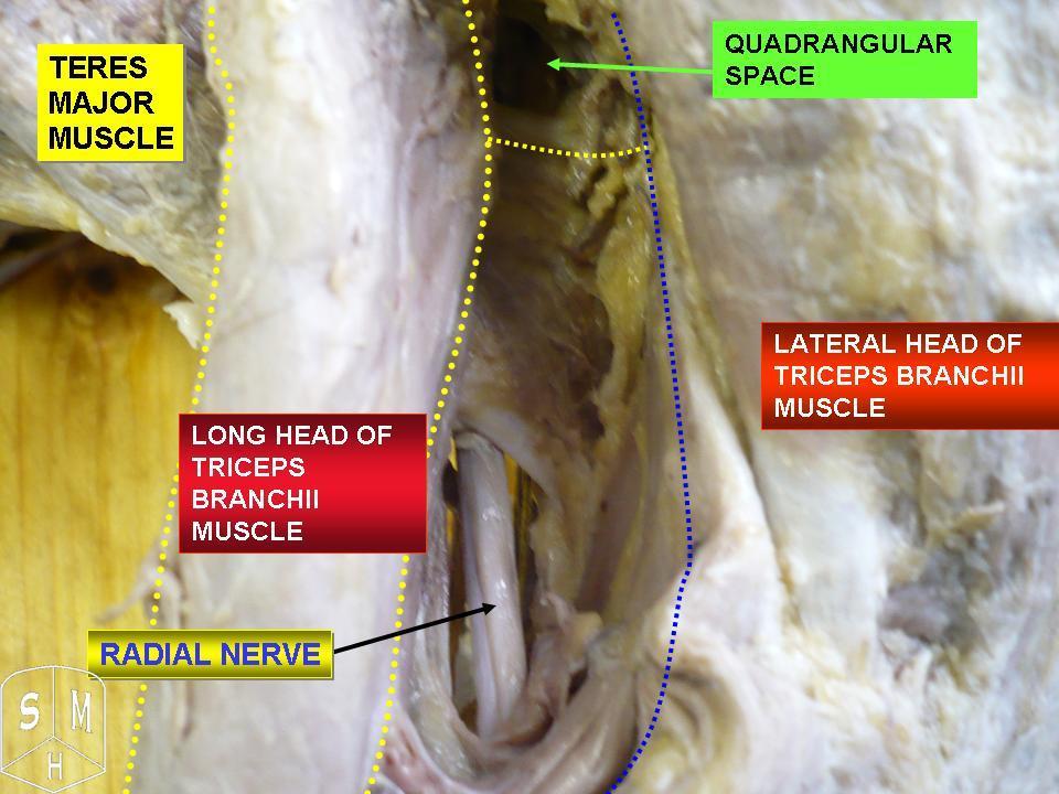
The radial nerve is a nerve in the human body that supplies the posterior portion of the upper limb. It innervates the medial and lateral heads of the triceps brachii muscle of the arm, as well as all 12 muscles in the Posterior compartment of the forearm, posterior osteofascial compartment of the forearm and the associated joints and overlying skin. It originates from the brachial plexus, carrying fibers from the posterior roots of spinal nerves C5, C6, C7, C8 and T1. The radial nerve and its branches provide Motor neuron, motor innervation to the dorsal arm muscles (the triceps brachii and the anconeus) and the extrinsic extensors of the wrists and hands; it also provides cutaneous Nerve supply to the skin, sensory innervation to most of the back of the hand, except for the back of the little finger and adjacent half of the ring finger (which are innervated by the ulnar nerve). The radial nerve divides into a deep branch, which becomes the posterior interosseous nerve, and a su ... [...More Info...] [...Related Items...] OR: [Wikipedia] [Google] [Baidu] |
Posterior Interosseous Nerve
The posterior interosseous nerve (or dorsal interosseous nerve/deep radial nerve) is a nerve in the forearm. It is the continuation of the deep branch of the radial nerve, after this has crossed the supinator muscle. It is considerably diminished in size compared to the deep branch of the radial nerve. The nerve fibers originate from cervical segments C7 and C8 in the spinal column. Structure Course It descends along the interosseous membrane, anterior to the extensor pollicis longus muscle, to the back of the carpus, where it presents a gangliform enlargement from which filaments are distributed to the ligaments and articulations of the carpus. Supply The posterior interosseous nerve supplies all the muscles of the posterior compartment of the forearm, except anconeus muscle, brachioradialis muscle, and extensor carpi radialis longus muscle. In other words, it supplies the following muscles: * Extensor carpi radialis brevis muscle — deep branch of radial nerve * Ext ... [...More Info...] [...Related Items...] OR: [Wikipedia] [Google] [Baidu] |
Deep Branch Of The Radial Nerve
The radial nerve divides into a superficial (sensory) and deep (motor) branch at the cubital fossa. The deep branch of the radial nerve winds to the back of the forearm around the lateral side of the radius between the two planes of fibers of the supinator In human anatomy, the supinator is a broad muscle in the posterior compartment of the forearm, curved around the upper third of the radius (bone), radius. Its function is to supination, supinate the forearm. Structure The supinator consists of tw ..., and is prolonged downward between the superficial and deep layers of muscles, to the middle of the forearm. The deep branch provides motor function to the muscles in the posterior compartment of the forearm, which is mostly the extensor muscles of the hand. The radial nerve arises from the posterior cord of the brachial plexus. The posterior cord takes nerves from the upper, lower, and middle trunk, so ultimately the radial nerve is formed from the anterior rami of C5 through T1. ... [...More Info...] [...Related Items...] OR: [Wikipedia] [Google] [Baidu] |
Radial Collateral Ligament (elbow)
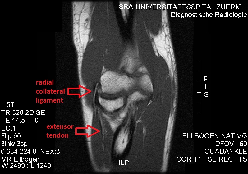
The radial collateral ligament (RCL), lateral collateral ligament (LCL), or external lateral ligamentAs opposed to the "internal lateral ligament", better known as the medial or ulnar collateral ligament is a ligament in the elbow on the side of the radius. Structure The composition of the triangular ligamentous structure on the lateral side of the elbow varies widely between individuals, see alsFigure 4/ref> and can be considered either a single ligament, in which case multiple distal attachments are generally mentioned and the annular ligament is described separately, or as several separate ligaments, in which case parts of those ligaments are often described as indistinguishable from each other. In the latter case, the ligaments are collectively referred to as the lateral collateral ligament complex (LCLC), consisting of four ligaments: * the radial collateral ligament roper(RCL), from the lateral epicondyle to the annular ligament deep to the common extensor tendon * the ... [...More Info...] [...Related Items...] OR: [Wikipedia] [Google] [Baidu] |
Deep Branch Of The Radial Nerve
The radial nerve divides into a superficial (sensory) and deep (motor) branch at the cubital fossa. The deep branch of the radial nerve winds to the back of the forearm around the lateral side of the radius between the two planes of fibers of the supinator In human anatomy, the supinator is a broad muscle in the posterior compartment of the forearm, curved around the upper third of the radius (bone), radius. Its function is to supination, supinate the forearm. Structure The supinator consists of tw ..., and is prolonged downward between the superficial and deep layers of muscles, to the middle of the forearm. The deep branch provides motor function to the muscles in the posterior compartment of the forearm, which is mostly the extensor muscles of the hand. The radial nerve arises from the posterior cord of the brachial plexus. The posterior cord takes nerves from the upper, lower, and middle trunk, so ultimately the radial nerve is formed from the anterior rami of C5 through T1. ... [...More Info...] [...Related Items...] OR: [Wikipedia] [Google] [Baidu] |
Lateral Epicondyle Of The Humerus
The lateral epicondyle of the humerus is a large, tuberculated eminence, curved a little forward, and giving attachment to the radial collateral ligament of the elbow joint, and to a tendon common to the origin of the supinator and some of the extensor muscles. Specifically, these extensor muscles include the anconeus muscle, the supinator, extensor carpi radialis brevis, extensor digitorum, extensor digiti minimi, and extensor carpi ulnaris. In birds, where the arm is somewhat rotated compared to other tetrapods, it is termed dorsal epicondyle of the humerus. In comparative anatomy, the term ''ectepicondyle'' is sometimes used. A common injury associated with the lateral epicondyle of the humerus is lateral epicondylitis also known as tennis elbow. Repetitive overuse of the forearm, as seen in tennis or other sports, can result in inflammation of "the tendons that join the forearm muscles on the outside of the elbow. The forearm muscles and tendons become damaged from overu ... [...More Info...] [...Related Items...] OR: [Wikipedia] [Google] [Baidu] |
Ulna
The ulna or ulnar bone (: ulnae or ulnas) is a long bone in the forearm stretching from the elbow to the wrist. It is on the same side of the forearm as the little finger, running parallel to the Radius (bone), radius, the forearm's other long bone. Longer and thinner than the radius, the ulna is considered to be the smaller long bone of the lower arm. The corresponding bone in the Human leg#Structure, lower leg is the fibula. Structure The ulna is a long bone found in the forearm that stretches from the elbow to the wrist, and when in standard anatomical position, is found on the Medial (anatomy), medial side of the forearm. It is broader close to the elbow, and narrows as it approaches the wrist. Close to the elbow, the ulna has a bony Process (anatomy), process, the olecranon process, a hook-like structure that fits into the olecranon fossa of the humerus. This prevents hyperextension and forms a hinge joint with the trochlea of the humerus. There is also a radial notch for ... [...More Info...] [...Related Items...] OR: [Wikipedia] [Google] [Baidu] |


