|
Superior Gluteal Artery
The superior gluteal artery is the terminal branch of the posterior division of the internal iliac artery. It exits the pelvis through the greater sciatic foramen before splitting into a superficial branch and a deep branch. Structure Origin The superior gluteal artery is the largest and final branch of the internal iliac artery. It branches from the posterior division of the internal iliac artery; it represents the continuation of the posterior division. Course, relations and branches It is a short artery. It passes posterior-ward between the lumbosacral trunk and the first sacral nerve (S1). Within the pelvis, it gives branches to the iliacus, piriformis, and obturator internus muscles. Just prior to exiting the pelvic cavity, it also gives off a nutrient artery which enters the ilium. It exits the pelvis through the greater sciatic foramen superior to the piriformis muscle, then promptly divides into a superficial branch and a deep branch. Superficial branch The superfi ... [...More Info...] [...Related Items...] OR: [Wikipedia] [Google] [Baidu] |
Sciatic Nerve
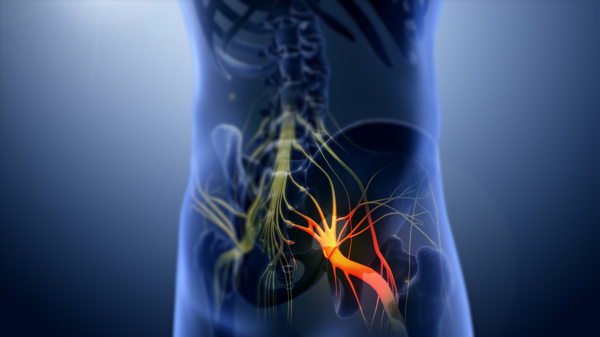
The sciatic nerve, also called the ischiadic nerve, is a large nerve in humans and other vertebrate animals. It is the largest branch of the sacral plexus and runs alongside the hip joint and down the right lower limb. It is the longest and widest single nerve in the human body, going from the top of the leg to the foot on the posterior aspect. The sciatic nerve has no cutaneous branches for the thigh. This nerve provides the connection to the nervous system for the skin of the lateral leg and the whole foot, the muscles of the back of the thigh, and those of the leg and foot. It is derived from Spinal nerve, spinal nerves Lumbar spinal nerve 4, L4 to Sacral spinal nerve 3, S3. It contains Axon, fibres from both the anterior and posterior divisions of the lumbosacral plexus. Structure In humans, the sciatic nerve is formed from the L4 to S3 segments of the sacral plexus, a collection of nerve fibres that emerge from the Sacrum, sacral part of the spinal cord. The lumbosacral trunk ... [...More Info...] [...Related Items...] OR: [Wikipedia] [Google] [Baidu] |
Inferior Gluteal Artery
The inferior gluteal artery (sciatic artery) is a terminal branch of the anterior trunk of the internal iliac artery. It exits the pelvis through the greater sciatic foramen. It is distributed chiefly to the buttock and the back of the thigh. Anatomy Origin It is the smaller of the two terminal branches of the anterior trunk of the internal iliac artery. Course It passes posterior-ward within parietal pelvic fascia. It travels in between the S1 nerve and S2 (or S2-S3) nerve(s). It descends upon the nerves of the sacral plexus and the piriformis muscle, posterior to the internal pudendal artery. It passes through the inferior part of the greater sciatic foramen. It exits the pelvis inferior to the piriformis muscle, between piriformis muscle and coccygeus muscle. It then descends in the interval between the greater trochanter of the femur and tuberosity of the ischium. It is accompanied by the sciatic nerve and the posterior femoral cutaneous nerves, and covered by the glu ... [...More Info...] [...Related Items...] OR: [Wikipedia] [Google] [Baidu] |
Medial Circumflex Femoral Artery
The medial circumflex femoral artery (internal circumflex artery, medial femoral circumflex artery) is an artery in the upper thigh that arises from the profunda femoris artery''.'' It supplies arterial blood to several muscles in the region, as well as the femoral head and neck. Damage to the artery following a femoral neck fracture may lead to avascular necrosis (ischemic) of the femoral neck/head. Structure Origin The medial femoral circumflex artery arises from the posteromedial aspect of the profunda femoris artery''.'' The medial femoral circumflex artery may occasionally arise directly from the femoral artery. Course and relations It winds around the medial side of the femur to pass along the posterior aspect of the femur. It first passes between the pectineus and the iliopsoas muscles, then between the obturator externus and the adductor brevis muscles. Branches At the upper border of the adductor brevis it gives off two branches: * The '' ascending branch'' * Th ... [...More Info...] [...Related Items...] OR: [Wikipedia] [Google] [Baidu] |
Hip-joint
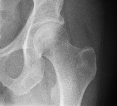
In vertebrate anatomy, the hip, or coxaLatin ''coxa'' was used by Celsus in the sense "hip", but by Pliny the Elder in the sense "hip bone" (Diab, p 77) (: ''coxae'') in medical terminology, refers to either an list of human anatomical regions, anatomical region or a joint on the outer (lateral) side of the pelvis. The hip region is located lateral (anatomy), lateral and anterior (anatomy), anterior to the Buttocks, gluteal region, inferior (anatomy), inferior to the iliac crest, and lateral to the obturator foramen, with muscle tendons and soft tissues overlying the greater trochanter of the femur. In adults, the three pelvic bones (ilium (bone), ilium, ischium and pubis (bone), pubis) have fused into one hip bone, which forms the superomedial/deep wall of the hip region. The hip joint, scientifically referred to as the acetabulofemoral joint (''art. coxae''), is the ball-and-socket joint between the pelvic acetabulum and the femoral head. Its primary function is to weight-bear ... [...More Info...] [...Related Items...] OR: [Wikipedia] [Google] [Baidu] |
Gluteal Muscles
The gluteal muscles, often called glutes, are a group of three muscles which make up the gluteal region commonly known as the buttocks: the gluteus maximus muscle, gluteus maximus, gluteus medius muscle, gluteus medius and gluteus minimus muscle, gluteus minimus. The three muscles originate from the ilium bone, ilium and sacrum and insert on the femur. The functions of the muscles include Extension (kinesiology), extension, Abduction (kinesiology), abduction, external rotation, and internal rotation of the hip joint. Structure The gluteus maximus is the largest and most wiktionary:superficial, superficial of the three gluteal muscles. It makes up a large part of the shape and appearance of the hips. It is a narrow and thick fleshy mass of a quadrilateral shape, and forms the prominence of the buttocks. The gluteus medius is a broad, thick, radiating muscle, situated on the outer surface of the pelvis. It lies profound to the gluteus maximus and its posterior third is covered by ... [...More Info...] [...Related Items...] OR: [Wikipedia] [Google] [Baidu] |
Lateral Femoral Circumflex Artery
The lateral circumflex femoral artery (also known as the lateral femoral circumflex artery or the external circumflex artery) is an artery in the upper thigh. It is usually a branch of the profunda femoris artery, and produces three branches. It is mostly distributed to the muscles of the lateral thigh, supplying arterial blood to muscles of the knee extensor group. Structure Origin The lateral femoral circumflex artery usually arises from the lateral side of the profunda femoris artery, but may occasionally arise directly from the femoral artery. It is the largest branch of the profunda femoris artery. Course and relations The lateral circumflex femoral artery usually courses anterior to the femoral neck. It passes horizontally between the divisions of the femoral nerve. It passes posterior to the sartorius muscle and rectus femoris muscle. It passes laterally across the hip joint capsule. It divides into ascending, transverse, and descending branches. Branches The lateral ... [...More Info...] [...Related Items...] OR: [Wikipedia] [Google] [Baidu] |
Deep Iliac Circumflex Artery
The deep circumflex iliac artery (or deep iliac circumflex artery) is an artery in the pelvis that travels along the iliac crest of the pelvic bone. Course The deep circumflex iliac artery arises from the lateral aspect of the external iliac artery nearly opposite the origin of the inferior epigastric artery. It ascends obliquely and laterally, posterior to the inguinal ligament, contained in a fibrous sheath formed by the junction of the transversalis fascia and iliac fascia. It travels to the anterior superior iliac spine, where it anastomoses with the ascending branch of the lateral femoral circumflex artery. It then pierces the transversalis fascia and passes medially along the inner lip of the crest of the ilium to a point where it perforates the transversus abdominis muscle. From there, it travels posteriorly between the transversus abdominis muscle and the internal oblique muscle to anastomose with the iliolumbar artery and the superior gluteal artery. Opposite the ant ... [...More Info...] [...Related Items...] OR: [Wikipedia] [Google] [Baidu] |
Anterior Superior Iliac Spine
The anterior superior iliac spine (ASIS) is a bony projection of the iliac bone, and an important landmark of surface anatomy. It refers to the anterior extremity of the iliac crest of the pelvis. It provides attachment for the inguinal ligament, and the sartorius muscle. The tensor fasciae latae muscle attaches to the lateral aspect of the superior anterior iliac spine, and also about 5 cm away at the iliac tubercle. Structure The anterior superior iliac spine refers to the anterior extremity of the iliac crest of the pelvis. This is a key surface landmark, and easily palpated. It provides attachment for the inguinal ligament, the sartorius muscle, and the tensor fasciae latae muscle. A variety of structures lie close to the anterior superior iliac spine, including the subcostal nerve, the femoral artery (which passes between it and the pubic symphysis), and the iliohypogastric nerve. Clinical significance The anterior superior iliac spine provides a clue i ... [...More Info...] [...Related Items...] OR: [Wikipedia] [Google] [Baidu] |
Tensor Fasciae Latae Muscle
The tensor fasciae latae (or tensor fasciæ latæ or, formerly, tensor vaginae femoris) is a muscle of the thigh. Together with the gluteus maximus, it acts on and is continuous with the iliotibial band, which attaches to the tibia. The muscle assists in keeping the balance of the pelvis while standing, walking, or running. Structure The tensor fasciae latae arises from the anterior part of the outer lip of the iliac crest; from the outer surface of the anterior superior iliac spine, and part of the outer border of the notch below it, between the gluteus medius and sartorius; and from the deep surface of the fascia lata. The tensor fasciae latae is inserted between the two layers of the iliotibial tract of the fascia lata about the junction of the middle and upper thirds of the thigh. It tautens the iliotibial tract and braces the knee, especially when the opposite foot is lifted.Saladin, Kenneth. Anatomy and Physiology. 6th ed. Mc-Graw Hill. 2010. The terminal insertion poin ... [...More Info...] [...Related Items...] OR: [Wikipedia] [Google] [Baidu] |
Gluteus Minimus
The gluteus minimus, or glutæus minimus, the smallest of the three gluteal muscles, is situated immediately beneath the gluteus medius. Structure It is fan-shaped, arising from the outer surface of the ilium, between the anterior and inferior gluteal lines, and behind, from the margin of the greater sciatic notch. The fibers converge to the deep surface of a radiated aponeurosis, and this ends in a tendon which is inserted into an impression on the anterior border of the greater trochanter, and gives an expansion to the capsule of the hip joint. Relations A bursa is interposed between the tendon and the greater trochanter. Between the gluteus medius and gluteus minimus are the deep branches of the superior gluteal vessels and the superior gluteal nerve. The deep surface of the gluteus minimus is in relation with the reflected tendon of the rectus femoris and the capsule of the hip joint. Variations The muscle may be divided into an anterior and a posterior pa ... [...More Info...] [...Related Items...] OR: [Wikipedia] [Google] [Baidu] |
Gluteus Medius
The gluteus medius, one of the three gluteal muscles, is a broad, thick, radiating muscle. It is situated on the outer surface of the pelvis. Its posterior third is covered by the gluteus maximus, its anterior two-thirds by the gluteal aponeurosis, which separates it from the superficial fascia and integument. Structure The gluteus medius muscle starts, or "originates", on the outer surface of the ilium between the iliac crest and the posterior gluteal line above, and the anterior gluteal line below; the gluteus medius also originates from its own fascia, the gluteal aponeurosis, that covers its outer surface. The fibers of the muscle converge into a strong flattened tendon that inserts on the lateral surface of the greater trochanter. More specifically, the muscle's tendon inserts into an oblique ridge that runs downward and forward on the lateral surface of the greater trochanter. Before the insertion the fibers cross from anterior to posterior and vice versa. Relations A ... [...More Info...] [...Related Items...] OR: [Wikipedia] [Google] [Baidu] |

