|
Suboccipital Triangle
The suboccipital triangle is a region of the neck bounded by the following three muscles of the suboccipital group of muscles: * Rectus capitis posterior major - above and medially * Obliquus capitis superior - above and laterally * Obliquus capitis inferior - below and laterally (Rectus capitis posterior minor is also in this region but does not form part of the triangle) It is covered by a layer of dense fibro-fatty tissue, situated beneath the semispinalis capitis. The floor is formed by the posterior atlantooccipital membrane, and the posterior arch of the atlas. In the deep groove on the upper surface of the posterior arch of the atlas are the vertebral artery and the first cervical or suboccipital nerve. In the past, the vertebral artery was accessed here in order to conduct angiography of the circle of Willis. Presently, formal angiography of the circle of Willis is performed via catheter angiography, with access usually being acquired at the common femoral artery. Alte ... [...More Info...] [...Related Items...] OR: [Wikipedia] [Google] [Baidu] |
Rectus Capitis Posterior Major Muscle
The rectus capitis posterior major (or rectus capitis posticus major) is a muscle in the upper back part of the neck. It is one of the suboccipital muscles. Its inferior attachment is at the spinous process of the axis (Second cervical vertebra); its superior attachment is onto the outer surface of the occipital bone on and around the side part of the inferior nuchal line. The muscle is innervated by the suboccipital nerve (the posterior ramus of cervical spinal nerve C1). The muscle acts to extend the head and rotate the head to its side. Anatomy The rectus capitis posterior major muscle is one of the suboccipital muscles. It forms the superomedial boundary of the suboccipital triangle. The muscle extends obliquely superiolaterally from its inferior attachment to its superior attachment. It becomes broader superiorly. Attachments Its inferior attachment is (via a pointed tendon) at (the external aspect of) the (bifid) spinous process of the axis (cervical vertebra C2) ... [...More Info...] [...Related Items...] OR: [Wikipedia] [Google] [Baidu] |
Suboccipital Nerve
The suboccipital nerve (first cervical dorsal ramus) is the dorsal primary ramus of the first cervical nerve (C1). It exits the spinal cord between the skull and the first cervical vertebra, the atlas. It lies within the suboccipital triangle along with the vertebral artery, where the artery enters the foramen magnum. It supplies muscles of the suboccipital triangle, the rectus capitis posterior major, obliquus capitis superior, and obliquus capitis inferior. The suboccipital nerve also innervates rectus capitis posterior minor. See also * Vertebral artery The vertebral arteries are major artery, arteries of the neck. Typically, the vertebral arteries originate from the subclavian arteries. Each vessel courses superiorly along each side of the neck, merging within the skull to form the single, m ... Additional images File:Gray792.png, Upper part of medulla spinalis and hind- and mid-brains; posterior aspect, exposed in situ. File:Suboccipital_triangle.PNG, Suboc ... [...More Info...] [...Related Items...] OR: [Wikipedia] [Google] [Baidu] |
Lesser Occipital Nerve
The lesser occipital nerve (or small occipital nerve) is a cutaneous spinal nerve of the cervical plexus. It arises from second cervical (spinal) nerve (C2) (along with the greater occipital nerve). It innervates the skin of the back of the upper neck and of the scalp posterior to the ear. Structure Origin It arises from the (lateral branch of the ventral ramus) of cervical spinal nerve C2; it (sources differ) receives or may also receive fibres from cervical spinal nerve C3. It originates between the atlas, and axis. The lesser occipital nerve is one of the four cutaneous branches of the cervical plexus. Course and relations It curves around the accessory nerve (CN XI) to come to course anterior to it. It then curves around and ascends along the posterior border of the sternocleidomastoid muscle; rarely, it may pierce the muscle. Near the cranium, it perforates the deep cervical fascia. It is continued upwards along the scalp posterior to the auricle. It divides in ... [...More Info...] [...Related Items...] OR: [Wikipedia] [Google] [Baidu] |
Greater Occipital Nerve
The greater occipital nerve is a nerve of the head. It is a spinal nerve, specifically the medial branch of the dorsal primary ramus of cervical spinal nerve 2. It arises from between the first and second cervical vertebrae, ascends, and then passes through the semispinalis muscle. It ascends further to supply the skin along the posterior part of the scalp to the vertex. It supplies sensation to the scalp at the top of the head, over the ear and over the parotid glands. Structure The greater occipital nerve is the medial branch of the dorsal primary ramus of cervical spinal nerve 2. It may also involve fibres from cervical spinal nerve 3. It arises from between the first and second cervical vertebrae, along with the lesser occipital nerve. It ascends after emerging from below the suboccipital triangle beneath the obliquus capitis inferior muscle. Just below the superior nuchal ridge, it pierces the fascia. It ascends further to supply the skin along the posterior part of ... [...More Info...] [...Related Items...] OR: [Wikipedia] [Google] [Baidu] |
Occipital Artery
The occipital artery is a branch of the external carotid artery that provides arterial supply to the back of the scalp, sternocleidomastoid muscles, and deep muscles of the back and neck. Structure Origin The occipital artery arises from (the posterior aspect of) the external carotid artery (some 2 cm distal to the origin of the external carotid artery). Course and relations At its origin, the hypoglossal nerve (CN XII) crosses artery superficially as the nerve passes posteroanteriorly. The artery passes superoposteriorly deep to the posterior belly of the digastricus muscle. It crosses the internal carotid artery and vein, the vagus nerve (CN X), accessory nerve (CN XI), and hypoglossal nerve (CN XII). It next ascends to the interval between the transverse process of the atlas and the mastoid process of the temporal bone, and passes horizontally backward, grooving the surface of the latter bone, being covered by the sternocleidomastoideus, splenius capitis, longi ... [...More Info...] [...Related Items...] OR: [Wikipedia] [Google] [Baidu] |
Suboccipital Muscles
The suboccipital muscles are a group of muscles defined by their location to the occiput. Suboccipital muscles are located below the occipital bone The occipital bone () is a neurocranium, cranial dermal bone and the main bone of the occiput (back and lower part of the skull). It is trapezoidal in shape and curved on itself like a shallow dish. The occipital bone lies over the occipital lob .... These are four paired muscles on the underside of the occipital bone; the two straight muscles (''rectus'') and the two oblique muscles (''obliquus''). The muscles are named *'' Rectus capitis posterior major'' goes from the spinous process of the axis (C2) to the occipital bone. *'' Rectus capitis posterior minor'' goes from the middle of the posterior arch of the atlas to the occiput. *'' Obliquus capitis superior'' goes from the transverse process of the atlas to the occiput. *'' Obliquus capitis inferior'' goes from the spine of the axis vertebra to the transverse process of the at ... [...More Info...] [...Related Items...] OR: [Wikipedia] [Google] [Baidu] |
Sternocleidomastoid
The sternocleidomastoid muscle is one of the largest and most superficial cervical muscles. The primary actions of the muscle are rotation of the head to the opposite side and flexion of the neck. The sternocleidomastoid is innervated by the accessory nerve. Etymology and location It is given the name ''sternocleidomastoid'' because it originates at the manubrium of the sternum (''sterno-'') and the clavicle (''cleido-'') and has an insertion at the mastoid process of the temporal bone of the skull. Structure The sternocleidomastoid muscle originates from two locations: the manubrium of the sternum and the clavicle, hence it is said to have two heads: sternal head and clavicular head. It travels obliquely across the side of the neck and inserts at the mastoid process of the temporal bone of the skull by a thin aponeurosis. The sternocleidomastoid is thick and narrow at its center, and broader and thinner at either end. The sternal head is a round fasciculus, tendinous in front ... [...More Info...] [...Related Items...] OR: [Wikipedia] [Google] [Baidu] |
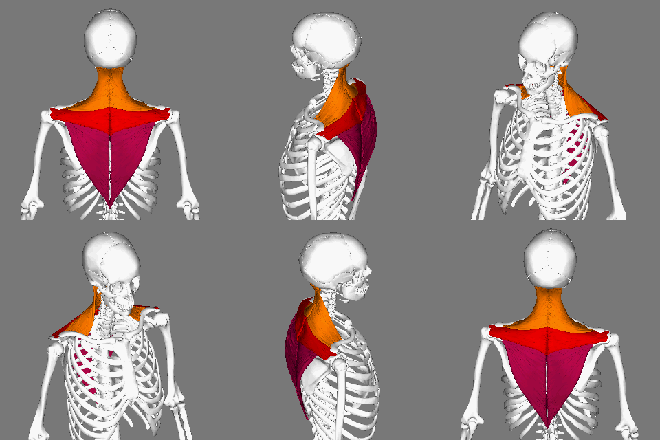
Trapezius
The trapezius is a large paired trapezoid-shaped surface muscle that extends longitudinally from the occipital bone to the lower thoracic vertebrae of the human spine, spine and laterally to the spine of the scapula. It moves the scapula and supports the arm. The trapezius has three functional parts: * an upper (descending) part which supports the weight of the arm; * a middle region (transverse), which retracts the scapula; and * a lower (ascending) part which medially rotates and depresses the scapula. Name and history The trapezius muscle resembles a trapezoid, trapezium, also known as a trapezoid, or diamond-shaped quadrilateral. The word "spinotrapezius" refers to the human trapezius, although it is not commonly used in modern texts. In other mammals, it refers to a portion of the analogous muscle. Structure The ''superior'' or ''upper'' (or descending) fibers of the trapezius originate from the spinous process of C7, the external occipital protuberance, the me ... [...More Info...] [...Related Items...] OR: [Wikipedia] [Google] [Baidu] |
Suboccipital Venous Plexus
The suboccipital venous plexus drains deoxygenated blood from the back of the head. It communicates with the external vertebral venous plexuses. The external vertebral venous plexuses travel inferiorly from this suboccipital region to drain into the brachiocephalic vein. The occipital vein joins in the formation of the plexus deep to the musculature of the back and from here drains into the external jugular vein. The plexus surrounds segments of the vertebral artery The vertebral arteries are major artery, arteries of the neck. Typically, the vertebral arteries originate from the subclavian arteries. Each vessel courses superiorly along each side of the neck, merging within the skull to form the single, m .... Veins of the head and neck {{circulatory-stub ... [...More Info...] [...Related Items...] OR: [Wikipedia] [Google] [Baidu] |
Circle Of Willis
The circle of Willis (also called Willis' circle, loop of Willis, cerebral arterial circle, and Willis polygon) is a circulatory anastomosis that supplies blood to the brain and surrounding structures in reptiles, birds and mammals, including humans. It is named after Thomas Willis (1621–1675), an English physician. Structure The circle of Willis is a part of the cerebral circulation and is composed of the following arteries: * Anterior cerebral artery (left and right) at their A1 segments * Anterior communicating artery * Internal carotid artery (left and right) at its distal tip (carotid terminus) * Posterior cerebral artery (left and right) at their P1 segments * Posterior communicating artery (left and right) The middle cerebral arteries, supplying the brain, are also considered part of the Circle of Willis Origin of arteries The left and right internal carotid arteries arise from the left and right common carotid arteries. The posterior communicating artery is given ... [...More Info...] [...Related Items...] OR: [Wikipedia] [Google] [Baidu] |
Angiography
Angiography or arteriography is a medical imaging technique used to visualize the inside, or lumen, of blood vessels and organs of the body, with particular interest in the arteries, veins, and the heart chambers. Modern angiography is performed by injecting a radio-opaque contrast agent into the blood vessel and imaging using X-ray based techniques such as fluoroscopy. With time-of-flight (TOF) magnetic resonance it is no longer necessary to use a contrast. The word itself comes from the Greek words ἀνγεῖον ''angeion'' 'vessel' and γράφειν ''graphein'' 'to write, record'. The film or image of the blood vessels is called an ''angiograph'', or more commonly an ''angiogram''. Though the word can describe both an arteriogram and a venogram, in everyday usage the terms angiogram and arteriogram are often used synonymously, whereas the term venogram is used more precisely. The term angiography has been applied to radionuclide angiography and newer vascular ima ... [...More Info...] [...Related Items...] OR: [Wikipedia] [Google] [Baidu] |
Vertebral Artery
The vertebral arteries are major artery, arteries of the neck. Typically, the vertebral arteries originate from the subclavian arteries. Each vessel courses superiorly along each side of the neck, merging within the skull to form the single, midline basilar artery. As the supplying component of the ''vertebrobasilar vascular system'', the vertebral arteries supply blood to the upper spinal cord, brainstem, cerebellum, and Cerebral circulation#Posterior cerebral circulation, posterior part of brain. Structure The vertebral arteries usually arise from the posterosuperior aspect of the central subclavian arteries on each side of the body, then enter deep to the transverse process at the level of the 6th cervical vertebrae (C6), or occasionally (in 7.5% of cases) at the level of C7. They then proceed superiorly, in the transverse foramen of each cervical vertebra. Once they have passed through the transverse foramen of C1 (also known as the Atlas (anatomy), atlas), the vertebral ... [...More Info...] [...Related Items...] OR: [Wikipedia] [Google] [Baidu] |


