|
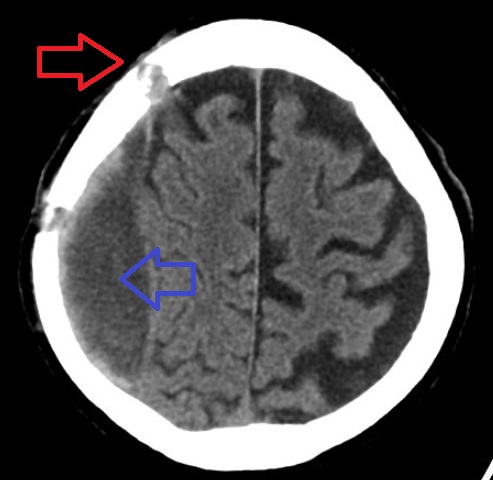
Subdural Hygroma
A subdural hygroma (SDG) is a collection of cerebrospinal fluid (CSF), without blood, located under the dura mater, dural membrane of the brain. Most Wiktionary:subdural, subdural hygromas are believed to be derived from chronic subdural hematomas. They are commonly seen in elderly people after minor trauma, but can also be seen in children following infection or trauma. One of the common causes of subdural hygroma is a sudden decrease in pressure as a result of placing a ventricular system, ventricular cerebral shunt, shunt. This can lead to leakage of CSF into the subdural space especially in cases with moderate to severe Cerebral atrophy, brain atrophy. In these cases, symptoms such as mild fever, headache, drowsiness and confusion can be seen, which can be relieved by draining this subdural fluid. Etiology and Pathophysiology Subdural hygromas require two conditions in order to occur. First, there must be a separation in the layers of the Meninges of the brain. Second, the r ... [...More Info...] [...Related Items...] OR: [Wikipedia] [Google] [Baidu] |
Cerebrospinal Fluid
Cerebrospinal fluid (CSF) is a clear, colorless Extracellular fluid#Transcellular fluid, transcellular body fluid found within the meninges, meningeal tissue that surrounds the vertebrate brain and spinal cord, and in the ventricular system, ventricles of the brain. CSF is mostly produced by specialized Ependyma, ependymal cells in the choroid plexuses of the ventricles of the brain, and absorbed in the arachnoid granulations. It is also produced by ependymal cells in the lining of the ventricles. In humans, there is about 125 mL of CSF at any one time, and about 500 mL is generated every day. CSF acts as a shock absorber, cushion or buffer, providing basic mechanical and immune system, immunological protection to the brain inside the Human skull, skull. CSF also serves a vital function in the cerebral autoregulation of cerebral blood flow. CSF occupies the subarachnoid space (between the arachnoid mater and the pia mater) and the ventricular system around and inside t ... [...More Info...] [...Related Items...] OR: [Wikipedia] [Google] [Baidu] |
Subdural Hematoma
A subdural hematoma (SDH) is a type of bleeding in which a collection of blood—usually but not always associated with a traumatic brain injury—gathers between the inner layer of the dura mater and the arachnoid mater of the meninges surrounding the brain. It usually results from rips in bridging veins that cross the subdural space. Subdural hematomas may cause an increase in the pressure inside the skull, which in turn can cause compression of and damage to delicate brain tissue. Acute subdural hematomas are often life-threatening. Chronic subdural hematomas have a better prognosis if properly managed. In contrast, epidural hematomas are usually caused by rips in arteries, resulting in a build-up of blood between the dura mater and the skull. The third type of brain hemorrhage, known as a subarachnoid hemorrhage (SAH), causes bleeding into the subarachnoid space between the arachnoid mater and the pia mater. SAH are often seen in trauma settings, or after rupture of in ... [...More Info...] [...Related Items...] OR: [Wikipedia] [Google] [Baidu] |
Gadolinium
Gadolinium is a chemical element; it has Symbol (chemistry), symbol Gd and atomic number 64. It is a silvery-white metal when oxidation is removed. Gadolinium is a malleable and ductile rare-earth element. It reacts with atmospheric oxygen or moisture slowly to form a black coating. Gadolinium below its Curie point of is ferromagnetism, ferromagnetic, with an attraction to a magnetic field higher than that of nickel. Above this temperature it is the most paramagnetism, paramagnetic element. It is found in nature only in an oxidized form. When separated, it usually has impurities of the other rare earths because of their similar chemical properties. Gadolinium was discovered in 1880 by Jean Charles Galissard de Marignac, Jean Charles de Marignac, who detected its oxide by using spectroscopy. It is named after the mineral gadolinite, one of the minerals in which gadolinium is found, itself named for the Finnish chemist Johan Gadolin. Pure gadolinium was first isolated by the chemis ... [...More Info...] [...Related Items...] OR: [Wikipedia] [Google] [Baidu] |
Magnetic Resonance Imaging
Magnetic resonance imaging (MRI) is a medical imaging technique used in radiology to generate pictures of the anatomy and the physiological processes inside the body. MRI scanners use strong magnetic fields, magnetic field gradients, and radio waves to form images of the organs in the body. MRI does not involve X-rays or the use of ionizing radiation, which distinguishes it from computed tomography (CT) and positron emission tomography (PET) scans. MRI is a medical application of nuclear magnetic resonance (NMR) which can also be used for imaging in other NMR applications, such as NMR spectroscopy. MRI is widely used in hospitals and clinics for medical diagnosis, staging and follow-up of disease. Compared to CT, MRI provides better contrast in images of soft tissues, e.g. in the brain or abdomen. However, it may be perceived as less comfortable by patients, due to the usually longer and louder measurements with the subject in a long, confining tube, although ... [...More Info...] [...Related Items...] OR: [Wikipedia] [Google] [Baidu] |
X-ray Computed Tomography
An X-ray (also known in many languages as Röntgen radiation) is a form of high-energy electromagnetic radiation with a wavelength shorter than those of ultraviolet rays and longer than those of gamma rays. Roughly, X-rays have a wavelength ranging from 10 nanometers to 10 picometers, corresponding to frequencies in the range of 30 petahertz to 30 exahertz ( to ) and photon energies in the range of 100 eV to 100 keV, respectively. X-rays were discovered in 1895 by the German scientist Wilhelm Conrad Röntgen, who named it ''X-radiation'' to signify an unknown type of radiation.Novelline, Robert (1997). ''Squire's Fundamentals of Radiology''. Harvard University Press. 5th edition. . X-rays can penetrate many solid substances such as construction materials and living tissue, so X-ray radiography is widely used in medical diagnostics (e.g., checking for broken bones) and materials science (e.g., identification of some chemical elements and ... [...More Info...] [...Related Items...] OR: [Wikipedia] [Google] [Baidu] |
Mass Effect (medicine)
In medicine, a mass effect is the effect of a growing mass that results in secondary pathological effects by pushing on or displacing surrounding tissue. In oncology, the mass typically refers to a tumor. For example, cancer of the thyroid gland may cause symptoms due to compressions of certain structures of the head and neck; pressure on the laryngeal nerves may cause voice changes, narrowing of the Vertebrate trachea, windpipe may cause stridor, pressure on the esophagus, gullet may cause dysphagia and so on. Surgery, Surgical removal or debulking is sometimes used to palliative care, palliate symptoms of the mass effect even if the underlying pathology is not curable. In neurology, a mass effect is the effect exerted by any mass, including, for example, hydrocephalus (cerebrospinal fluid buildup) or an evolving intracranial hemorrhage (bleeding within the skull) presenting with a clinically significant hematoma. The hematoma can exert a mass effect on the brain, increasing ... [...More Info...] [...Related Items...] OR: [Wikipedia] [Google] [Baidu] |
Iodinated Contrast
Iodinated contrast is a form of water-soluble, intravenous radiocontrast agent containing iodine, which enhances the visibility of vascular structures and organs during radiography, radiographic procedures. Some pathologies, such as cancer, have particularly improved visibility with iodinated contrast. The radiodensity of iodinated contrast is 25–30 Hounsfield units (HU) per milligram of iodine per milliliter at a tube voltage of 100–120 kVp. Types Iodine-based contrast media are usually classified as ionic or nonionic. Both types are used most commonly in radiology due to their relatively harmless interaction with the body and their solubility. Contrast media are primarily used to visualize vessels and changes in tissues on radiography and CT Scan, CT (computerized tomography). Contrast media can also be used for tests of the urinary tract, uterus and fallopian tubes. It may cause the patient to feel as if they have had urinary incontinence. It also puts a metallic taste in t ... [...More Info...] [...Related Items...] OR: [Wikipedia] [Google] [Baidu] |
Connective Tissue Disease
Connective tissue diseases (also termed connective tissue disorders, or collagen vascular diseases), are medical conditions that affect connective tissue. Connective tissues protect, support, and provide structure for the body's other tissues and structures. They hold the body's structures together. Connective tissues consist of two distinct proteins: elastin and collagen. Tendons, ligaments, skin, cartilage, bone, and blood vessels are all made of collagen. Skin and ligaments also contain elastin. These proteins and the surrounding tissues may suffer damage when the connective tissues become inflamed. The two main categories of connective tissue diseases are (1) a set of relatively rare genetic disorders affecting the primary structure of connective tissue, and (2) a variety of acquired diseases where the connective tissues are the site of multiple, more or less distinct immunological and inflammatory reactions. Diseases in which inflammation or weakness of collagen tends to ... [...More Info...] [...Related Items...] OR: [Wikipedia] [Google] [Baidu] |
Lymphoma
Lymphoma is a group of blood and lymph tumors that develop from lymphocytes (a type of white blood cell). The name typically refers to just the cancerous versions rather than all such tumours. Signs and symptoms may include enlarged lymph nodes, fever, drenching sweats, unintended weight loss, itching, and constantly feeling tired. The enlarged lymph nodes are usually painless. The sweats are most common at night. Many subtypes of lymphomas are known. The two main categories of lymphomas are the non-Hodgkin lymphoma (NHL) (90% of cases) and Hodgkin lymphoma (HL) (10%). Lymphomas, leukemias and myelomas are a part of the broader group of tumors of the hematopoietic and lymphoid tissues. Risk factors for Hodgkin lymphoma include infection with Epstein–Barr virus and a history of the disease in the family. Risk factors for common types of non-Hodgkin lymphomas include autoimmune diseases, HIV/AIDS, infection with human T-lymphotropic virus, immunosuppressant medicat ... [...More Info...] [...Related Items...] OR: [Wikipedia] [Google] [Baidu] |
Neurosurgery
Neurosurgery or neurological surgery, known in common parlance as brain surgery, is the specialty (medicine), medical specialty that focuses on the surgical treatment or rehabilitation of disorders which affect any portion of the nervous system including the Human brain, brain, spinal cord, peripheral nervous system, and cerebrovascular system. Neurosurgery as a medical specialty also includes non-surgical management of some neurological conditions. Education and context In different countries, there are different requirements for an individual to legally practice neurosurgery, and there are varying methods through which they must be educated. In most countries, neurosurgeon training requires a minimum period of seven years after graduating from medical school. United Kingdom In the United Kingdom, students must gain entry into medical school. The MBBS qualification (Bachelor of Medicine, Bachelor of Surgery) takes four to six years depending on the student's route. The newly qu ... [...More Info...] [...Related Items...] OR: [Wikipedia] [Google] [Baidu] |
Parenchyma
upright=1.6, Lung parenchyma showing damage due to large subpleural bullae. Parenchyma () is the bulk of functional substance in an animal organ such as the brain or lungs, or a structure such as a tumour. In zoology, it is the tissue that fills the interior of flatworms. In botany, it is some layers in the cross-section of the leaf. Etymology The term ''parenchyma'' is Neo-Latin from the Ancient Greek word meaning 'visceral flesh', and from meaning 'to pour in' from 'beside' + 'in' + 'to pour'. Originally, Erasistratus and other anatomists used it for certain human tissues. Later, it was also applied to plant tissues by Nehemiah Grew. Structure The parenchyma is the ''functional'' parts of an organ, or of a structure such as a tumour in the body. This is in contrast to the stroma, which refers to the ''structural'' tissue of organs or of structures, namely, the connective tissues. Brain The brain parenchyma refers to the functional tissue in th ... [...More Info...] [...Related Items...] OR: [Wikipedia] [Google] [Baidu] |


