|
Striosome
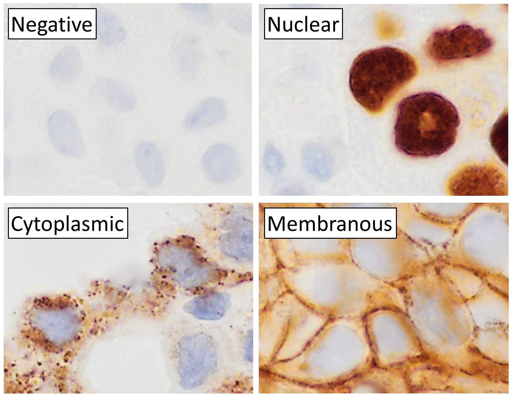
The striosomes (also referred to as striatal patches) are one of two complementary chemical compartments within the striatum (the other compartment is known as the matrix) that can be visualized by staining for immunocytochemical markers such as mu opioid receptors, acetylcholinesterase, enkephalin, substance P, limbic system-associated membrane protein (LAMP), AMPA receptor subunit 1 (GluR1), dopamine receptor subunits, and calcium binding proteins. Striosomal abnormalities have been associated with neurological disorders, such as mood dysfunction in Huntington's disease, though their precise function remains unknown. Recently studies have identified the presence of "exo-patch" neurons that are biochemically and genetically the same as striosomal neurons, but reside in the matrix compartment. This study also characterized the different input and output connections of the striosome and matrix compartments, revealing that both regions have direct inputs to dopamine neurons ( ... [...More Info...] [...Related Items...] OR: [Wikipedia] [Google] [Baidu] |
Striatum
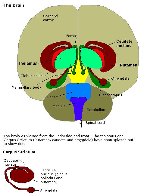
The striatum (: striata) or corpus striatum is a cluster of interconnected nuclei that make up the largest structure of the subcortical basal ganglia. The striatum is a critical component of the motor and reward systems; receives glutamatergic and dopaminergic inputs from different sources; and serves as the primary input to the rest of the basal ganglia. Functionally, the striatum coordinates multiple aspects of cognition, including both motor and action planning, decision-making, motivation, reinforcement, and reward perception. The striatum is made up of the caudate nucleus and the lentiform nucleus. However, some authors believe it is made up of caudate nucleus, putamen, and ventral striatum. The lentiform nucleus is made up of the larger putamen, and the smaller globus pallidus. Strictly speaking the globus pallidus is part of the striatum. It is common practice, however, to implicitly exclude the globus pallidus when referring to striatal structures. In pr ... [...More Info...] [...Related Items...] OR: [Wikipedia] [Google] [Baidu] |
Ann Graybiel
Ann Martin Graybiel (born 1942) is an Institute Professor and a faculty member in the Department of Brain and Cognitive Sciences at the Massachusetts Institute of Technology. She is also an investigator at the McGovern Institute for Brain Research. She is an expert on the basal ganglia and the neurophysiology of habit formation, implicit learning, and her work is relevant to Parkinson's disease, Huntington's disease, obsessive–compulsive disorder, substance abuse and other disorders that affect the basal ganglia. Research For much of her career, Graybiel has focused on the physiology of the striatum, a basal ganglia structure implicated in the control of movement, cognition, habit formation, and decision-making. In the late 1970s, Graybiel discovered that while striatal neurons appeared to be an amorphous mass, they were in fact organized into chemical compartments, which she termed striosomes. Later research revealed links between striosomal abnormalities and neurological ... [...More Info...] [...Related Items...] OR: [Wikipedia] [Google] [Baidu] |
Mood (psychology)
In psychology, a mood is an affective state. In contrast to emotions or feelings, moods are less specific, less intense and less likely to be provoked or instantiated by a particular stimulus or event. Moods are typically described as having either a positive or negative valence. In other words, people usually talk about being in a good mood or a bad mood. There are many different factors that influence mood, and these can lead to positive or negative effects on mood. Mood also differs from temperament or personality traits which are even longer-lasting. Nevertheless, personality traits such as optimism and neuroticism predispose certain types of moods. Long-term disturbances of mood such as clinical depression and bipolar disorder are considered mood disorders. Mood is an internal, subjective state, but it often can be inferred from posture and other behaviors. "We can be sent into a mood by an unexpected event, from the happiness of seeing an old friend to the anger of d ... [...More Info...] [...Related Items...] OR: [Wikipedia] [Google] [Baidu] |
Histochemistry
Immunohistochemistry is a form of immunostaining. It involves the process of selectively identifying antigens in cells and tissue, by exploiting the principle of antibodies binding specifically to antigens in biological tissues. Albert Hewett Coons, Ernest Berliner, Norman Jones and Hugh J Creech was the first to develop immunofluorescence in 1941. This led to the later development of immunohistochemistry. Immunohistochemical staining is widely used in the diagnosis of abnormal cells such as those found in cancerous tumors. In some cancer cells certain tumor antigens are expressed which make it possible to detect. Immunohistochemistry is also widely used in basic research, to understand the distribution and localization of biomarkers and differentially expressed proteins in different parts of a biological tissue. Sample preparation Immunohistochemistry can be performed on tissue that has been fixed and embedded in paraffin, but also cryopreservated (frozen) tissue. Based on ... [...More Info...] [...Related Items...] OR: [Wikipedia] [Google] [Baidu] |
Autoradiography
An autoradiograph is an image on an X-ray film or nuclear emulsion produced by the pattern of decay emissions (e.g., beta particles or gamma rays) from a distribution of a radioactive substance. Alternatively, the autoradiograph is also available as a digital image (digital autoradiography), due to the recent development of scintillation gas detectors or rare-earth phosphorimaging systems. The film or emulsion is apposed to the labeled tissue section to obtain the autoradiograph (also called an autoradiogram). The '' auto-'' prefix indicates that the radioactive substance is within the sample, as distinguished from the case of historadiography or microradiography, in which the sample is marked using an external source. Some autoradiographs can be examined microscopically for localization of silver grains (such as on the interiors or exteriors of cells or organelles) in which the process is termed micro-autoradiography. For example, micro-autoradiography was used to examine whet ... [...More Info...] [...Related Items...] OR: [Wikipedia] [Google] [Baidu] |
Bed Nucleus Of The Stria Terminalis
The stria terminalis (or terminal stria) is a structure in the brain consisting of a band of fibers running along the lateral margin of the ventricular surface of the thalamus. Serving as a major output pathway of the amygdala, the stria terminalis runs from its centromedial division to the ventromedial nucleus of the hypothalamus. Anatomy The stria terminalis covers the superior thalamostriate vein, marking a line of separation between the thalamus and the caudate nucleus as seen upon gross dissection of the ventricles of the brain, viewed from the superior aspect. The stria terminalis extends from the region of the interventricular foramina to the temporal horn of the lateral ventricle, carrying fibers from the amygdala to the septal nuclei, hypothalamic, and thalamic areas of the brain. It also carries fibers projecting from these areas back to the amygdala. Bed nucleus of the stria terminalis The bed nucleus of the stria terminalis (BNST) is a collection of nuclei a ... [...More Info...] [...Related Items...] OR: [Wikipedia] [Google] [Baidu] |
Amygdala
The amygdala (; : amygdalae or amygdalas; also '; Latin from Greek language, Greek, , ', 'almond', 'tonsil') is a paired nucleus (neuroanatomy), nuclear complex present in the Cerebral hemisphere, cerebral hemispheres of vertebrates. It is considered part of the limbic system. In Primate, primates, it is located lateral and medial, medially within the temporal lobes. It consists of many nuclei, each made up of further subnuclei. The subdivision most commonly made is into the Basolateral amygdala, basolateral, Central nucleus of the amygdala, central, cortical, and medial nuclei together with the intercalated cells of the amygdala, intercalated cell clusters. The amygdala has a primary role in the processing of memory, decision making, decision-making, and emotions, emotional responses (including fear, anxiety, and aggression). The amygdala was first identified and named by Karl Friedrich Burdach in 1822. Structure Thirteen Nucleus (neuroanatomy), nuclei have been identif ... [...More Info...] [...Related Items...] OR: [Wikipedia] [Google] [Baidu] |
Huntington's Disease
Huntington's disease (HD), also known as Huntington's chorea, is an incurable neurodegenerative disease that is mostly Genetic disorder#Autosomal dominant, inherited. It typically presents as a triad of progressive psychiatric, cognitive, and motor symptoms. The earliest symptoms are often subtle problems with mood or mental/psychiatric abilities, which precede the motor symptoms for many people. The definitive physical symptoms, including a general Ataxia, lack of coordination and an unsteady human gait, gait, eventually follow. Over time, the basal ganglia region of the brain gradually Basal ganglia disease#Huntington's disease, becomes damaged. The disease is primarily characterized by a distinctive hyperkinesia, hyperkinetic movement disorder known as ''chorea.'' Chorea classically presents as uncoordinated, involuntary, "dance-like" body movements that become more apparent as the disease advances. Physical abilities gradually worsen until Motor coordination, coordinated mo ... [...More Info...] [...Related Items...] OR: [Wikipedia] [Google] [Baidu] |
Neurological Disorders
Neurological disorders represent a complex array of medical conditions that fundamentally disrupt the functioning of the nervous system. These Disorder of consciousness, disorders affect the brain, spinal cord, and nerve networks, presenting unique diagnosis, treatment, and patient care challenges. At their core, they represent disruptions to the intricate communication systems within the nervous system, stemming from genetic predispositions, environmental factors, infections, structural abnormalities, or degenerative processes. The impact of neurological disorders is profound and far-reaching. Conditions like epilepsy create recurring seizures through abnormal electrical brain activity, while multiple sclerosis damages the protective myelin covering of nerve fibers, interrupting communication between the brain and body. Parkinson's disease progressively affects movement through the loss of dopamine-producing nerve cells, and strokes can cause immediate and potentially permanent neu ... [...More Info...] [...Related Items...] OR: [Wikipedia] [Google] [Baidu] |
Calcium Binding Protein
Calcium-binding proteins are proteins that participate in calcium cell signaling pathways by binding to Ca2+, the calcium ion that plays an important role in many cellular processes. Calcium-binding proteins have specific domains that bind to calcium and are known to be heterogeneous. One of the functions of calcium binding proteins is to regulate the amount of free (unbound) Ca2+ in the cytosol of the cell. The cellular regulation of calcium is known as calcium homeostasis. Types Many different calcium-binding proteins exist, with different cellular and tissue distribution and involvement in specific functions. Calcium binding proteins also serve an important physiological role for cells. The most ubiquitous Ca2+-sensing protein, found in all eukaryotic organisms including yeasts, is calmodulin. Intracellular storage and release of Ca2+ from the sarcoplasmic reticulum is associated with the high-capacity, low-affinity calcium-binding protein calsequestrin. Calretinin is another t ... [...More Info...] [...Related Items...] OR: [Wikipedia] [Google] [Baidu] |
Dopamine Receptor
Dopamine receptors are a class of G protein-coupled receptors that are prominent in the vertebrate central nervous system (CNS). Dopamine receptors activate different effectors through not only G-protein coupling, but also signaling through different protein (dopamine receptor-interacting proteins) interactions. The neurotransmitter dopamine is the primary endogenous ligand for dopamine receptors. Dopamine receptors are implicated in many neurological processes, including motivational and incentive salience, cognition, memory, learning, and fine motor control, as well as modulation of neuroendocrine signaling. Abnormal dopamine receptor signaling and dopaminergic nerve function is implicated in several neuropsychiatric disorders. Thus, dopamine receptors are common neurologic drug targets; antipsychotics are often dopamine receptor antagonists while psychostimulants are typically indirect agonists of dopamine receptors. Subtypes The existence of multiple types of receptors for ... [...More Info...] [...Related Items...] OR: [Wikipedia] [Google] [Baidu] |


