|
Sinoatrial
The sinoatrial node (also known as the sinuatrial node, SA node, sinus node or Keith–Flack node) is an oval shaped region of special cardiac muscle in the upper back wall of the right atrium made up of cells known as pacemaker cells. The sinus node is approximately 15 mm long, 3 mm wide, and 1 mm thick, located directly below and to the side of the superior vena cava. These cells produce an electrical impulse known as a cardiac action potential that travels through the electrical conduction system of the heart, causing it to contract. In a healthy heart, the SA node continuously produces action potentials, setting the rhythm of the heart (sinus rhythm), and so is known as the heart's natural pacemaker. The rate of action potentials produced (and therefore the heart rate) is influenced by the nerves that supply it. Structure The sinoatrial node is an oval-shaped structure that is approximately 15 mm long, 3 mm wide, and 1 mm thick, located directly below ... [...More Info...] [...Related Items...] OR: [Wikipedia] [Google] [Baidu] |
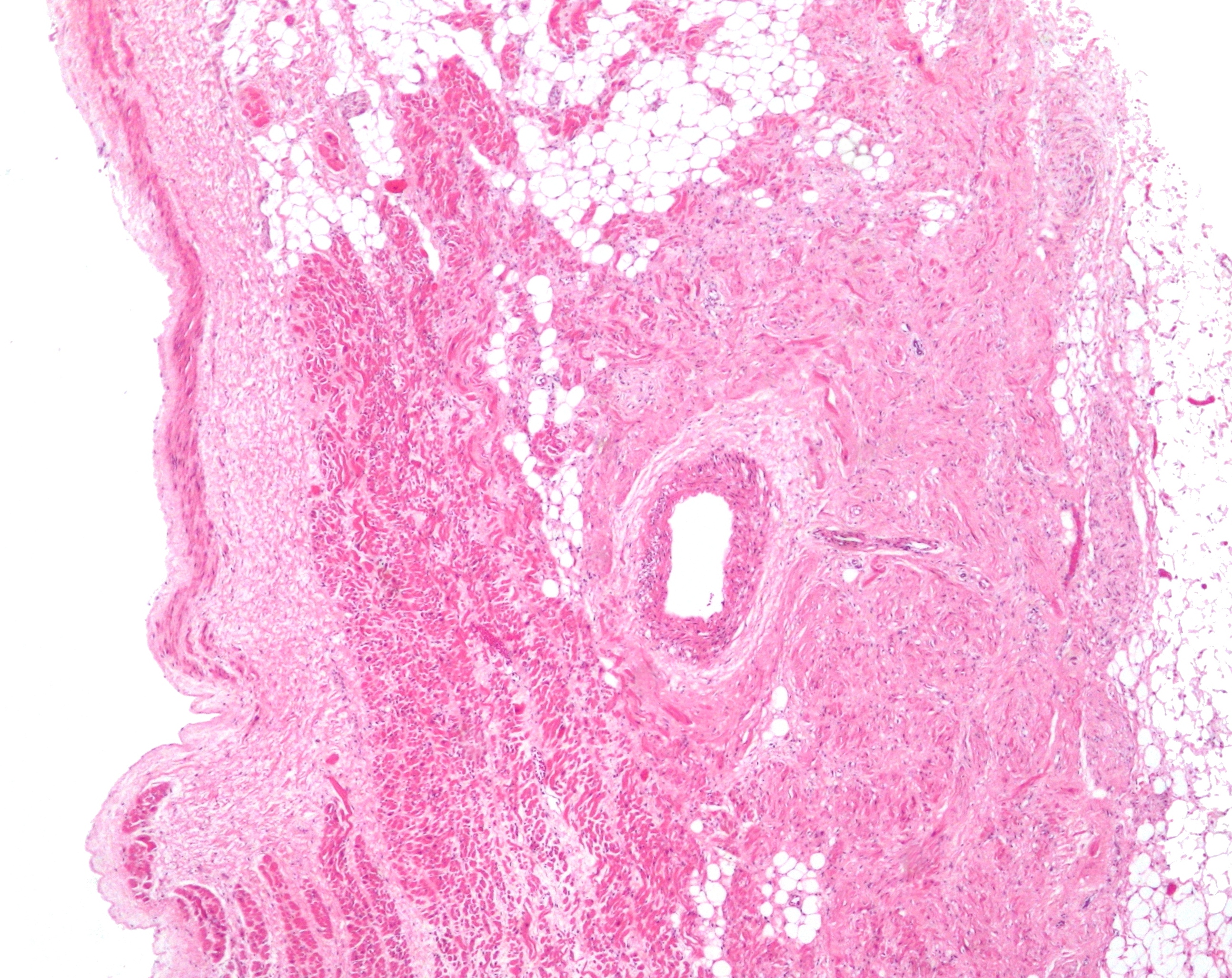
Sinoatrial Node 2 Low Mag
The sinoatrial node (also known as the sinuatrial node, SA node, sinus node or Keith–Flack node) is an oval shaped region of special cardiac muscle in the upper back wall of the right atrium made up of cells known as pacemaker cells. The sinus node is approximately 15 mm long, 3 mm wide, and 1 mm thick, located directly below and to the side of the superior vena cava. These cells produce an electrical impulse known as a cardiac action potential that travels through the electrical conduction system of the heart, causing it to contract. In a healthy heart, the SA node continuously produces action potentials, setting the rhythm of the heart ( sinus rhythm), and so is known as the heart's natural pacemaker. The rate of action potentials produced (and therefore the heart rate) is influenced by the nerves that supply it. Structure The sinoatrial node is an oval-shaped structure that is approximately 15 mm long, 3 mm wide, and 1 mm thick, located directly bel ... [...More Info...] [...Related Items...] OR: [Wikipedia] [Google] [Baidu] |
Heart
The heart is a muscular Organ (biology), organ found in humans and other animals. This organ pumps blood through the blood vessels. The heart and blood vessels together make the circulatory system. The pumped blood carries oxygen and nutrients to the tissue, while carrying metabolic waste such as carbon dioxide to the lungs. In humans, the heart is approximately the size of a closed fist and is located between the lungs, in the middle compartment of the thorax, chest, called the mediastinum. In humans, the heart is divided into four chambers: upper left and right Atrium (heart), atria and lower left and right Ventricle (heart), ventricles. Commonly, the right atrium and ventricle are referred together as the right heart and their left counterparts as the left heart. In a healthy heart, blood flows one way through the heart due to heart valves, which prevent cardiac regurgitation, backflow. The heart is enclosed in a protective sac, the pericardium, which also contains a sma ... [...More Info...] [...Related Items...] OR: [Wikipedia] [Google] [Baidu] |
Sinoatrial Nodal Artery
The sinoatrial nodal artery, sinoatrial nodal artery or sinoatrial artery is an artery of the heart which supplies the sinoatrial node, the natural pacemaker center of the heart. It is usually a branch of the right coronary artery. It passes between the right atrium, and the opening of the superior vena cava. Anatomy Origin It arises from the right coronary artery in around 60% of individuals, from the left circumflex coronary artery in about 40% of individuals, and in less than 1% of humans, the artery has an anomalous origin directly from the coronary sinus, descending aorta, or distal right coronary artery. The origin of the sinoatrial node artery is not related to coronary artery dominance, which means the side (right or left) that provides the circulation to the back of the heart. In contrast, the atrioventricular nodal branch, that is the artery that brings blood to the atrioventricular node, depends on coronary artery dominance. Course In more than 50% of human hea ... [...More Info...] [...Related Items...] OR: [Wikipedia] [Google] [Baidu] |
Cardiac Action Potential
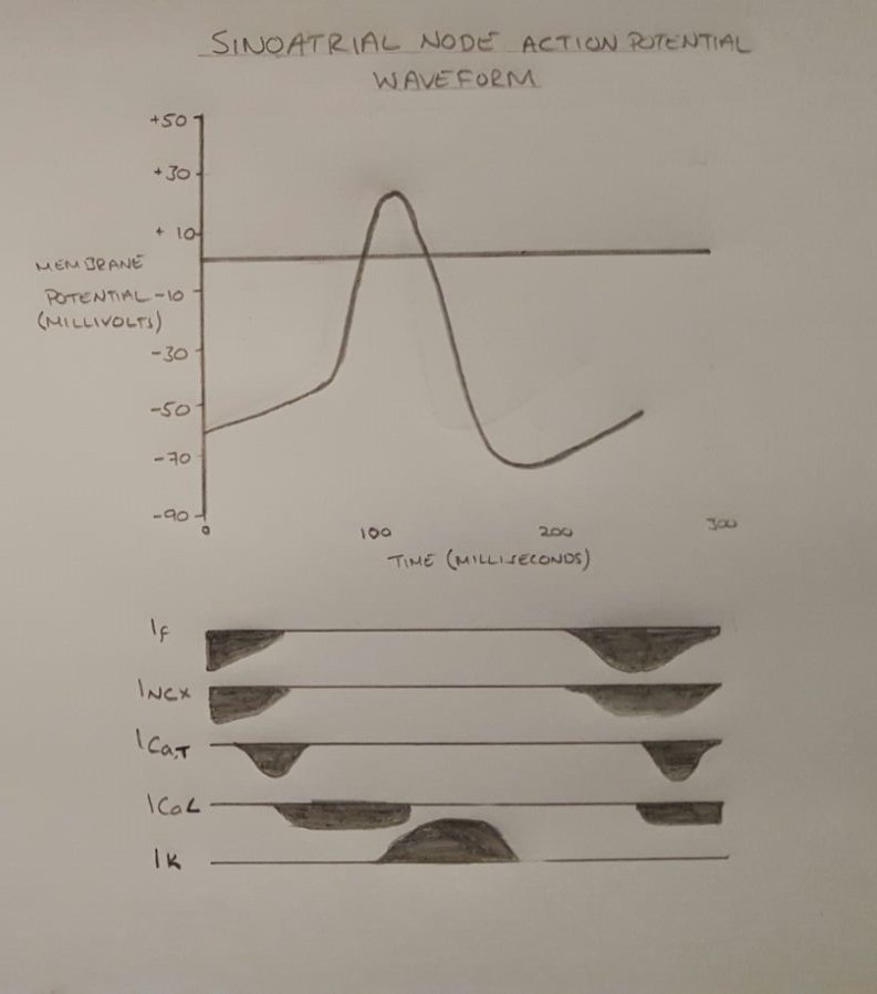
Unlike the action potential in skeletal muscle cells, the cardiac action potential is not initiated by nervous activity. Instead, it arises from a group of specialized cells known as pacemaker cells, that have automatic action potential generation capability. In healthy hearts, these cells form the cardiac pacemaker and are found in the sinoatrial node in the right atrium. They produce roughly 60–100 action potentials every minute. The action potential passes along the cell membrane causing the cell to contract, therefore the activity of the sinoatrial node results in a resting heart rate of roughly 60–100 beats per minute. All cardiac muscle cells are electrically linked to one another, by intercalated discs which allow the action potential to pass from one cell to the next. This means that all atrial cells can contract together, and then all ventricular cells. Rate dependence of the action potential is a fundamental property of cardiac cells and alterations can lead to se ... [...More Info...] [...Related Items...] OR: [Wikipedia] [Google] [Baidu] |
Heart Rate
Heart rate is the frequency of the cardiac cycle, heartbeat measured by the number of contractions of the heart per minute (''beats per minute'', or bpm). The heart rate varies according to the body's Human body, physical needs, including the need to absorb oxygen and excrete carbon dioxide. It is also modulated by numerous factors, including (but not limited to) genetics, physical fitness, Psychological stress, stress or psychological status, diet, drugs, hormonal status, environment, and disease/illness, as well as the interaction between these factors. It is usually equal or close to the pulse rate measured at any peripheral point. The American Heart Association states the normal resting adult human heart rate is 60–100 bpm. An ultra-trained athlete would have a resting heart rate of 37–38 bpm. ''Tachycardia'' is a high heart rate, defined as above 100 bpm at rest. ''Bradycardia'' is a low heart rate, defined as below 60 bpm at rest. When a human sleeps, a heartbeat with ra ... [...More Info...] [...Related Items...] OR: [Wikipedia] [Google] [Baidu] |
Cardiac Pacemaker
image:ConductionsystemoftheheartwithouttheHeart-en.svg, 350px, Image showing the cardiac pacemaker or SA node, the primary pacemaker within the electrical conduction system of the heart The cardiac pacemaker is the heart's natural rhythm generator. It employs pacemaker Cell (biology), cells that produce electrical impulses, known as Cardiac action potential, cardiac action potentials, which control the rate of contraction of the cardiac muscle, that is, the heart rate. In most humans, these cells are concentrated in the sinoatrial (SA) node, the primary pacemaker, which regulates the heart’s sinus rhythm. Sometimes a secondary pacemaker sets the pace, if the SA node is damaged or if the electrical conduction system of the heart has problems. Cardiac arrhythmias can cause heart block, in which the contractions lose their rhythm. In humans, and sometimes in other animals, a mechanical device called an artificial pacemaker (or simply "pacemaker") may be used after damage to the ... [...More Info...] [...Related Items...] OR: [Wikipedia] [Google] [Baidu] |
Pacemaker Cell
350px, Image showing the cardiac pacemaker or SA node, the primary pacemaker within the electrical conduction system of the heart The cardiac pacemaker is the heart's natural rhythm generator. It employs pacemaker Cell (biology), cells that produce electrical impulses, known as Cardiac action potential, cardiac action potentials, which control the rate of contraction of the cardiac muscle, that is, the heart rate. In most humans, these cells are concentrated in the sinoatrial (SA) node, the primary pacemaker, which regulates the heart’s sinus rhythm. Sometimes a secondary pacemaker sets the pace, if the SA node is damaged or if the electrical conduction system of the heart has problems. Cardiac arrhythmias can cause heart block, in which the contractions lose their rhythm. In humans, and sometimes in other animals, a mechanical device called an artificial pacemaker (or simply "pacemaker") may be used after damage to the body's intrinsic conduction system to produce these i ... [...More Info...] [...Related Items...] OR: [Wikipedia] [Google] [Baidu] |
Electrical Conduction System Of The Heart
The cardiac conduction system (CCS, also called the electrical conduction system of the heart) transmits the Cardiac action potential, signals generated by the sinoatrial node – the heart's Cardiac pacemaker, pacemaker, to cause the heart muscle to Muscle contraction, contract, and pump blood through the body's circulatory system. The Cardiac pacemaker, pacemaking signal travels through the right atrium to the atrioventricular node, along the bundle of His, and through the bundle branches to Purkinje fibers in the Ventricle (heart), walls of the ventricles. The Purkinje fibers transmit the signals more rapidly to stimulate contraction of the ventricles. The conduction system consists of specialized Cardiomyocyte, heart muscle cells, situated within the myocardium. There is a cardiac skeleton, skeleton of fibrous tissue that surrounds the conduction system which can be seen on an ECG. Dysfunction of the conduction system can cause Heart arrhythmia, irregular heart rhythms includ ... [...More Info...] [...Related Items...] OR: [Wikipedia] [Google] [Baidu] |
Action Potential
An action potential (also known as a nerve impulse or "spike" when in a neuron) is a series of quick changes in voltage across a cell membrane. An action potential occurs when the membrane potential of a specific Cell (biology), cell rapidly rises and falls. This depolarization then causes adjacent locations to similarly depolarize. Action potentials occur in several types of Membrane potential#Cell excitability, excitable cells, which include animal cells like neurons and myocyte, muscle cells, as well as some plant cells. Certain endocrine cells such as pancreatic beta cells, and certain cells of the anterior pituitary gland are also excitable cells. In neurons, action potentials play a central role in cell–cell interaction, cell–cell communication by providing for—or with regard to saltatory conduction, assisting—the propagation of signals along the neuron's axon toward axon terminal, synaptic boutons situated at the ends of an axon; these signals can then connect wit ... [...More Info...] [...Related Items...] OR: [Wikipedia] [Google] [Baidu] |
Cardiac Muscle
Cardiac muscle (also called heart muscle or myocardium) is one of three types of vertebrate muscle tissues, the others being skeletal muscle and smooth muscle. It is an involuntary, striated muscle that constitutes the main tissue of the wall of the heart. The cardiac muscle (myocardium) forms a thick middle layer between the outer layer of the heart wall (the pericardium) and the inner layer (the endocardium), with blood supplied via the coronary circulation. It is composed of individual cardiac muscle cells joined by intercalated discs, and encased by collagen fibers and other substances that form the extracellular matrix. Cardiac muscle contracts in a similar manner to skeletal muscle, although with some important differences. Electrical stimulation in the form of a cardiac action potential triggers the release of calcium from the cell's internal calcium store, the sarcoplasmic reticulum. The rise in calcium causes the cell's myofilaments to slide past each other i ... [...More Info...] [...Related Items...] OR: [Wikipedia] [Google] [Baidu] |
Crista Terminalis
The crista terminalis (also known as the terminal crest, or crista terminalis of His) is a vertical ridge on the posterolateral inner surface of the adult right atrium extending between the superior vena cava, and the inferior vena cava. The crista terminalis denotes where the junction of the embryologic sinus venosus and the right atrium occurred during embryonic development. It forms a boundary between the rough trabecular portion and the smooth, sinus venosus-derived portion (sinus venarum) of the internal surface of the right atrium. The sinoatrial node is located within the crista terminalis. Anatomy The crista terminalis generally takes the form of a smooth-surfaced, crescent-shaped thickened portion of heart muscle at the opening into the right atrial appendage. It consists of fibromuscular tissue. Features On the external aspect of the right atrium, corresponding to the crista terminalis, is a groove - the terminal sulcus. The crista terminalis provides the origin ... [...More Info...] [...Related Items...] OR: [Wikipedia] [Google] [Baidu] |


