|
Scleral Diseases
The sclera, also known as the white of the eye or, in older literature, as the tunica albuginea oculi, is the opaque, fibrous, protective outer layer of the eye containing mainly collagen and some crucial elastic fiber. In the development of the embryo, the sclera is derived from the neural crest. In children, it is thinner and shows some of the underlying pigment, appearing slightly blue. In the elderly, fatty deposits on the sclera can make it appear slightly yellow. People with dark skin can have naturally darkened sclerae, the result of melanin pigmentation. In humans, and some other vertebrates, the whole sclera is white or pale, contrasting with the coloured iris. The cooperative eye hypothesis suggests that the pale sclera evolved as a method of nonverbal communication that makes it easier for one individual to identify where another individual is looking. Other mammals with white or pale sclera include chimpanzees, many orangutans, some gorillas, and bonobos. Structure ... [...More Info...] [...Related Items...] OR: [Wikipedia] [Google] [Baidu] |
Cornea
The cornea is the transparency (optics), transparent front part of the eyeball which covers the Iris (anatomy), iris, pupil, and Anterior chamber of eyeball, anterior chamber. Along with the anterior chamber and Lens (anatomy), lens, the cornea Refraction, refracts light, accounting for approximately two-thirds of the eye's total optical power. In humans, the refractive power of the cornea is approximately 43 dioptres. The cornea can be reshaped by surgical procedures such as LASIK. While the cornea contributes most of the eye's focusing power, its Focus (optics), focus is fixed. Accommodation (eye), Accommodation (the refocusing of light to better view near objects) is accomplished by changing the geometry of the lens. Medical terms related to the cornea often start with the prefix "''wikt:kerat-, kerat-''" from the Ancient Greek, Greek word κέρας, ''horn''. Structure The cornea has myelinated, unmyelinated nerve endings sensitive to touch, temperature and chemicals; a to ... [...More Info...] [...Related Items...] OR: [Wikipedia] [Google] [Baidu] |
Orangutan
Orangutans are great apes native to the rainforests of Indonesia and Malaysia. They are now found only in parts of Borneo and Sumatra, but during the Pleistocene they ranged throughout Southeast Asia and South China. Classified in the genus ''Pongo'', orangutans were originally considered to be one species. In 1996, they were divided into two species: the Bornean orangutan (''P. pygmaeus'', with three subspecies) and the Sumatran orangutan (''P. abelii''); a third species, the Tapanuli orangutan (''P. tapanuliensis''), was identified definitively in 2017. The orangutans are the only surviving members of the subfamily Ponginae, which diverged genetically from the other hominids (gorillas, chimpanzees, and humans) between 19.3 and 15.7 million years ago. The most arboreal of the great apes, orangutans spend most of their time in trees. They have proportionally long arms and short legs, and have reddish-brown hair covering their bodies. Adult males weigh about , while female ... [...More Info...] [...Related Items...] OR: [Wikipedia] [Google] [Baidu] |
Nerve Fascicle
A nerve fascicle is a bundle of nerve fibers belonging to a nerve in the peripheral nervous system. A nerve fascicle is also called a fasciculus, as is a nerve tract in the central nervous system. A nerve fascicle is enclosed by perineurium, a layer of fascial connective tissue. Each enclosed nerve fiber in the fascicle is enclosed by a connective tissue layer of endoneurium. Bundles of nerve fascicles are called fasciculi and are constituents of a nerve trunk. A main nerve trunk may contain a great many fascicles enclosing many thousands of axons. In the central nervous system a bundle of nerve fibers is called a nerve tract, and in neuroanatomy different tracts in the spinal cord are bundled into fasciculi such as the medial longitudinal fasciculus. In the spinal cord fasciculi are bundled into columns called funiculi such as the anterior funiculus. See also * Epineurium * Nervous tissue Nervous tissue, also called neural tissue, is the main tissue component of the ne ... [...More Info...] [...Related Items...] OR: [Wikipedia] [Google] [Baidu] |
Lamina Cribrosa Sclerae
The nerve fibers forming the optic nerve exit the eye posteriorly through a hole in the sclera that is occupied by a mesh-like structure called the lamina cribrosa. It is formed by a multilayered network of collagen fibers that extend from the scleral canal wall. The nerve fibers that comprise the optic nerve run through pores formed by these collagen beams. In humans, a central retinal artery is located slightly off-center in the nasal direction. The lamina cribrosa is thought to help support the retinal ganglion cell axons as they traverse the scleral canal. Being structurally weaker than the much thicker and denser sclera, the lamina cribrosa is more sensitive to changes in the intraocular pressure and tends to react to increased pressure through posterior displacement. This is thought to be one of the causes of nerve damage in glaucoma Glaucoma is a group of eye diseases that can lead to damage of the optic nerve. The optic nerve transmits visual information from the eye ... [...More Info...] [...Related Items...] OR: [Wikipedia] [Google] [Baidu] |
Choroid
The choroid, also known as the choroidea or choroid coat, is a part of the uvea, the vascular layer of the eye. It contains connective tissues, and lies between the retina and the sclera. The human choroid is thickest at the far extreme rear of the eye (at 0.2 mm), while in the outlying areas it narrows to 0.1 mm. The choroid provides oxygen and nourishment to the outer layers of the retina. Along with the ciliary body and iris, the choroid forms the uveal tract. The structure of the choroid is generally divided into four layers (classified in order of furthest away from the retina to closest): *Haller's layer – outermost layer of the choroid consisting of larger diameter blood vessels; * Sattler's layer – layer of medium diameter blood vessels; * Choriocapillaris – layer of capillaries; and * Bruch's membrane (synonyms: Lamina basalis, Complexus basalis, Lamina vitra) – innermost layer of the choroid. Blood supply There are two circulations of the eye: ... [...More Info...] [...Related Items...] OR: [Wikipedia] [Google] [Baidu] |
Optic Disc
The optic disc or optic nerve head is the point of exit for ganglion cell axons leaving the eye. Because there are no rods or cones overlying the optic disc, it corresponds to a small blind spot in each eye. The ganglion cell axons form the optic nerve after they leave the eye. The optic disc represents the beginning of the optic nerve and is the point where the axons of retinal ganglion cells come together. The optic disc in a normal human eye carries 1–1.2 million afferent nerve fibers from the eye toward the brain. The optic disc is also the entry point for the major arteries that supply the retina with blood, and the exit point for the veins from the retina. Structure The optic disc is located 3 to 4 mm to the nasal side of the fovea. It is a vertical oval, with average dimensions of 1.76mm horizontally by 1.92mm vertically. There is a central depression, of variable size, called the optic cup. This depression can be a variety of shapes from a shallow indent ... [...More Info...] [...Related Items...] OR: [Wikipedia] [Google] [Baidu] |
Optic Nerve
In neuroanatomy, the optic nerve, also known as the second cranial nerve, cranial nerve II, or simply CN II, is a paired cranial nerve that transmits visual system, visual information from the retina to the brain. In humans, the optic nerve is derived from optic stalks during the seventh week of development and is composed of retinal ganglion cell axons and glial cells; it extends from the optic disc to the optic chiasma and continues as the optic tract to the lateral geniculate nucleus, Pretectal area, pretectal nuclei, and superior colliculus. Structure The optic nerve has been classified as the second of twelve paired cranial nerves, but it is technically a myelinated tract of the central nervous system, rather than a classical nerve of the peripheral nervous system because it is derived from an out-pouching of the diencephalon (optic stalks) during embryonic development. As a consequence, the fibers of the optic nerve are covered with myelin produced by oligodendrocytes, r ... [...More Info...] [...Related Items...] OR: [Wikipedia] [Google] [Baidu] |
Extraocular Muscle
The extraocular muscles, or extrinsic ocular muscles, are the seven extrinsic muscles of the eye in humans and other animals. Six of the extraocular muscles, the four recti muscles, and the superior and inferior oblique muscles, control movement of the eye. The other muscle, the levator palpebrae superioris, controls eyelid elevation. The actions of the six muscles responsible for eye movement depend on the position of the eye at the time of muscle contraction. The ciliary muscle, pupillary sphincter muscle and pupillary dilator muscle sometimes are called intrinsic ocular muscles or intraocular muscles. Structure Since only a small part of the eye called the fovea provides sharp vision, the eye must move to follow a target. Eye movements must be precise and fast. This is seen in scenarios like reading, where the reader must shift gaze constantly. Although under voluntary control, most eye movement is accomplished without conscious effort. Precisely how the integration betwe ... [...More Info...] [...Related Items...] OR: [Wikipedia] [Google] [Baidu] |
Globe (human Eye)
The human eye is a sensory organ in the visual system that reacts to visible light allowing eyesight. Other functions include maintaining the circadian rhythm, and keeping balance. The eye can be considered as a living optical device. It is approximately spherical in shape, with its outer layers, such as the outermost, white part of the eye (the sclera) and one of its inner layers (the pigmented choroid) keeping the eye essentially light tight except on the eye's optic axis. In order, along the optic axis, the optical components consist of a first lens (the cornea—the clear part of the eye) that accounts for most of the optical power of the eye and accomplishes most of the focusing of light from the outside world; then an aperture (the pupil) in a diaphragm (the iris—the coloured part of the eye) that controls the amount of light entering the interior of the eye; then another lens (the crystalline lens) that accomplishes the remaining focusing of light into imag ... [...More Info...] [...Related Items...] OR: [Wikipedia] [Google] [Baidu] |
Connective Tissue
Connective tissue is one of the four primary types of animal tissue, a group of cells that are similar in structure, along with epithelial tissue, muscle tissue, and nervous tissue. It develops mostly from the mesenchyme, derived from the mesoderm, the middle embryonic germ layer. Connective tissue is found in between other tissues everywhere in the body, including the nervous system. The three meninges, membranes that envelop the brain and spinal cord, are composed of connective tissue. Most types of connective tissue consists of three main components: elastic and collagen fibers, ground substance, and cells. Blood and lymph are classed as specialized fluid connective tissues that do not contain fiber. All are immersed in the body water. The cells of connective tissue include fibroblasts, adipocytes, macrophages, mast cells and leukocytes. The term "connective tissue" (in German, ) was introduced in 1830 by Johannes Peter Müller. The tissue was already recognized as ... [...More Info...] [...Related Items...] OR: [Wikipedia] [Google] [Baidu] |
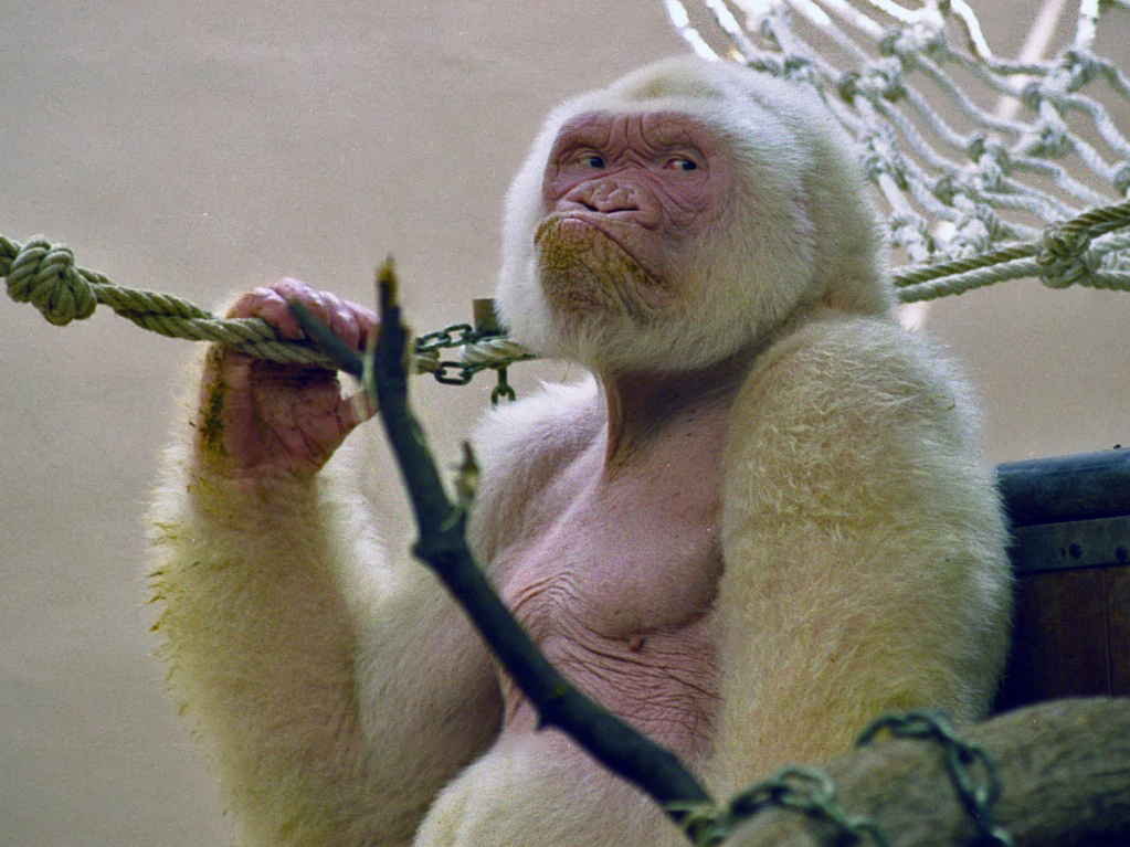
Western Lowland Gorilla
The western lowland gorilla (''Gorilla gorilla gorilla'') is one of two Critically Endangered subspecies of the western gorilla (''Gorilla gorilla'') that lives in Montane ecosystems#Montane forests, montane, Old-growth forest, primary and secondary forest, secondary forest and lowland swampland in central Africa in Angola (Cabinda Province), Cameroon, Central African Republic, Republic of the Congo, Democratic Republic of the Congo, Equatorial Guinea and Gabon. It is the nominate subspecies of the western gorilla, and the smallest of the four gorilla subspecies. The western lowland gorilla is the only subspecies kept in zoos with the exception of Amahoro (gorilla), Amahoro, a female eastern lowland gorilla at Antwerp Zoo, and a few mountain gorillas kept captive in the Democratic Republic of the Congo. Description The western lowland gorilla is the smallest subspecies of gorilla but still has exceptional size and strength. This species of gorillas exhibits pronounced sexual ... [...More Info...] [...Related Items...] OR: [Wikipedia] [Google] [Baidu] |


