|
Scar Free Healing
Scar free healing is the process by which significant injuries can heal without permanent damage to the tissue the injury has affected. In most healing, scars form due to the fibrosis and wound contraction, however in scar free healing, tissue is completely regenerated. During the 1990s, published research on the subject increased; it is a relatively recent term in the literature. Scar free healing occurs in foetal life but the ability progressively diminishes into adulthood. In other animals such as amphibians, however, tissue regeneration occurs, for example as skin regeneration in the adult axolotl. Scarring versus scar free healing Scarring takes place in response to damaged or missing tissue following injury due to biological processes or wounding: it is a process that occurs in order to replace the lost tissue. The process of scarring is complex, it involves the inflammatory response and remodelling amongst other cell activities. Many growth factors and cytokines are als ... [...More Info...] [...Related Items...] OR: [Wikipedia] [Google] [Baidu] |
Wound Healing
Wound healing refers to a living organism's replacement of destroyed or damaged tissue by newly produced tissue. In undamaged skin, the epidermis (surface, epithelial layer) and dermis (deeper, connective layer) form a protective barrier against the external environment. When the barrier is broken, a regulated sequence of biochemical events is set into motion to repair the damage. This process is divided into predictable phases: blood clotting (hemostasis), inflammation, tissue growth ( cell proliferation), and tissue remodeling (maturation and cell differentiation). Blood clotting may be considered to be part of the inflammation stage instead of a separate stage. The wound-healing process is not only complex but fragile, and it is susceptible to interruption or failure leading to the formation of non-healing chronic wounds. Factors that contribute to non-healing chronic wounds are diabetes, venous or arterial disease, infection, and metabolic deficiencies of old age.Enoch, ... [...More Info...] [...Related Items...] OR: [Wikipedia] [Google] [Baidu] |
Inflammatory Mediators
{{Disambig ...
Inflammatory may refer to: * Inflammation, a biological response to harmful stimuli * The word ''inflammatory'' is also used to refer literally to fire and flammability, and figuratively in relation to comments that are provocative and arouse passions and emotions. * An objection (United States law) In the law of the United States of America, an objection is a formal protest to evidence, argument, or questions that are in violation of the rules of evidence or other procedural law. Objections are often raised in court during a trial to dis ... [...More Info...] [...Related Items...] OR: [Wikipedia] [Google] [Baidu] |
Scarless Wound Healing
Wound healing refers to a living organism's replacement of destroyed or damaged tissue by newly produced tissue. In undamaged skin, the epidermis (surface, epithelial layer) and dermis (deeper, connective tissue, connective layer) form a protective barrier against the external environment. When the barrier is broken, a regulated sequence of biochemical events is set into motion to repair the damage. This process is divided into predictable phases: blood clotting (hemostasis), inflammation, tissue growth (cell proliferation), and tissue remodeling (maturation and cell differentiation). Blood clotting may be considered to be part of the inflammation stage instead of a separate stage. The wound-healing process is not only complex but fragile, and it is susceptible to interruption or failure leading to the formation of non-healing chronic wounds. Factors that contribute to non-healing chronic wounds are diabetes, Venous disease, venous or arterial disease, infection, and metabolic d ... [...More Info...] [...Related Items...] OR: [Wikipedia] [Google] [Baidu] |
Embryo
An embryo ( ) is the initial stage of development for a multicellular organism. In organisms that reproduce sexually, embryonic development is the part of the life cycle that begins just after fertilization of the female egg cell by the male sperm cell. The resulting fusion of these two cells produces a single-celled zygote that undergoes many cell divisions that produce cells known as blastomeres. The blastomeres (4-cell stage) are arranged as a solid ball that when reaching a certain size, called a morula, (16-cell stage) takes in fluid to create a cavity called a blastocoel. The structure is then termed a blastula, or a blastocyst in mammals. The mammalian blastocyst hatches before implantating into the endometrial lining of the womb. Once implanted the embryo will continue its development through the next stages of gastrulation, neurulation, and organogenesis. Gastrulation is the formation of the three germ layers that will form all of the different parts of t ... [...More Info...] [...Related Items...] OR: [Wikipedia] [Google] [Baidu] |
Thymic Epithelial Cell
Thymic epithelial cells (TECs) are specialized cells with high degree of anatomic, phenotypic and functional heterogeneity that are located in the outer layer (epithelium) of the thymic stroma. The thymus, as a primary lymphoid organ, mediates T cell development and maturation. The thymic microenvironment is established by TEC network filled with thymocytes (blood cell precursors of T cells) in different developing stages. TECs and thymocytes are the most important components in the thymus, that are necessary for production of functionally competent T lymphocytes and self tolerance. Dysfunction of TECs causes several immunodeficiencies and autoimmune diseases. They are also called epithelial reticular cells, or epithelioreticular cells (ERC). Groups The final anatomical location of the thymic gland is reached at 6 weeks in the fetus. TECs originate from non-hematopoietic cells that are characterized by negative expression of CD45 and positive expression of EpCAM. Then TECs are ... [...More Info...] [...Related Items...] OR: [Wikipedia] [Google] [Baidu] |
Liver Regeneration
Liver regeneration is the process by which the liver is able to replace damaged or lost liver tissue. The liver is the only visceral organ with the capacity to regenerate. The liver can regenerate after partial hepatectomy or injury due to hepatotoxic agents such as certain medications, toxins, or chemicals. Only 10% of the original liver mass is required for the organ to regenerate back to full size. The phenomenon of liver regeneration is seen in all vertebrates, from humans to fish. The liver manages to restore any lost mass and adjust its size to that of the organism, while at the same time providing full support for body homeostasis during the entire regenerative process. The process of regeneration in mammals is mainly compensatory growth or hyperplasia because while the lost mass of the liver is replaced, it does not regain its original shape. During compensatory hyperplasia, the remaining liver tissue becomes larger so that the organ can continue to function. In lower speci ... [...More Info...] [...Related Items...] OR: [Wikipedia] [Google] [Baidu] |
Microarray
A microarray is a multiplex (assay), multiplex lab-on-a-chip. Its purpose is to simultaneously detect the expression of thousands of biological interactions. It is a two-dimensional array on a Substrate (materials science), solid substrate—usually a glass slide or silicon thin-film cell—that assays (tests) large amounts of biotic material, biological material using high-throughput screening miniaturized, multiplexed and parallel processing and detection methods. The concept and methodology of microarrays was first introduced and illustrated in antibody microarrays (also referred to as antibody matrix) by Tse Wen Chang in 1983 in a scientific publication and a series of patents. The "gene chip" industry started to grow significantly after the 1995 ''Science Magazine'' article by the Ron Davis and Pat Brown labs at Stanford University. With the establishment of companies, such as Affymetrix, Agilent, Applied Microarrays, Arrayjet, Illumina (company), Illumina, and others, the te ... [...More Info...] [...Related Items...] OR: [Wikipedia] [Google] [Baidu] |
Tumor Necrosis Factor Superfamily
The tumor necrosis factor (TNF) superfamily is a protein superfamily of type II transmembrane proteins containing TNF homology domain and forming trimers. Members of this superfamily can be released from the cell membrane by extracellular proteolytic cleavage and function as a cytokine. These proteins are expressed predominantly by immune cells and they regulate diverse cell functions, including immune response and inflammation, but also proliferation, differentiation, apoptosis and embryogenesis An embryo ( ) is the initial stage of development for a multicellular organism. In organisms that reproduce sexually, embryonic development is the part of the life cycle that begins just after fertilization of the female egg cell by the male .... The superfamily contains 19 members that bind to 29 members of TNF receptor superfamily. An occurrence of orthologs in invertebrates hints at ancient origin of this superfamily in evolution. The PROSITE pattern of this superfam ... [...More Info...] [...Related Items...] OR: [Wikipedia] [Google] [Baidu] |
Interleukin-1 Family
The Interleukin-1 family (IL-1 family) is a group of 11 cytokines that plays a central role in the regulation of immune and inflammatory responses to infections or sterile insults. Discovery Discovery of these cytokines began with studies on the pathogenesis of fever. The studies were performed by Eli Menkin and Paul Beeson in 1943–1948 on the fever-producing properties of proteins released from rabbit peritoneal exudate cells. These studies were followed by contributions of several investigators, who were primarily interested in the link between fever and infection/inflammation. The basis for the term "interleukin" was to streamline the growing number of biological properties attributed to soluble factors from macrophages and lymphocytes. IL-1 was the name given to the macrophage product, whereas IL-2 was used to define the lymphocyte product. At the time of the assignment of these names, there was no amino acid sequence analysis known and the terms were used to define bi ... [...More Info...] [...Related Items...] OR: [Wikipedia] [Google] [Baidu] |
Hyaluronic Acid
Hyaluronic acid (; abbreviated HA; conjugate base hyaluronate), also called hyaluronan, is an anionic, nonsulfated glycosaminoglycan distributed widely throughout connective, epithelial, and neural tissues. It is unique among glycosaminoglycans as it is non-sulfated, forms in the plasma membrane instead of the Golgi apparatus, and can be very large: human synovial HA averages about per molecule, or about 20,000 disaccharide monomers, while other sources mention . Medically, hyaluronic acid is used to treat osteoarthritis of the knee and dry eye, for wound repair, and as a cosmetic filler. The average 70 kg (150 lb) person has roughly 15 grams of hyaluronan in the body, one third of which is turned over (i.e., degraded and synthesized) per day. As one of the chief components of the extracellular matrix, it contributes significantly to cell proliferation and migration, and is involved in the progression of many malignant tumors. Hyaluronic acid is also a ... [...More Info...] [...Related Items...] OR: [Wikipedia] [Google] [Baidu] |
Tenascin
Tenascins are extracellular matrix glycoproteins. They are abundant in the extracellular matrix of developing vertebrate embryos and they reappear around healing wounds and in the stroma of some tumors. Types There are four members of the tenascin gene family: tenascin-C, tenascin-R, tenascin-X and tenascin-W. * Tenascin-C is the founding member of the gene family. In the embryo it is made by migrating cells like the neural crest; it is also abundant in developing tendons, bone and cartilage. * Tenascin-R is found in the developing and adult nervous system. * Tenascin-X is found primarily in loose connective tissue; mutations in the human tenascin-X gene can lead to a form of Ehlers-Danlos syndrome. * Tenascin-W is found in the kidney and in developing bone. The basic structure is 14 EGF-like repeats towards the N-terminal end, and 8 or more fibronectin-III domains which vary upon species and variant. Tenascin-C is the most intensely studied member of the family. It h ... [...More Info...] [...Related Items...] OR: [Wikipedia] [Google] [Baidu] |
Fibronectin
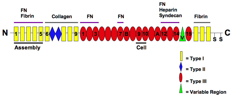
Fibronectin is a high- molecular weight (~500-~600 kDa) glycoprotein of the extracellular matrix that binds to membrane-spanning receptor proteins called integrins. Fibronectin also binds to other extracellular matrix proteins such as collagen, fibrin, and heparan sulfate proteoglycans (e.g. syndecans). Fibronectin exists as a protein dimer, consisting of two nearly identical monomers linked by a pair of disulfide bonds. The fibronectin protein is produced from a single gene, but alternative splicing of its pre-mRNA leads to the creation of several isoforms. Two types of fibronectin are present in vertebrates: * soluble plasma fibronectin (formerly called "cold-insoluble globulin", or CIg) is a major protein component of blood plasma (300 μg/ml) and is produced in the liver by hepatocytes. * insoluble cellular fibronectin is a major component of the extracellular matrix. It is secreted by various cells, primarily fibroblasts, as a soluble protein dimer and is ... [...More Info...] [...Related Items...] OR: [Wikipedia] [Google] [Baidu] |


