|
Roof Plate
The alar plate (or alar lamina) is a neural structure in the embryonic nervous system, part of the dorsal side of the neural tube, that involves the communication of general somatic and general visceral sensory impulses. The caudal part later becomes the sensory axon part of the spinal cord. The alar plate specifically later on becomes the dorsal gray of the spinal cord, and also develops into the sensory nuclei of cranial nerves V, VII, VIII, IX, and X. The inferior olivary nucleus The inferior olivary nucleus (ION) is a structure found in the medulla oblongata underneath the superior olivary nucleus.Gado, Thomas A. Woolsey; Joseph Hanaway; Mokhtar H. (2003). The brain atlas a visual guide to the human central nervous syste ..., mesencephalic nucleus of V, and main sensory nucleus of V are also developed from this plate. The cerebellum also develops from the alar plate, particularly from the rhombic lip. This is considered an exception to the general differentiation ... [...More Info...] [...Related Items...] OR: [Wikipedia] [Google] [Baidu] |
Neural Tube
In the developing chordate (including vertebrates), the neural tube is the embryonic precursor to the central nervous system, which is made up of the brain and spinal cord. The neural groove gradually deepens as the neural folds become elevated, and ultimately the folds meet and coalesce in the middle line and convert the groove into the closed neural tube. In humans, neural tube closure usually occurs by the fourth week of pregnancy (the 28th day after conception). Development The neural tube develops in two ways: primary neurulation and secondary neurulation. Primary neurulation divides the ectoderm into three cell types: * The internally located neural tube * The externally located epidermis * The neural crest cells, which develop in the region between the neural tube and epidermis but then migrate to new locations # Primary neurulation begins after the neural plate forms. The edges of the neural plate start to thicken and lift upward, forming the neural folds. The center ... [...More Info...] [...Related Items...] OR: [Wikipedia] [Google] [Baidu] |
Neural Structure
Neuroanatomy is the study of the structure and organization of the nervous system. In contrast to animals with radial symmetry, whose nervous system consists of a distributed network of cells, animals with bilateral symmetry have segregated, defined nervous systems. Their neuroanatomy is therefore better understood. In vertebrates, the nervous system is segregated into the internal structure of the brain and spinal cord (together called the central nervous system, or CNS) and the series of nerves that connect the CNS to the rest of the body (known as the peripheral nervous system, or PNS). Breaking down and identifying specific parts of the nervous system has been crucial for figuring out how it operates. For example, much of what neuroscientists have learned comes from observing how damage or "lesions" to specific brain areas affects behavior or other neural functions. For information about the composition of non-human animal nervous systems, see nervous system. For information ab ... [...More Info...] [...Related Items...] OR: [Wikipedia] [Google] [Baidu] |
Dorsal Gray
Dorsal (from Latin ''dorsum'' ‘back’) may refer to: * Dorsal (anatomy), an anatomical term of location referring to the back or upper side of an organism or parts of an organism * Dorsal, positioned on top of an aircraft's fuselage * Dorsal consonant, a consonant articulated with the back of the tongue * Dorsal fin A dorsal fin is a fin on the back of most marine and freshwater vertebrates. Dorsal fins have evolved independently several times through convergent evolution adapting to marine environments, so the fins are not all homologous. They are found ..., the fin located on the back of a fish or aircraft * Dorsal transcription factor, a maternally synthesized transcription factor {{disambig ... [...More Info...] [...Related Items...] OR: [Wikipedia] [Google] [Baidu] |
Caudal (anatomical Term)
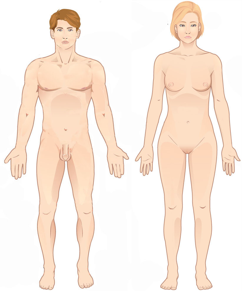
Standard anatomical terms of location are used to describe unambiguously the anatomy of humans and other animals. The terms, typically derived from Latin or Greek roots, describe something in its standard anatomical position. This position provides a definition of what is at the front ("anterior"), behind ("posterior") and so on. As part of defining and describing terms, the body is described through the use of anatomical planes and axes. The meaning of terms that are used can change depending on whether a vertebrate is a biped or a quadruped, due to the difference in the neuraxis, or if an invertebrate is a non-bilaterian. A non-bilaterian has no anterior or posterior surface for example but can still have a descriptor used such as proximal or distal in relation to a body part that is nearest to, or furthest from its middle. International organisations have determined vocabularies that are often used as standards for subdisciplines of anatomy. For example, '' Terminologia ... [...More Info...] [...Related Items...] OR: [Wikipedia] [Google] [Baidu] |
General Visceral Afferent Fibers
The general visceral afferent (GVA) fibers conduct sensory impulses (usually pain or reflex sensations) from the internal organs, glands, and blood vessels to the central nervous system. They are considered to be part of the visceral nervous system, which is closely related to the autonomic nervous system, but 'visceral nervous system' and 'autonomic nervous system' are not direct synonyms and care should be taken when using these terms. Unlike the efferent fibers of the autonomic nervous system, the afferent fibers are not classified as either sympathetic or parasympathetic. GVA fibers create referred pain by activating general somatic afferent fibers where the two meet in the posterior grey column. The cranial nerves that contain GVA fibers include the glossopharyngeal nerve (CN IX) and the vagus nerve (CN X). Generally, they are insensitive to cutting, crushing or burning; however, excessive tension in smooth muscle and some pathological conditions produce visceral pa ... [...More Info...] [...Related Items...] OR: [Wikipedia] [Google] [Baidu] |
General Somatic Afferent Fibers
The general somatic afferent fibers (GSA or somatic sensory fibers) are afferent fibers that arise from neurons in sensory ganglia and are found in all the spinal nerves, except occasionally the first cervical. General somatic afferents conduct impulses of Pain#Nociceptive, pain, touch and temperature from the surface of the body through the dorsal roots to the spinal cord, and impulses of muscle sense, tendon sense and joint sense from the deeper structures. See also * Afferent nerve * General visceral afferent fiber (GVA) * Special somatic afferent fiber (SSA) * Special visceral afferent fiber (SVA) References Spinal cord {{Portal bar, Anatomy ... [...More Info...] [...Related Items...] OR: [Wikipedia] [Google] [Baidu] |
Neural Tube
In the developing chordate (including vertebrates), the neural tube is the embryonic precursor to the central nervous system, which is made up of the brain and spinal cord. The neural groove gradually deepens as the neural folds become elevated, and ultimately the folds meet and coalesce in the middle line and convert the groove into the closed neural tube. In humans, neural tube closure usually occurs by the fourth week of pregnancy (the 28th day after conception). Development The neural tube develops in two ways: primary neurulation and secondary neurulation. Primary neurulation divides the ectoderm into three cell types: * The internally located neural tube * The externally located epidermis * The neural crest cells, which develop in the region between the neural tube and epidermis but then migrate to new locations # Primary neurulation begins after the neural plate forms. The edges of the neural plate start to thicken and lift upward, forming the neural folds. The center ... [...More Info...] [...Related Items...] OR: [Wikipedia] [Google] [Baidu] |
Dorsal (anatomy)
Standard anatomical terms of location are used to describe unambiguously the anatomy of humans and other animals. The terms, typically derived from Latin or Greek language, Greek roots, describe something in its standard anatomical position. This position provides a definition of what is at the front ("anterior"), behind ("posterior") and so on. As part of defining and describing terms, the body is described through the use of anatomical planes and anatomical axes, axes. The meaning of terms that are used can change depending on whether a vertebrate is a biped or a quadruped, due to the difference in the neuraxis, or if an invertebrate is a non-bilaterian. A non-bilaterian has no anterior or posterior surface for example but can still have a descriptor used such as proximal or distal in relation to a body part that is nearest to, or furthest from its middle. International organisations have determined vocabularies that are often used as standards for subdisciplines of anatomy. ... [...More Info...] [...Related Items...] OR: [Wikipedia] [Google] [Baidu] |
Embryonic Nervous System
The development of the nervous system, or neural development (neurodevelopment), refers to the processes that generate, shape, and reshape the nervous system of animals, from the earliest stages of embryonic development to adulthood. The field of neural development draws on both neuroscience and developmental biology to describe and provide insight into the cellular and molecular mechanisms by which complex nervous systems develop, from nematodes and fruit flies to mammals. Defects in neural development can lead to malformations such as holoprosencephaly, and a wide variety of neurological disorders including limb paresis and paralysis, balance and vision disorders, and seizures, and in humans other disorders such as Rett syndrome, Down syndrome and intellectual disability. Vertebrate brain development The vertebrate central nervous system (CNS) is derived from the ectoderm—the outermost germ layer of the embryo. A part of the dorsal ectoderm becomes specified to neur ... [...More Info...] [...Related Items...] OR: [Wikipedia] [Google] [Baidu] |
Mesencephalic Nucleus Of Trigeminal Nerve
The mesencephalic nucleus of trigeminal nerve is one of the sensory nuclei of the Trigeminal nerve, trigeminal nerve (cranial nerve V). It is located in the brainstem. It receives Proprioception, proprioceptive sensory information from the muscles of mastication and other muscles of the head and neck. It is involved in processing information about the position of the jaw/teeth. It is functionally responsible for preventing excessive biting that may damage the dentition, regulating tooth pain perception, and mediating the jaw jerk reflex (by means of projecting to the motor nucleus of the trigeminal nerve). The axons of the neuron cell bodies of this nucleus provide sensory innervation to target tissues directly, whereas other sensory nuclei of the trigeminal nerve receive their sensory inputs by synapsing with Primary sensory neuron, primary sensory neurons in the trigeminal ganglion. Anatomy The MNTN is located in the brainstem, more specifically (sources vary) spanning the leng ... [...More Info...] [...Related Items...] OR: [Wikipedia] [Google] [Baidu] |
Dorsal Gray
Dorsal (from Latin ''dorsum'' ‘back’) may refer to: * Dorsal (anatomy), an anatomical term of location referring to the back or upper side of an organism or parts of an organism * Dorsal, positioned on top of an aircraft's fuselage * Dorsal consonant, a consonant articulated with the back of the tongue * Dorsal fin A dorsal fin is a fin on the back of most marine and freshwater vertebrates. Dorsal fins have evolved independently several times through convergent evolution adapting to marine environments, so the fins are not all homologous. They are found ..., the fin located on the back of a fish or aircraft * Dorsal transcription factor, a maternally synthesized transcription factor {{disambig ... [...More Info...] [...Related Items...] OR: [Wikipedia] [Google] [Baidu] |
Inferior Olivary Nucleus
The inferior olivary nucleus (ION) is a structure found in the medulla oblongata underneath the superior olivary nucleus.Gado, Thomas A. Woolsey; Joseph Hanaway; Mokhtar H. (2003). The brain atlas a visual guide to the human central nervous system (2nd ed.). Hoboken, NJ: Wiley. p. 206. . In vertebrates, the ION is known to coordinate signals from the spinal cord to the cerebellum to regulate motor coordination and learning.Schweighofer N, Lang EJ, Kawato M. Role of the olivo-cerebellar complex in motor learning and control. ''Frontiers in Neural Circuits''. 2013;7:94. . These connections have been shown to be tightly associated, as degeneration of either the cerebellum or the ION results in degeneration of the other. Neurons of the ION are glutamatergic and receive inhibitory input via GABA receptors. There are two distinct GABAα receptor populations that are spatially organized within each neuron present in the ION. The GABAα receptor make-up varies based on where the rec ... [...More Info...] [...Related Items...] OR: [Wikipedia] [Google] [Baidu] |


