|
Rhomboid Minor
In human anatomy, the rhomboid minor is a small skeletal muscle of the back that connects the scapula to the vertebrae of the spinal column. It arises from the nuchal ligament, the 7th cervical and 1st thoracic vertebrae and intervening supraspinous ligaments; it inserts onto the medial border of the scapula, and is innervated by the dorsal scapular nerve. It acts together with the rhomboid major to keep the scapula pressed against the thoracic wall. Anatomy Origin The rhomboid minor arises from the inferior border of the nuchal ligament, from the spinous processes of the vertebrae C7–T1, and from the intervening supraspinous ligaments. Insertion It inserts onto a small area of the medial border of the scapula at the level of the scapular spine. Innervation It is innervated by the dorsal scapular nerve (a branch of the brachial plexus), with most of its fibers derived from the C5 nerve root and only minor contribution from C4 or C6., p. 4 Blood supply The rhom ... [...More Info...] [...Related Items...] OR: [Wikipedia] [Google] [Baidu] |
Nuchal Ligaments
The nuchal ligament is a ligament at the back of the neck that is continuous with the supraspinous ligament. Structure The nuchal ligament extends from the external occipital protuberance on the skull and median nuchal line to the spinous process of the seventh cervical vertebra in the lower part of the neck. From the anterior border of the nuchal ligament, a fibrous lamina is given off. This is attached to the posterior tubercle of the atlas, and to the spinous processes of the cervical vertebrae, and forms a septum between the muscles on either side of the neck. The trapezius and splenius capitis muscle attach to the nuchal ligament. Function It is a tendon-like structure that has developed independently in humans and other animals well adapted for running. In some four-legged animals, particularly ungulates and canids, the nuchal ligament serves to sustain the weight of the head. Clinical significance In Chiari malformation treatment, decompression and duraplasty with ... [...More Info...] [...Related Items...] OR: [Wikipedia] [Google] [Baidu] |
Nuchal Ligament
The nuchal ligament is a ligament at the back of the neck that is continuous with the supraspinous ligament. Structure The nuchal ligament extends from the external occipital protuberance on the skull and median nuchal line to the spinous process of the seventh cervical vertebra in the lower part of the neck. From the anterior border of the nuchal ligament, a fibrous lamina is given off. This is attached to the posterior tubercle of the atlas, and to the spinous processes of the cervical vertebrae, and forms a septum between the muscles on either side of the neck. The trapezius and splenius capitis muscle attach to the nuchal ligament. Function It is a tendon-like structure that has developed independently in humans and other animals well adapted for running. In some four-legged animals, particularly ungulates and canids, the nuchal ligament serves to sustain the weight of the head. Clinical significance In Chiari malformation treatment, decompression and duraplas ... [...More Info...] [...Related Items...] OR: [Wikipedia] [Google] [Baidu] |
Gray's Anatomy
''Gray's Anatomy'' is a reference book of human anatomy written by Henry Gray, illustrated by Henry Vandyke Carter and first published in London in 1858. It has had multiple revised editions, and the current edition, the 42nd (October 2020), remains a standard reference, often considered "the doctors' bible". Earlier editions were called ''Anatomy: Descriptive and Surgical'', ''Anatomy of the Human Body'' and ''Gray's Anatomy: Descriptive and Applied'', but the book's name is commonly shortened to, and later editions are titled, ''Gray's Anatomy''. The book is widely regarded as an extremely influential work on the subject. Publication history Origins The English anatomist Henry Gray was born in 1827. He studied the development of the endocrine glands and spleen and in 1853 was appointed Lecturer on Anatomy at St George's Hospital Medical School in London. In 1855, he approached his colleague Henry Vandyke Carter with his idea to produce an inexpensive and access ... [...More Info...] [...Related Items...] OR: [Wikipedia] [Google] [Baidu] |
Trapezius Muscle
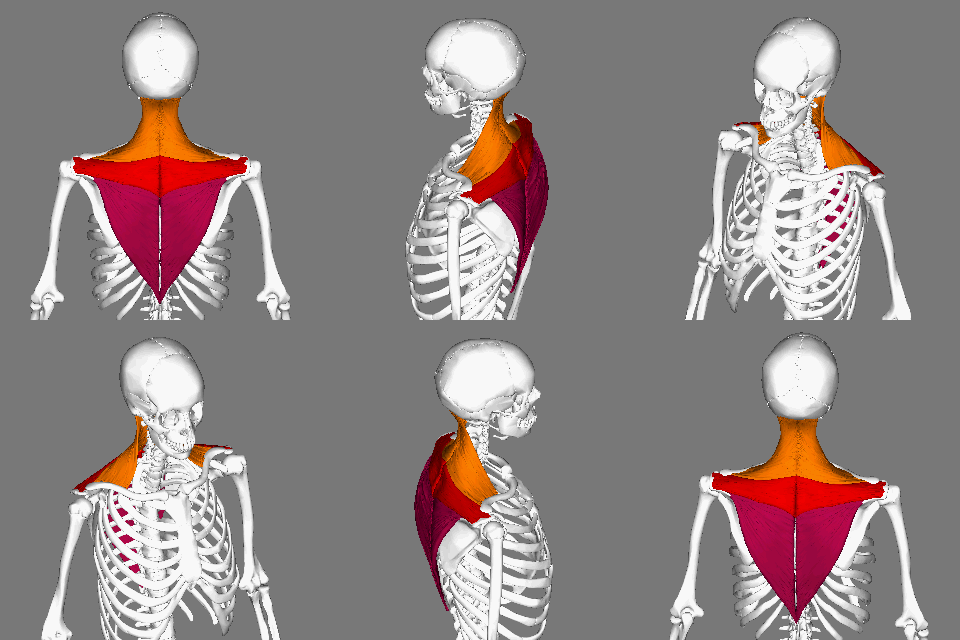
The trapezius is a large paired trapezoid-shaped surface muscle that extends longitudinally from the occipital bone to the lower thoracic vertebrae of the human spine, spine and laterally to the spine of the scapula. It moves the scapula and supports the arm. The trapezius has three functional parts: * an upper (descending) part which supports the weight of the arm; * a middle region (transverse), which retracts the scapula; and * a lower (ascending) part which medially rotates and depresses the scapula. Name and history The trapezius muscle resembles a trapezoid, trapezium, also known as a trapezoid, or diamond-shaped quadrilateral. The word "spinotrapezius" refers to the human trapezius, although it is not commonly used in modern texts. In other mammals, it refers to a portion of the analogous muscle. Structure The ''superior'' or ''upper'' (or descending) fibers of the trapezius originate from the spinous process of C7, the external occipital protuberance, the me ... [...More Info...] [...Related Items...] OR: [Wikipedia] [Google] [Baidu] |
Levator Scapulae Muscle
The levator scapulae is a slender skeletal muscle Skeletal muscle (commonly referred to as muscle) is one of the three types of vertebrate muscle tissue, the others being cardiac muscle and smooth muscle. They are part of the somatic nervous system, voluntary muscular system and typically are a ... situated at the back and side of the neck. It originates from the transverse processes of the four uppermost cervical vertebrae; it inserts onto the upper portion of the medial border of the scapula. It is innervated by the cervical nerves C3-C4, and frequently also by the dorsal scapular nerve. As the Latin name suggests, its main function is to lift the scapula. Anatomy Attachments The muscle descends diagonally from its origin to its insertion. Origin The levator scapulae originates from the posterior tubercles of the transverse processes of cervical vertebrae C1-4. At its origin, it attaches via tendinous slips. Insertion It inserts onto the medial border of th ... [...More Info...] [...Related Items...] OR: [Wikipedia] [Google] [Baidu] |
Dorsal Scapular Artery
The transverse cervical artery (transverse artery of neck or transversa colli artery) is an artery in the neck and a branch of the thyrocervical trunk, running at a higher level than the suprascapular artery. Structure It passes transversely below the inferior belly of the omohyoid muscle to the anterior margin of the trapezius, beneath which it divides into a superficial and a deep branch. It crosses in front of the phrenic nerve and the scalene muscles, and in front of or between the divisions of the brachial plexus, and is covered by the platysma and sternocleidomastoid muscles, and crossed by the omohyoid and trapezius. The transverse cervical artery originates from the thyrocervical trunk, it passes through the posterior triangle of the neck to the anterior border of the levator scapulae muscle, where it divides into deep and superficial branches. * Superficial branch ** Ascending branch ** Descending branch (also known as superficial cervical artery, which suppli ... [...More Info...] [...Related Items...] OR: [Wikipedia] [Google] [Baidu] |
Artery
An artery () is a blood vessel in humans and most other animals that takes oxygenated blood away from the heart in the systemic circulation to one or more parts of the body. Exceptions that carry deoxygenated blood are the pulmonary arteries in the pulmonary circulation that carry blood to the lungs for oxygenation, and the umbilical arteries in the fetal circulation that carry deoxygenated blood to the placenta. It consists of a multi-layered artery wall wrapped into a tube-shaped channel. Arteries contrast with veins, which carry deoxygenated blood back towards the heart; or in the pulmonary and fetal circulations carry oxygenated blood to the lungs and fetus respectively. Structure The anatomy of arteries can be separated into gross anatomy, at the macroscopic scale, macroscopic level, and histology, microanatomy, which must be studied with a microscope. The arterial system of the human body is divided into systemic circulation, systemic arteries, carrying blood from the ... [...More Info...] [...Related Items...] OR: [Wikipedia] [Google] [Baidu] |
Brachial Plexus
The brachial plexus is a network of nerves (nerve plexus) formed by the anterior rami of the lower four Spinal nerve#Cervical nerves, cervical nerves and first Spinal nerve#Thoracic nerves, thoracic nerve (cervical spinal nerve 5, C5, Cervical spinal nerve 6, C6, cervical spinal nerve 7, C7, cervical spinal nerve 8, C8, and thoracic spinal nerve 1, T1). This plexus extends from the spinal cord, through the cervicoaxillary canal in the neck, over the first rib, and into the axilla, armpit, it supplies Afferent nerve fiber, afferent and efferent nerve fibers to the chest, shoulder, arm, forearm, and hand. Structure The brachial plexus is divided into five ''roots'', three ''trunks'', six ''divisions'' (three anterior and three posterior), three ''cords'', and five ''branches''. There are five "terminal" branches and numerous other "pre-terminal" or "collateral" branches, such as the subscapular nerve, the thoracodorsal nerve, and the long thoracic nerve, that leave the plexus at vari ... [...More Info...] [...Related Items...] OR: [Wikipedia] [Google] [Baidu] |
Spine Of Scapula
The spine of the scapula or scapular spine is a prominent plate of bone, which crosses obliquely the medial four-fifths of the scapula at its upper part, and separates the supra- from the infraspinatous fossa. Structure It begins at the vertical (vertebral or medial) border by a smooth, triangular area over which the tendon of insertion of the lower part of the Trapezius glides. Gradually becoming more elevated, it ends in the acromion, which overhangs the shoulder-joint. The spine is triangular, and flattened from above downward, its apex being directed toward the vertebral border. Root The ''root of the spine'' of the scapula is the most medial part of the scapular spine. It is termed "triangular area of the spine of scapula", based on its triangular shape giving it distinguishable visible shape on x-ray images. The root of the spine is on a level with the tip of the spinous process of the third thoracic vertebra.Gray's Anatomy (1918)p.1306/ref> File:Root of spine - lef ... [...More Info...] [...Related Items...] OR: [Wikipedia] [Google] [Baidu] |
Spinous Process
Each vertebra (: vertebrae) is an irregular bone with a complex structure composed of bone and some hyaline cartilage, that make up the vertebral column or spine, of vertebrates. The proportions of the vertebrae differ according to their spinal segment and the particular species. The basic configuration of a vertebra varies; the vertebral body (also ''centrum'') is of bone and bears the load of the vertebral column. The upper and lower surfaces of the vertebra body give attachment to the intervertebral discs. The posterior part of a vertebra forms a vertebral arch, in eleven parts, consisting of two pedicles (pedicle of vertebral arch), two laminae, and seven processes. The laminae give attachment to the ligamenta flava (ligaments of the spine). There are vertebral notches formed from the shape of the pedicles, which form the intervertebral foramina when the vertebrae articulate. These foramina are the entry and exit conduits for the spinal nerves. The body of the vertebra ... [...More Info...] [...Related Items...] OR: [Wikipedia] [Google] [Baidu] |
Rhomboid Major Muscle
The rhomboid major is a skeletal muscle of the back that connects the scapula with the vertebrae of the spinal column. It originates from the spinous processes of the thoracic vertebrae T2–T5 and supraspinous ligament; it inserts onto the lower portion of the Medial border of scapula, medial border of the scapula. It acts together with the Rhomboid minor muscle, rhomboid minor to keep the scapula pressed against thoracic wall and to retract the scapula toward the vertebral column. As the word ''rhomboid'' suggests, the rhomboid major is diamond-shaped. The ''major'' in its name indicates that it is the larger of the two rhomboids. Structure Origin The rhomboid major arises from the spinous processes of the thoracic vertebrae T2–T5 as well as the supraspinous ligament. Insertion It inserts on the Medial border of scapula, medial border of the scapula, from about the level of the scapular spine to the scapula's inferior angle. Innervation The rhomboid major, like the ... [...More Info...] [...Related Items...] OR: [Wikipedia] [Google] [Baidu] |
Dorsal Scapular Nerve
The dorsal scapular nerve is a branch of the brachial plexus, usually derived from the ventral ramus of cervical nerve C5. It provides motor innervation to the rhomboid major muscle, rhomboid minor muscle, and levator scapulae muscle. Dorsal scapular nerve syndrome can cause a winged scapula, with Shoulder problem, pain and limited motion. Structure Origin The dorsal scapular nerve arises from the brachial plexus, usually from the plexus root (Ventral ramus, anterior (ventral) ramus) of cervical nerve C5. Course and relations Once the nerve leaves C5 it commonly pierces the Scalene muscles, middle scalene muscle. It continues deep to levator scapulae muscle and the rhomboids (minor superior to major). The nerve is accompanied by dorsal scapular artery. Function The dorsal scapular nerve provides motor innervation to the two rhomboid muscles, and the levator scapulae muscle. Clinical significance Injury to the dorsal scapular nerve is usually apparent on inspecti ... [...More Info...] [...Related Items...] OR: [Wikipedia] [Google] [Baidu] |


