|
Rete Canalis Hypoglossi
The venous plexus of hypoglossal canal is a small venous plexus surrounding the hypoglossal nerve (cranial nerve XII) as it passes through the hypoglossal canal. The plexus connects with the occipital sinus (intercranially), inferior petrosal sinus (intercranially), internal jugular vein (extracranially), condylar vein, and paravertebral venous plexus. Anatomy Anatomical studies have demonstrated that the venous plexus is the dominant structure of the hypoglossal canal. Research has shown that the size of the hypoglossal canal varies in relation to an individual's skull size, suggesting that the venous plexus functions as an essential emissary veinous structure. Variation Occasionally, it may be a single vein rather than a venous plexus. Clinical significance A case report In medicine, a case report is a detailed report of the symptoms, signs, diagnosis, treatment, and follow-up of an individual patient. Case reports may contain a demographic profile of the patient, but ... [...More Info...] [...Related Items...] OR: [Wikipedia] [Google] [Baidu] |
Terminologia Anatomica
''Terminologia Anatomica'' (commonly abbreviated TA) is the international standard for human anatomy, human anatomical terminology. It is developed by the Federative International Programme on Anatomical Terminology (FIPAT) a program of the International Federation of Associations of Anatomists (IFAA). History The sixth edition of the previous standard, ''Nomina Anatomica'', was released in 1989. The first edition of ''Terminologia Anatomica'', superseding Nomina Anatomica, was developed by the Federative Committee on Anatomical Terminology (FCAT) and the International Federation of Associations of Anatomists (IFAA) and released in 1998. In April 2011, this edition was published online by the Federative International Programme on Anatomical Terminologies (FIPAT), the successor of FCAT. The first edition contained 7635 Latin items. The second edition was released online by FIPAT in 2019 and approved and adopted by the IFAA General Assembly in 2020. The latest errata is dated Au ... [...More Info...] [...Related Items...] OR: [Wikipedia] [Google] [Baidu] |
Venous Plexus
In vertebrates, a venous plexus is a normal congregation anywhere in the body of multiple veins. A list of venous plexuses: * Basilar plexus * Batson venous plexus * Epidural venous plexus * External vertebral venous plexuses * Internal vertebral venous plexuses * Pampiniform venous plexus * Prostatic venous plexus * Pterygoid plexus * Rectal venous plexus * Soleal venous plexus * Submucosal venous plexus of the nose * Suboccipital venous plexus * Uterine venous plexus * Vaginal venous plexus * Venous plexus of hypoglossal canal * Vesical venous plexus The vesical venous plexus is a venous plexus situated at the fundus of the urinary bladder. It collects venous blood from the urinary bladder in both sexes, from the accessory sex glands in males, and from the corpora cavernosa of clitoris in fem ... References Veins {{Circulatory-stub ... [...More Info...] [...Related Items...] OR: [Wikipedia] [Google] [Baidu] |
Hypoglossal Nerve
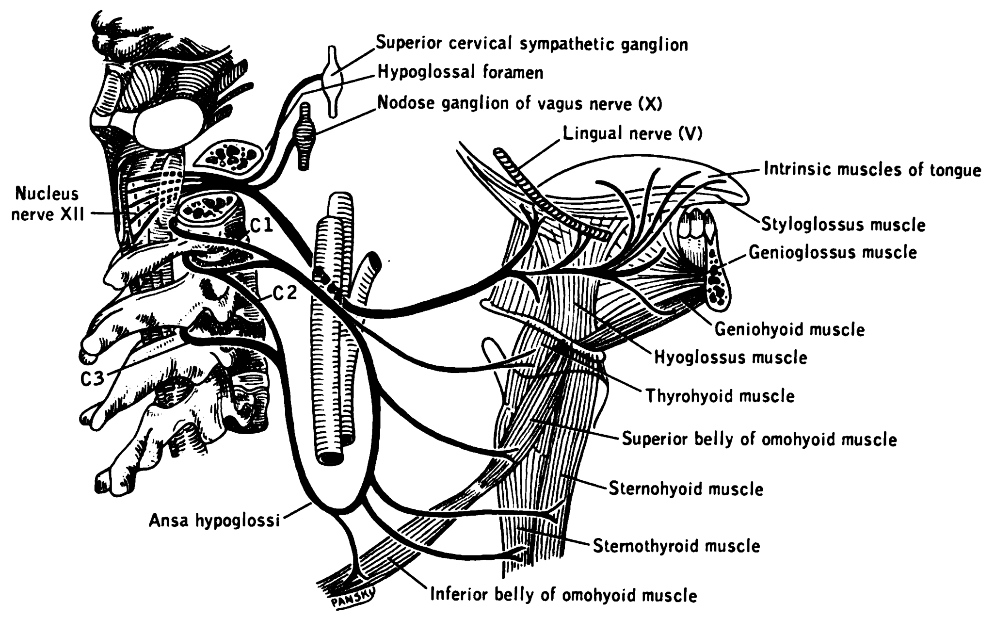
The hypoglossal nerve, also known as the twelfth cranial nerve, cranial nerve XII, or simply CN XII, is a cranial nerve that innervates all the extrinsic and intrinsic muscles of the tongue except for the palatoglossus, which is innervated by the vagus nerve. CN XII is a nerve with a sole motor function. The nerve arises from the hypoglossal nucleus in the medulla as a number of small rootlets, pass through the hypoglossal canal and down through the neck, and eventually passes up again over the tongue muscles it supplies into the tongue. The nerve is involved in controlling tongue movements required for speech and swallowing, including sticking out the tongue and moving it from side to side. Damage to the nerve or the neural pathways which control it can affect the ability of the tongue to move and its appearance, with the most common sources of damage being injury from trauma or surgery, and motor neuron disease. The first recorded description of the nerve was by Her ... [...More Info...] [...Related Items...] OR: [Wikipedia] [Google] [Baidu] |
Hypoglossal Canal
The hypoglossal canal is a foramen in the occipital bone of the skull. It is hidden medially and superiorly to each occipital condyle. It transmits the hypoglossal nerve. Structure The hypoglossal canal lies in the epiphyseal junction between the basiocciput and the jugular process of the occipital bone. Variation Embryonic variants sometimes lead to the presence of more than two canals as the occipital bone is formed. Development The hypoglossal canal is formed during the embryological stage of development in mammals. Function The hypoglossal canal transmits the hypoglossal nerve from its point of entry near the medulla oblongata to its exit from the base of the skull near the jugular foramen. Clinical significance Study of the hypoglossal canal aids in the diagnosis of a variety of tumors found at the base of the skull. Benign tumors involving the hypoglossal nerve and canal include large glomus jugulare neoplasms. Malignant tumors revolving around the hypogloss ... [...More Info...] [...Related Items...] OR: [Wikipedia] [Google] [Baidu] |
Occipital Sinus
The occipital sinus is the smallest of the dural venous sinuses. It is usually unpaired, and is sometimes altogether absent. It is situated in the attached margin of the falx cerebelli. It commences near the foramen magnum, and ends by draining into the confluence of sinuses. Occipital sinuses were discovered by Guichard Joseph Duverney. Anatomy The occipital sinus is present in around 65% of individuals. It is usually single, but occasionally paired. It is situated in the attached margin of the falx cerebelli. Course The occipital sinus commences around the margin of the foramen magnum by several small venous channels (one of which joins the terminal part of the sigmoid sinus The sigmoid sinuses (sigma- or s-shaped hollow curve), also known as the , are paired dural venous sinuses within the skull that receive blood from posterior transverse sinuses. Structure The sigmoid sinus is a dural venous sinus situated withi ...). It terminates by draining into the confluenc ... [...More Info...] [...Related Items...] OR: [Wikipedia] [Google] [Baidu] |
Inferior Petrosal Sinus
The inferior petrosal sinuses are two small sinuses situated on the inferior border of the petrous part of the temporal bone, one on each side. Each inferior petrosal sinus drains the cavernous sinus into the internal jugular vein. Structure The inferior petrosal sinus is situated in the inferior petrosal sulcus, formed by the junction of the petrous part of the temporal bone with the basilar part of the occipital bone. It begins below and behind the cavernous sinus and, passing through the anterior part of the jugular foramen, ends in the superior bulb of the internal jugular vein. Function The inferior petrosal sinus receives the internal auditory veins and also veins from the medulla oblongata, pons, and under surface of the cerebellum. Additional images File:Gray568.png, Sagittal section of the skull, showing the sinuses of the dura. See also * Dural venous sinuses The dural venous sinuses (also called dural sinuses, cerebral sinuses, or cranial sinuses) are veno ... [...More Info...] [...Related Items...] OR: [Wikipedia] [Google] [Baidu] |
Internal Jugular Vein
The internal jugular vein is a paired jugular vein that collects blood from the brain and the superficial parts of the face and neck. This vein runs in the carotid sheath with the common carotid artery and vagus nerve. It begins in the posterior compartment of the jugular foramen, at the base of the skull. It is somewhat dilated at its origin, which is called the ''superior bulb''. This vein also has a common trunk into which drains the anterior branch of the retromandibular vein, the facial vein, and the lingual vein. It runs down the side of the neck in a vertical direction, being at one end lateral to the internal carotid artery, and then lateral to the common carotid artery, and at the root of the neck, it unites with the subclavian vein to form the brachiocephalic vein (innominate vein); a little above its termination is a second dilation, the ''inferior bulb''. Above, it lies upon the rectus capitis lateralis, behind the internal carotid artery and the nerves pa ... [...More Info...] [...Related Items...] OR: [Wikipedia] [Google] [Baidu] |
Emissary Veins
The emissary veins connect the extracranial venous system with the intracranial venous sinuses. They connect the veins outside the cranium to the venous sinuses inside the cranium. They drain from the scalp, through the skull, into the larger meningeal veins and dural venous sinuses. They may also connect to diploic veins within the skull. Emissary veins have an important role in selective cooling of the head. They also serve as routes where infections are carried into the cranial cavity from the extracranial veins to the intracranial veins. There are several types of emissary veins including the posterior condyloid, mastoid, occipital and parietal emissary veins. Structure There are also emissary veins passing through the foramen ovale, jugular foramen, foramen lacerum, and hypoglossal canal. Function Because the emissary veins are valveless, they are an important part in selective brain cooling through bidirectional flow of cooler blood from the evaporating surface of t ... [...More Info...] [...Related Items...] OR: [Wikipedia] [Google] [Baidu] |
Case Report
In medicine, a case report is a detailed report of the symptoms, signs, diagnosis, treatment, and follow-up of an individual patient. Case reports may contain a demographic profile of the patient, but usually describe an unusual or novel occurrence. Some case reports also contain a literature review of other reported cases. Case reports are professional narratives that provide feedback on clinical practice guidelines and offer a framework for early signals of effectiveness, adverse events, and cost. They can be shared for medical, scientific, or educational purposes. Types Most case reports are on one of six topics: * An unexpected association between diseases or symptoms. * An unexpected event in the course of observing or treating a patient. * Findings that shed new light on the possible pathogenesis of a disease or an adverse effect. * Unique or rare features of a disease. * Unique therapeutic approaches. * A positional or quantitative variation of the anatomical structure ... [...More Info...] [...Related Items...] OR: [Wikipedia] [Google] [Baidu] |
Cerebellomedullary Cistern
The cisterna magna (posterior cerebellomedullary cistern, or cerebellomedullary cistern) is the largest of the subarachnoid cisterns. It occupies the space created by the angle between the caudal/inferior surface of the cerebellum, and the dorsal/posterior surface of the medulla oblongata (it is created by the arachnoidea that bridges this angle). The fourth ventricle communicates with the cistern via the unpaired midline median aperture. It is continuous inferiorly with the subarachnoid space of the spinal canal. The cisterna magna contains the two vertebral arteries, the origins of the two posterior inferior cerebellar arteries, the glossopharyngeal nerve (CN IX), vagus nerve (CN X), accessory nerve (CN XI), hypoglossal nerve (XII), and choroid plexus. The vertebral artery and posterior inferior cerebellar artery of either side pass traverse either lateral portion of the cistern. Etymology The ''Terminologia Anatomica'' classifies the terms ''cisterna magna'' and ''poste ... [...More Info...] [...Related Items...] OR: [Wikipedia] [Google] [Baidu] |
Saunders (imprint)
Saunders is an American academic publisher based in the United States. It is currently an imprint of Elsevier. Formerly independent, the W. B. Saunders company was acquired by CBS in 1968, who added it to their publishing division Holt, Rinehart & Winston. When CBS left the publishing field in 1986, it sold the academic publishing units to Harcourt Brace Jovanovich. Harcourt was acquired by Reed Elsevier in 2001. . Northern Illinois University Libraries. Retrieved May 2, 2015. W. B. Saunders published the Kinsey Reports
[...More Info...] [...Related Items...] OR: [Wikipedia] [Google] [Baidu] |

