|
Rectal
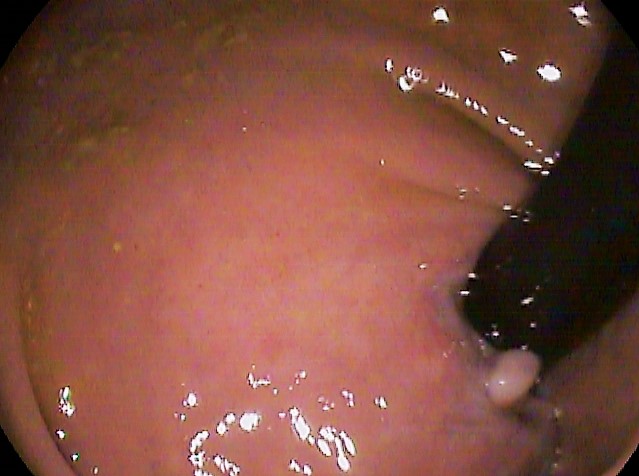
The rectum (: rectums or recta) is the final straight portion of the large intestine in humans and some other mammals, and the Gastrointestinal tract, gut in others. Before expulsion through the anus or cloaca, the rectum stores the feces temporarily. The adult human rectum is about long, and begins at the rectosigmoid junction (the end of the sigmoid colon) at the level of the third sacral vertebra or the sacral promontory depending upon what definition is used. Its diameter is similar to that of the sigmoid colon at its commencement, but it is dilated near its termination, forming the rectal ampulla. It terminates at the level of the anorectal ring (the level of the puborectalis sling) or the dentate line, again depending upon which definition is used. In humans, the rectum is followed by the anal canal, which is about long, before the gastrointestinal tract terminates at the anal verge. The word rectum comes from the Latin ''Wikt:rectum, rēctum Wikt:intestinum, intestīn ... [...More Info...] [...Related Items...] OR: [Wikipedia] [Google] [Baidu] [Amazon] |
Rectum
The rectum (: rectums or recta) is the final straight portion of the large intestine in humans and some other mammals, and the gut in others. Before expulsion through the anus or cloaca, the rectum stores the feces temporarily. The adult human rectum is about long, and begins at the rectosigmoid junction (the end of the sigmoid colon) at the level of the third sacral vertebra or the sacral promontory depending upon what definition is used. Its diameter is similar to that of the sigmoid colon at its commencement, but it is dilated near its termination, forming the rectal ampulla. It terminates at the level of the anorectal ring (the level of the puborectalis sling) or the dentate line, again depending upon which definition is used. In humans, the rectum is followed by the anal canal, which is about long, before the gastrointestinal tract terminates at the anal verge. The word rectum comes from the Latin '' rēctum intestīnum'', meaning ''straight intestine''. Struc ... [...More Info...] [...Related Items...] OR: [Wikipedia] [Google] [Baidu] [Amazon] |
Puborectalis
The levator ani is a broad, thin muscle group, situated on either side of the pelvis. It is formed from three muscle components: the pubococcygeus, the iliococcygeus, and the puborectalis. It is attached to the inner surface of each side of the lesser pelvis, and these unite to form the greater part of the pelvic floor. The coccygeus muscle completes the pelvic floor, which is also called the ''pelvic diaphragm''. It supports the viscera in the pelvic cavity, and surrounds the various structures that pass through it. The levator ani is the main pelvic floor muscle and contracts rhythmically during female orgasm, and painfully during vaginismus. Structure The levator ani is made up of 3 parts: * Iliococcygeus muscle * Pubococcygeus muscle * Puborectalis muscle The iliococcygeus arises from the inner side of the ischium (the lower and back part of the hip bone) and from the posterior part of the tendinous arch of the obturator fascia, and is attached to the coccyx and anoco ... [...More Info...] [...Related Items...] OR: [Wikipedia] [Google] [Baidu] [Amazon] |
Middle Rectal Artery
The middle rectal artery is an artery in the pelvis that supplies blood to the rectum. Structure The middle rectal artery usually arises from the internal iliac artery. It is distributed to the rectum above the pectinate line. It anastomoses with the inferior vesical artery, superior rectal artery, and inferior rectal artery. In males, the middle rectal artery may give off branches to the prostate and the seminal vesicles. In females, the middle rectal artery gives off branches to the vagina. Function The middle rectal artery supplies the rectum and the anal canal inferior to the pectinate line The pectinate line (dentate line) is a line which divides the upper two-thirds and lower third of the anal canal. Developmentally, this line represents the hindgut- proctodeum junction. It is an important anatomical landmark in humans, and fo .... Pathology The middle rectal artery may be embolized to treat patients with symptomatic internal hemorrhoids in a procedure cal ... [...More Info...] [...Related Items...] OR: [Wikipedia] [Google] [Baidu] [Amazon] |
Large Intestine
The large intestine, also known as the large bowel, is the last part of the gastrointestinal tract and of the Digestion, digestive system in tetrapods. Water is absorbed here and the remaining waste material is stored in the rectum as feces before being removed by defecation. The Colon (anatomy), colon (progressing from the ascending colon to the transverse colon, transverse, the descending colon, descending and finally the sigmoid colon) is the longest portion of the large intestine, and the terms "large intestine" and "colon" are often used interchangeably, but most sources define the large intestine as the combination of the cecum, colon, rectum, and anal canal. Some other sources exclude the anal canal. In humans, the large intestine begins in the right iliac region of the pelvis, just at or below the waist, where it is joined to the end of the small intestine at the cecum, via the ileocecal valve. It then continues as the colon ascending colon, ascending the abdomen, across t ... [...More Info...] [...Related Items...] OR: [Wikipedia] [Google] [Baidu] [Amazon] |
Dentate Line
The pectinate line (dentate line) is a line which divides the upper two-thirds and lower third of the anal canal. Developmentally, this line represents the hindgut- proctodeum junction. It is an important anatomical landmark in humans, and forms the boundary between the anal canal and the rectum according to the anatomic definition. Colorectal surgeons instead define the anal canal as the zone from the anal verge to the anorectal ring (palpable structure formed by the external anal sphincter and the puborectalis muscle The levator ani is a broad, thin muscle group, situated on either side of the pelvis. It is formed from three muscle components: the pubococcygeus, the iliococcygeus, and the puborectalis. It is attached to the inner surface of each side of the ...). Several distinctions can be made based upon the location of a structure relative to the pectinate line: Additional images File:Rectoanal jxn.JPG, Microscopic cross section of the anorectal junction File:A ... [...More Info...] [...Related Items...] OR: [Wikipedia] [Google] [Baidu] [Amazon] |
Superior Rectal Artery
The superior rectal artery (superior hemorrhoidal artery) is an artery that descends into the pelvis to supply blood to the rectum. Structure The superior rectal artery is the continuation of the inferior mesenteric artery. It descends into the pelvis between the layers of the mesentery of the sigmoid colon, crossing the left common iliac artery and vein. It divides, opposite the third sacral vertebra into two branches, which descend one on either side of the rectum. About 10 or 12 cm from the anus, these branches break up into several small branches. These pierce the muscular coat of the bowel and run downward, as straight vessels, placed at regular intervals from each other in the wall of the gut between its muscular and mucous coats, to the level of the internal anal sphincter; here they form a series of loops around the lower end of the rectum, and communicate with the middle rectal artery (from the internal iliac artery) and with the inferior rectal artery (from ... [...More Info...] [...Related Items...] OR: [Wikipedia] [Google] [Baidu] [Amazon] |
Middle Rectal Veins
The middle rectal veins (or middle hemorrhoidal vein) take origin in the hemorrhoidal plexus and receive tributaries from the bladder, prostate, and seminal vesicle. They run lateralward on the pelvic surface of the levator ani The levator ani is a broad, thin muscle group, situated on either side of the pelvis. It is formed from three muscle components: the pubococcygeus, the iliococcygeus, and the puborectalis. It is attached to the inner surface of each side of the ... to end in the internal iliac vein. Veins superior to the middle rectal vein in the colon and rectum drain via the portal system to the liver. Veins inferior, and including, the middle rectal vein drain into systemic circulation and are returned to the heart, bypassing the liver. References Veins of the torso {{circulatory-stub ... [...More Info...] [...Related Items...] OR: [Wikipedia] [Google] [Baidu] [Amazon] |
Anal Canal
The anal canal is the part that connects the rectum to the anus, located below the level of the pelvic diaphragm. It is located within the anal triangle of the perineum, between the right and left ischioanal fossa. As the final functional segment of the bowel, it functions to regulate release of excrement by two muscular sphincter complexes. The anus is the aperture at the terminal portion of the anal canal. Structure In humans, the anal canal is approximately long, from the anorectal junction to the anus. It is directed downwards and backwards. It is surrounded by inner involuntary and outer voluntary sphincters which keep the lumen closed in the form of an anteroposterior slit. The canal is differentiated from the rectum by a transition along the internal surface from endodermal to skin-like ectodermal tissue. The anal canal is traditionally divided into two segments, upper and lower, separated by the pectinate line (also known as the dentate line): * upper zo ... [...More Info...] [...Related Items...] OR: [Wikipedia] [Google] [Baidu] [Amazon] |
Anal Verge
The anal canal is the part that connects the rectum to the anus, located below the level of the pelvic diaphragm. It is located within the anal triangle of the perineum, between the right and left ischioanal fossa. As the final functional segment of the bowel, it functions to regulate release of excrement by two muscular sphincter complexes. The anus is the aperture at the terminal portion of the anal canal. Structure In humans, the anal canal is approximately long, from the anorectal junction to the anus. It is directed downwards and backwards. It is surrounded by inner involuntary and outer voluntary sphincters which keep the lumen closed in the form of an anteroposterior slit. The canal is differentiated from the rectum by a transition along the internal surface from endodermal to skin-like ectodermal tissue. The anal canal is traditionally divided into two segments, upper and lower, separated by the pectinate line (also known as the dentate line): * upper zone (zona c ... [...More Info...] [...Related Items...] OR: [Wikipedia] [Google] [Baidu] [Amazon] |
Defecation
Defecation (or defaecation) follows digestion and is the necessary biological process by which organisms eliminate a solid, semisolid, or liquid metabolic waste, waste material known as feces (or faeces) from the digestive tract via the anus or cloaca. The act has a variety of names, ranging from the technical (e.g. bowel movement), to the common (like pooping or crapping), to the obscene (''Shit, shitting''), to the euphemistic ("doing number two", "dropping a deuce" or "taking a dump"), to the juvenile ("going poo-poo" or "making doo-doo"). The topic, usually avoided in polite company, forms the basis of scatological humor. human feces, Humans expel feces with a frequency varying from a few times daily to a few times weekly. Waves of muscle, muscular contraction (known as ''peristalsis'') in the walls of the colon (anatomy), colon move fecal matter through the digestive tract towards the rectum. Flatus may also be expulsed. Undigested food may also be expelled within the fec ... [...More Info...] [...Related Items...] OR: [Wikipedia] [Google] [Baidu] [Amazon] |
Superior Rectal Veins
The inferior mesenteric vein begins in the rectum as the superior rectal vein (superior hemorrhoidal vein), which has its origin in the hemorrhoidal plexus, and through this plexus communicates with the middle and inferior hemorrhoidal veins. The superior rectal vein leaves the lesser pelvis and crosses the left common iliac vessels with the superior rectal artery, and is continued upward as the inferior mesenteric vein In human anatomy, the inferior mesenteric vein (IMV) is a blood vessel that drains blood from the large intestine. It usually terminates when reaching the splenic vein, which goes on to form the portal vein with the superior mesenteric vein (SMV) .... References Veins of the torso Rectum {{circulatory-stub ... [...More Info...] [...Related Items...] OR: [Wikipedia] [Google] [Baidu] [Amazon] |


