|
Pulmonary Artery Agenesis
Pulmonary artery agenesis refers to a rare congenital absence of pulmonary artery due to a malformation in the sixth aortic arch. It can occur bilaterally, with both left and right pulmonary arteries being absent, or unilaterally, the absence of either left or right pulmonary artery (UAPA). About 67% of UAPA occurs isolated in the right lung. The absence of pulmonary artery can be an isolated disorder, or accompanied by other related lesions, most commonly Tetralogy of Fallot. Back in 1868, Fraentzel was the first to report isolated unilateral absence of pulmonary artery (IUAPA) in literature. Subsequently, literature has documented a total of 420 cases. The estimated prevalence of IUAPA is 1 in 200,000 adults. No sex preference is observed. Patients with severe complications are usually diagnosed early in age while adult patients are mainly asymptomatic. The overall mortality rate reaches 7%. Individuals may exhibit a variety of symptoms, or they may not exhibit any symptoms at a ... [...More Info...] [...Related Items...] OR: [Wikipedia] [Google] [Baidu] [Amazon] |
Respiratory Failure
Respiratory failure results from inadequate gas exchange by the respiratory system, meaning that the arterial oxygen, carbon dioxide, or both cannot be kept at normal levels. A drop in the oxygen carried in the blood is known as hypoxemia; a rise in arterial carbon dioxide levels is called hypercapnia. Respiratory failure is classified as either Type 1 or Type 2, based on whether there is a high carbon dioxide level, and can be acute or chronic. In clinical trials, the definition of respiratory failure usually includes increased respiratory rate, abnormal blood gases (hypoxemia, hypercapnia, or both), and evidence of increased work of breathing. Respiratory failure causes an altered state of consciousness due to ischemia in the brain. The typical partial pressure reference values are oxygen Pa more than 80 mmHg (11 kPa) and carbon dioxide Pa less than 45 mmHg (6.0 kPa). Cause A variety of conditions that can potentially result in respiratory failure. The etiologie ... [...More Info...] [...Related Items...] OR: [Wikipedia] [Google] [Baidu] [Amazon] |
Trachea
The trachea (: tracheae or tracheas), also known as the windpipe, is a cartilaginous tube that connects the larynx to the bronchi of the lungs, allowing the passage of air, and so is present in almost all animals' lungs. The trachea extends from the larynx and branches into the two primary bronchi. At the top of the trachea, the cricoid cartilage attaches it to the larynx. The trachea is formed by a number of horseshoe-shaped rings, joined together vertically by overlying annular ligaments of trachea, ligaments, and by the trachealis muscle at their ends. The epiglottis closes the opening to the larynx during swallowing. The trachea begins to form in the second month of embryo development, becoming longer and more fixed in its position over time. Its epithelium is lined with columnar epithelium, column-shaped cells that have hair-like extensions called cilia, with scattered goblet cells that produce protective mucins. The trachea can be affected by inflammation or infection, usua ... [...More Info...] [...Related Items...] OR: [Wikipedia] [Google] [Baidu] [Amazon] |
Mediastinum
The mediastinum (from ;: mediastina) is the central compartment of the thoracic cavity. Surrounded by loose connective tissue, it is a region that contains vital organs and structures within the thorax, mainly the heart and its vessels, the esophagus, the trachea, the vagus nerve, vagus, phrenic nerve, phrenic and cardiac nerves, the thoracic duct, the thymus and the lymph nodes of the central chest. Anatomy The mediastinum lies within the thorax and is enclosed on the right and left by pulmonary pleurae, pleurae. It is surrounded by the chest wall in front, the lungs to the sides and the Spine (anatomy), spine at the back. It extends from the sternum in front to the vertebral column behind. It contains all the organs of the thorax except the lungs. It is continuous with the loose connective tissue of the neck. The mediastinum can be divided into an upper (or superior) and lower (or inferior) part: * The superior mediastinum starts at the superior thoracic aperture and ends ... [...More Info...] [...Related Items...] OR: [Wikipedia] [Google] [Baidu] [Amazon] |
Thorax
The thorax (: thoraces or thoraxes) or chest is a part of the anatomy of mammals and other tetrapod animals located between the neck and the abdomen. In insects, crustaceans, and the extinct trilobites, the thorax is one of the three main divisions of the body, each in turn composed of multiple segments. The human thorax includes the thoracic cavity and the thoracic wall. It contains organs including the heart, lungs, and thymus gland, as well as muscles and various other internal structures. The chest may be affected by many diseases, of which the most common symptom is chest pain. Etymology The word thorax comes from the Greek θώραξ ''thṓrax'' " breastplate, cuirass, corslet" via . Humans Structure In humans and other hominids, the thorax is the chest region of the body between the neck and the abdomen, along with its internal organs and other contents. It is mostly protected and supported by the rib cage, spine, and shoulder girdle. Contents The ... [...More Info...] [...Related Items...] OR: [Wikipedia] [Google] [Baidu] [Amazon] |
Chest Radiograph
A chest radiograph, chest X-ray (CXR), or chest film is a projection radiograph of the chest used to diagnose conditions affecting the chest, its contents, and nearby structures. Chest radiographs are the most common film taken in medicine. Like all methods of radiography, chest radiography employs ionizing radiation in the form of X-rays to generate images of the chest. The mean radiation dose to an adult from a chest radiograph is around 0.02 mSv (2 mrem) for a front view (PA, or posteroanterior) and 0.08 mSv (8 mrem) for a side view (LL, or latero-lateral). Together, this corresponds to a background radiation equivalent time of about 10 days. Medical uses Conditions commonly identified by chest radiography * Pneumonia * Pneumothorax * Interstitial lung disease * Heart failure * Bone fracture * Hiatal hernia * Pulmonary tuberculosis Chest radiographs are used to diagnose many conditions involving the chest wall, including its bones, and also structures contained ... [...More Info...] [...Related Items...] OR: [Wikipedia] [Google] [Baidu] [Amazon] |
Electrocardiography
Electrocardiography is the process of producing an electrocardiogram (ECG or EKG), a recording of the heart's electrical activity through repeated cardiac cycles. It is an electrogram of the heart which is a graph of voltage versus time of the electrical activity of the heart using electrodes placed on the skin. These electrodes detect the small electrical changes that are a consequence of cardiac muscle depolarization followed by repolarization during each cardiac cycle (heartbeat). Changes in the normal ECG pattern occur in numerous cardiac abnormalities, including: * Cardiac rhythmicity, Cardiac rhythm disturbances, such as atrial fibrillation and ventricular tachycardia; * Inadequate coronary artery blood flow, such as myocardial ischemia and myocardial infarction; * and electrolyte disturbances, such as hypokalemia. Traditionally, "ECG" usually means a 12-lead ECG taken while lying down as discussed below. However, other devices can record the electrical activity of ... [...More Info...] [...Related Items...] OR: [Wikipedia] [Google] [Baidu] [Amazon] |
Circulatory System
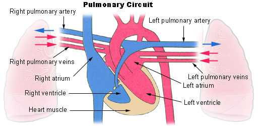
In vertebrates, the circulatory system is a system of organs that includes the heart, blood vessels, and blood which is circulated throughout the body. It includes the cardiovascular system, or vascular system, that consists of the heart and blood vessels (from Greek meaning ''heart'', and Latin meaning ''vessels''). The circulatory system has two divisions, a systemic circulation or circuit, and a pulmonary circulation or circuit. Some sources use the terms ''cardiovascular system'' and ''vascular system'' interchangeably with ''circulatory system''. The network of blood vessels are the great vessels of the heart including large elastic arteries, and large veins; other arteries, smaller arterioles, capillaries that join with venules (small veins), and other veins. The circulatory system is closed in vertebrates, which means that the blood never leaves the network of blood vessels. Many invertebrates such as arthropods have an open circulatory system with a he ... [...More Info...] [...Related Items...] OR: [Wikipedia] [Google] [Baidu] [Amazon] |
Arteriole
An arteriole is a small-diameter blood vessel in the microcirculation that extends and branches out from an artery and leads to capillary, capillaries. Arterioles have vascular smooth muscle, muscular walls (usually only one to two layers of smooth muscle cells) and are the primary site of vascular resistance. The greatest change in blood pressure and velocity of blood flow occurs at the transition of arterioles to capillaries. This function is extremely important because it prevents the thin, one-layer capillaries from exploding upon pressure. The arterioles achieve this decrease in pressure, as they are the site with the highest resistance (a large contributor to total peripheral resistance) which translates to a large decrease in the pressure. Structure In a healthy vascular system, the endothelium lines all blood-contacting surfaces, including arteries, arterioles, veins, venules, capillaries, and heart chambers. This healthy condition is promoted by the ample production of n ... [...More Info...] [...Related Items...] OR: [Wikipedia] [Google] [Baidu] [Amazon] |
Endothelin 1
Endothelin 1 (ET-1), also known as preproendothelin-1 (PPET1), is a potent vasoconstrictor peptide produced by vascular endothelial cells, as well as by cells in the heart (affecting contractility) and kidney (affecting sodium handling). The protein encoded by this gene ''EDN1'' is proteolytically processed to release endothelin 1. Endothelin 1 is one of three isoforms of human endothelin. Sources Preproendothelin is precursor of the peptide ET-1. Endothelial cells convert preproendothelin to proendothelin and subsequently to mature endothelin, which the cells release. Clinical significance Patients with salt-sensitive hypertension have higher plasma ET-1. Endothelin-1 receptor antagonists (Bosentan) are used in the treatment of pulmonary hypertension. Use of these antagonists prevents pulmonary arterial constriction and thus inhibits pulmonary hypertension. As of 2020, the role of endothelin-1 in affecting lipid metabolism and insulin resistance in obesity mechanisms wa ... [...More Info...] [...Related Items...] OR: [Wikipedia] [Google] [Baidu] [Amazon] |
Vasoconstriction
Vasoconstriction is the narrowing of the blood vessels resulting from contraction of the muscular wall of the vessels, in particular the large arteries and small arterioles. The process is the opposite of vasodilation, the widening of blood vessels. The process is particularly important in controlling hemorrhage and reducing acute blood loss. When blood vessels constrict, the flow of blood is restricted or decreased, thus retaining body heat or increasing vascular resistance. This makes the skin turn paler because less blood reaches the surface, reducing the radiation of heat. On a larger level, vasoconstriction is one mechanism by which the body regulates and maintains mean arterial pressure. Medications causing vasoconstriction, also known as vasoconstrictors, are one type of medicine used to raise blood pressure. Generalized vasoconstriction usually results in an increase in systemic blood pressure, but it may also occur in specific tissues, causing a localized reduction in b ... [...More Info...] [...Related Items...] OR: [Wikipedia] [Google] [Baidu] [Amazon] |


